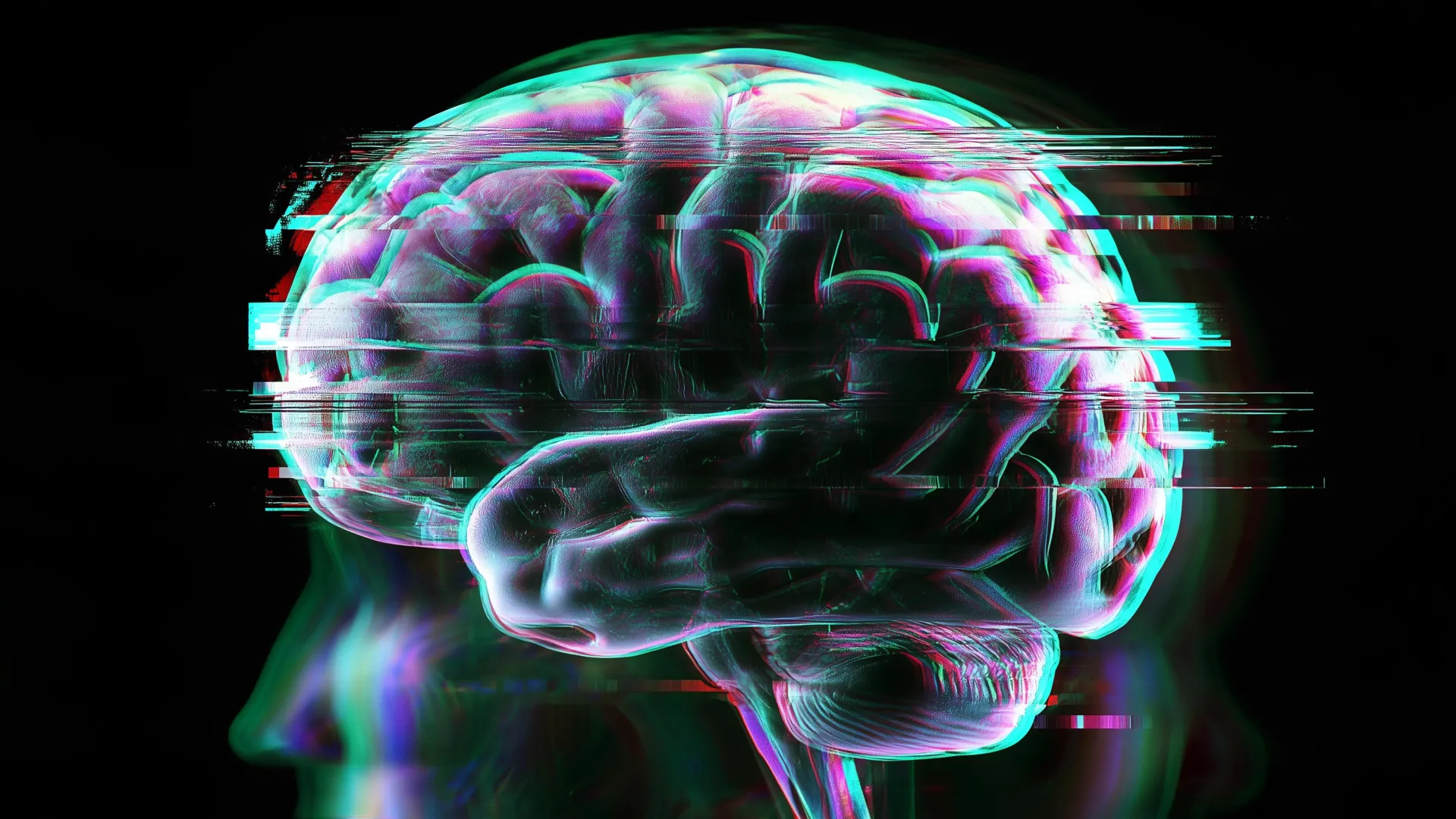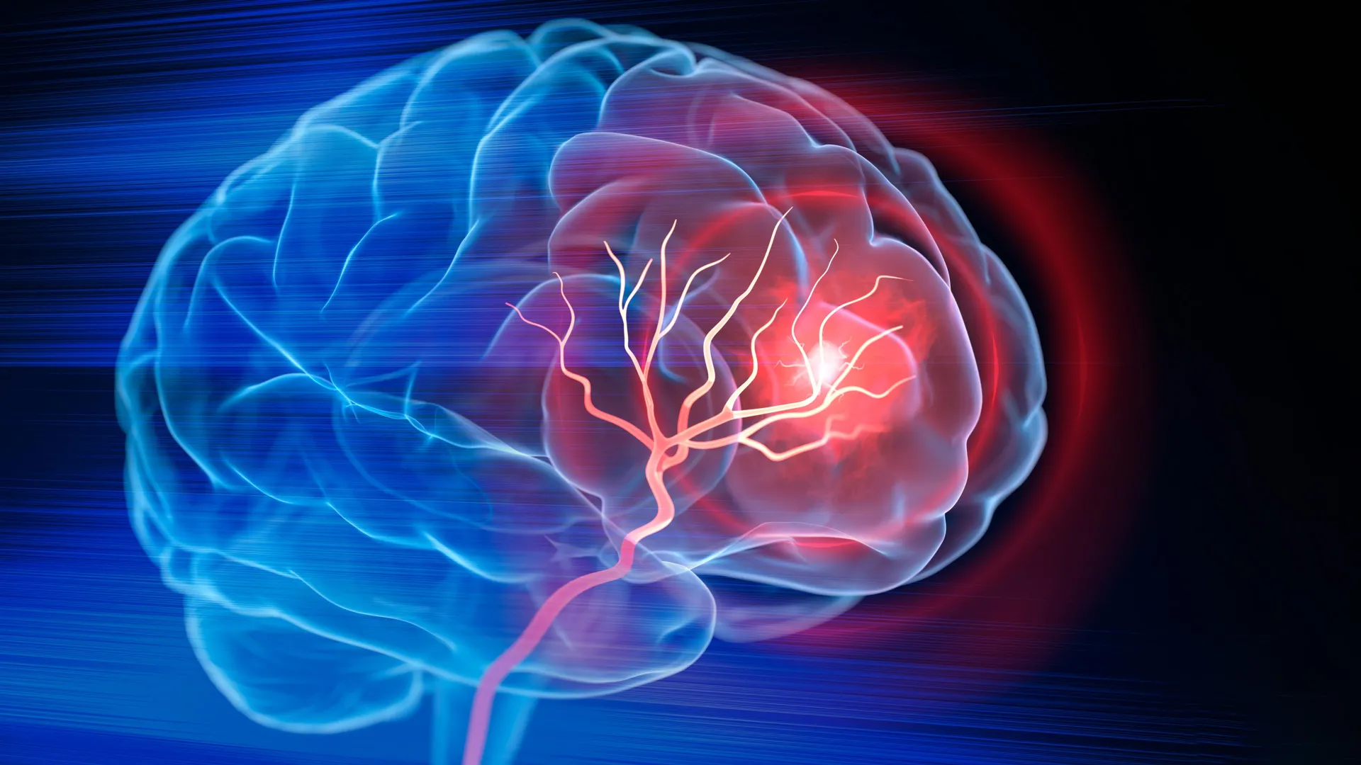
The ability to focus on a single task while ignoring a multitude of environmental distractions is a fundamental survival mechanism, yet the neurological origins of this process have long remained a subject of intense scientific debate. Researchers at Johns Hopkins University have recently published a landmark study in the journal Nature Communications, identifying a specific cluster of neurons located in the brainstem—an evolutionarily ancient region of the brain—that acts as a primary filter for selective spatial attention. This discovery challenges the long-held scientific consensus that high-level cognitive focus is almost exclusively the domain of the prefrontal cortex, the most recently evolved part of the brain. By pinpointing these neurons in mice, scientists have uncovered a conserved vertebrate system that likely exists in humans, offering a potential breakthrough in the understanding and treatment of neurodevelopmental conditions such as Attention-Deficit/Hyperactivity Disorder (ADHD) and Autism Spectrum Disorder (ASD).
The Evolutionary Mystery of Attention
For decades, the prevailing theory in neuroscience suggested that the prefrontal cortex was the "commander-in-chief" of attention. Because the prefrontal cortex is exceptionally well-developed in humans and primates, it was assumed that complex tasks, such as filtering out background noise or focusing on a specific visual target in a crowded environment, were products of this advanced neural architecture. However, this theory faced a significant logical hurdle: many "lower" vertebrates, including fish, birds, and reptiles, exhibit sophisticated attentional capabilities despite lacking a highly developed prefrontal cortex.
Lead author Ninad Kothari, a postdoctoral fellow in the Johns Hopkins University Department of Psychological and Brain Sciences, noted that the ability to focus must have much deeper evolutionary roots. The research team hypothesized that if birds and fish can selectively attend to prey or predators amidst chaos, the mechanism for doing so must reside in a more primitive part of the brain shared across the vertebrate lineage. Their investigation led them to the brainstem, a structure that regulates basic life-sustaining functions such as heart rate and breathing, but which now appears to house the fundamental "engine" of cognitive selection.
Experimental Methodology: Testing the Focus Filter
To test their hypothesis, the research team designed a rigorous behavioral experiment using mice. The study sought to replicate the "selective spatial attention" tasks often used in human psychology trials. Mice were placed in a controlled environment where they were required to respond to visual cues displayed on a screen directly in front of them. When the mice correctly identified and responded to these primary cues, they were rewarded.
The complexity of the task was increased by introducing "distractors"—visual signals that appeared off to the side or in the periphery. A mouse with a functioning attentional system would ignore these peripheral flashes and remain focused on the central task. To isolate the role of the brainstem neurons, the researchers utilized advanced neuro-engineering techniques to temporarily "silence" or inactivate this specific group of inhibitory neurons.
The results were immediate and profound. When the neurons were active, the mice performed with high precision, successfully filtering out even intense distractions. However, once the neurons were chemically or light-inactivated, the mice became "hyper-distractable." Even the faintest, most irrelevant peripheral flicker would cause the animals to lose focus on the primary task. Interestingly, the researchers observed that when the neurons were "turned back on" the following day, the mice regained their full capacity for focus, proving that the neurons were the direct cause of the attentional control.
The Role of Inhibitory Neurons as a Selection Engine
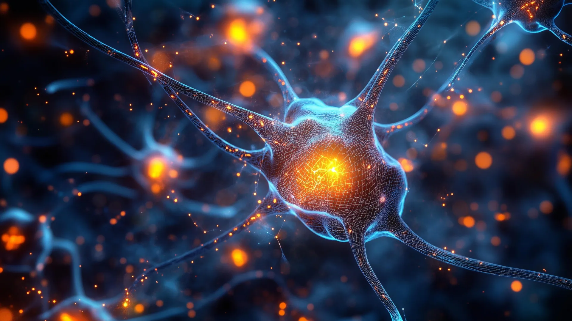
The specific cells identified in the study are inhibitory neurons. In the context of the brain, inhibitory neurons do not "excite" or trigger activity; rather, they suppress it. This is a critical distinction in understanding how attention works. Instead of simply "turning up the volume" on important information, these brainstem neurons work by "muting" the noise.
Senior author Shreesh Mysore, a neuroscientist specializing in neural circuits and behavior, described this region as an "attentional selection engine." The brain is constantly bombarded by more sensory data than it can process simultaneously—visual movements, sounds, physical sensations, and internal thoughts. The brainstem’s inhibitory network acts as a gatekeeper, comparing the strength and relevance of various signals. When it identifies the most important information, it suppresses the competing signals, allowing the higher-order regions of the brain to process the priority data without interference.
This mechanism explains how a person can maintain a conversation in a crowded restaurant (the "cocktail party effect") or how a predator can track a single animal in a moving herd. By silencing the "distractors," the brainstem provides a clear channel for the cortex to operate.
Supporting Data and Chronology of Research
The Johns Hopkins study is the culmination of years of cross-species research. The timeline of this discovery began with earlier observations in non-mammalian species. Dr. Mysore and his colleagues had previously investigated similar neural circuits in birds, frogs, and turtles. These animals demonstrated a remarkable ability to prioritize sensory input, which pointed toward a subcortical (below the cortex) mechanism.
The transition to studying mice was a pivotal step in the research chronology. Because mice are mammals, their brain structure is more closely aligned with that of humans, yet they still retain the ancient brainstem architecture found in earlier vertebrates. The study’s findings, which were federally funded and subsequently selected as an "editorial highlight" by Nature Communications, provide the most definitive evidence to date that the brainstem is not merely a relay station for sensory data, but an active processor of cognitive priority.
Data from the experiments ruled out other potential causes for the mice’s failure during neuron inactivation. The researchers conducted control tests to ensure the mice weren’t suffering from impaired vision or motor coordination. The mice could still see the cues and move their bodies perfectly; they simply could no longer "choose" what to look at. This data confirmed that the impairment was purely cognitive and specific to the process of selective attention.
Clinical Implications for ADHD and Autism
The discovery of this "ancient" attention center has significant implications for the medical community’s approach to neurodevelopmental disorders. Currently, Attention-Deficit/Hyperactivity Disorder (ADHD) is often treated with medications that affect broad neurotransmitter systems, such as dopamine and norepinephrine, across the entire brain. While effective for many, these treatments can have widespread side effects and do not work for all patients.
"A hallmark of ADHD is that even faint distractors draw attention away," Mysore explained. The behavior of the mice with silenced brainstem neurons provided a near-perfect mirror of the clinical symptoms observed in humans with ADHD. If the same neurons are responsible for attention in humans, it suggests that ADHD may not be solely a "frontal lobe" disorder, but may involve malfunctions in the brainstem’s filtering mechanism.
Similarly, individuals on the autism spectrum often experience sensory overload, where the brain is unable to filter out background stimuli, leading to distress and difficulty focusing. If the brainstem’s "selection engine" is underactive or miswired, the result would be a flood of competing information that the conscious mind cannot manage.
The identification of a specific, localized group of neurons opens the door for the development of "precision medicine." Future therapies could potentially target these specific brainstem circuits, either through more localized pharmacological agents or through non-invasive neuromodulation techniques, providing relief for those who do not respond to traditional ADHD or autism therapies.
Analysis of Broader Impacts on Neuroscience
The shift in focus from the prefrontal cortex to the brainstem represents a paradigm shift in the field of neuroscience. It suggests that the "higher" functions we associate with being human—such as focus, willpower, and cognitive selection—are actually built upon a foundation that has been refined over hundreds of millions of years of evolution.
This "bottom-up" model of attention suggests that the prefrontal cortex might act more like a fine-tuner or a director, while the brainstem provides the essential machinery. Without the brainstem’s filter, the prefrontal cortex is overwhelmed by data. This understanding could lead to a re-evaluation of how other cognitive functions, such as memory and decision-making, are integrated across different levels of the brain’s evolutionary hierarchy.
Furthermore, the study highlights the importance of comparative biology. By looking at what humans share with fish and birds, researchers were able to find a solution to a problem that had remained elusive when looking at the human brain in isolation.
Conclusion and Future Directions
The research team at Johns Hopkins, which includes contributors Arunima Banerjee, Qingcheng (Jessica) Zhang, and Wen-Kai You, plans to move forward by investigating whether these neurons function identically in humans. While the biological evidence strongly suggests these cells are present in the human brainstem, confirming their role in human selective attention will require sophisticated imaging and clinical studies.
"All the evidence to date suggests that these neurons exist in humans too," said Mysore. "But are they responsible for selective spatial attention in humans? An exciting hypothesis is that they play a crucial role."
Upcoming research may involve studying the activity of these brainstem circuits in patients with diagnosed attention disorders. If a correlation is found between neuron dysfunction and symptom severity, it would validate the brainstem as a primary site for clinical intervention. For now, the discovery serves as a powerful reminder that to understand the complexities of the modern human mind, science must sometimes look back into our ancient biological past.