A groundbreaking study led by researchers at Baylor College of Medicine has fundamentally challenged the traditional understanding of the human brain’s activity during states of deep unconsciousness. Published in the journal Nature, the research demonstrates that the human brain can continue to perform sophisticated language tasks, including the identification of specific parts of speech and the prediction of upcoming words, even when a patient is under the influence of clinical-grade general anesthesia. The findings suggest that the boundary between the conscious and unconscious mind is far more porous than previously believed, opening new avenues for the development of neural prosthetics and the treatment of consciousness disorders.
For decades, the prevailing medical consensus was that general anesthesia acted as a "functional disconnect," effectively silencing the higher-order cognitive processes responsible for language and complex perception. While it was known that the brain could still respond to basic sensory stimuli—such as a loud noise or a physical touch—the capacity for the brain to parse the nuances of human grammar and anticipate narrative flow was thought to be exclusively a feature of the conscious, waking state. The Baylor team’s discovery reveals that the hippocampus, a region primarily associated with memory formation, remains highly active and linguistically capable during periods of induced unconsciousness.
Advanced Methodology: High-Resolution Recording in the Hippocampus
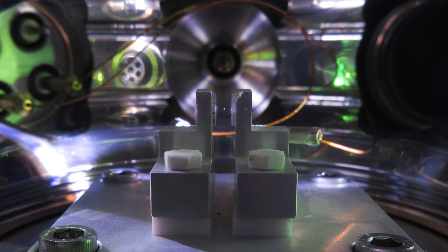
The research was made possible through a rare clinical opportunity involving patients undergoing surgical treatment for epilepsy. Because these patients already required the implantation of electrodes to locate the source of their seizures, researchers were able to utilize Neuropixels probes—a sophisticated neurotechnology capable of recording the activity of hundreds of individual neurons simultaneously. This study marks one of the first instances where such high-resolution technology was deployed within the human hippocampus to observe the effects of anesthesia.
Dr. Sameer Sheth, professor and Cullen Foundation Endowed chair of neurosurgery at Baylor and a McNair Scholar, emphasized the significance of the technological approach. According to Dr. Sheth, the ability to observe individual neural firing patterns allowed the team to see that the brain is far more active and capable during unconsciousness than previously thought. Even when patients were fully anesthetized and unresponsive to external commands, their hippocampal neurons continued to analyze the linguistic structure of the environment.
The patients were administered propofol, a common general anesthetic that induces a state of deep sedation. While in this state, they were exposed to two distinct auditory experiments. The first was a simple "oddball" paradigm, where a series of repeating tones was interrupted by occasional, unexpected sounds. The second, more complex experiment involved playing short narrative stories to the anesthetized patients while the Neuropixels probes captured the real-time response of the brain.
Evidence of Neural Plasticity and Predictive Coding
In the initial tone experiment, the researchers observed that neurons in the hippocampus consistently fired in response to the unexpected "oddball" tones. More importantly, the neural response to these tones evolved over the course of the session. As the patients were exposed to more sequences, the hippocampus became more efficient at recognizing the patterns, suggesting that neural plasticity—the brain’s ability to learn and adapt—remains functional even under general anesthesia.
The second phase of the study, involving narrative stories, yielded even more startling results. The researchers used machine learning algorithms to decode the neural signals and found that the hippocampus was distinguishing between different linguistic categories. The brain activity patterns differed significantly depending on whether the patient was hearing a noun, a verb, or an adjective. This indicates that the unconscious brain is not merely "hearing" sound, but is actively categorizing the grammatical components of language.
Furthermore, the study identified a phenomenon known as "predictive coding." In a waking state, the human brain constantly tries to stay ahead of the environment by predicting what will happen next. In the context of language, this means the brain anticipates the next word in a sentence based on context. The Baylor study found that even under anesthesia, the hippocampus generated signals that could predict upcoming words before they were actually played in the audio recording.
Dr. Benjamin Hayden, professor of neurosurgery at Baylor, noted that this type of predictive coding is traditionally associated with being awake and attentive. The fact that it occurs during a state of deep anesthesia suggests that prediction is a fundamental, perhaps automatic, operation of the mammalian brain that does not require the "light" of conscious awareness.
Parallelisms with Artificial Intelligence
The researchers drew a direct comparison between the brain’s unconscious predictive capabilities and the architecture of modern artificial intelligence, specifically Large Language Models (LLMs) like those used in chatbots. These AI systems function by predicting the most statistically likely next word in a sequence. The discovery that the human hippocampus performs a similar statistical prediction while the "self" is unconscious provides a biological parallel to how AI processes information.
This comparison has significant implications for both neuroscience and computer science. By understanding how the biological brain handles language prediction without consciousness, developers may be able to refine AI models to be more efficient. Conversely, the mathematical frameworks used to build AI are now providing neuroscientists with new tools to interpret the complex firing patterns of the human brain.
Clinical Implications for Speech Prosthetics
One of the most promising applications of this research lies in the field of medical technology, specifically for patients who have lost the ability to speak due to stroke, traumatic brain injury, or neurodegenerative diseases like ALS (Amyotrophic Lateral Sclerosis). Current speech prosthetics often rely on recording activity from the motor cortex—the part of the brain that controls the muscles of the mouth and throat. However, if the hippocampus is shown to be a robust center for language processing and prediction, it could serve as a secondary or even primary source of data for brain-computer interfaces (BCIs).
Dr. Vigi Katlowitz, a neurosurgery resident at Baylor and the study’s first author, raised the possibility of using these hippocampal signals to run speech prosthetics. If the brain is already "pre-calculating" language in the hippocampus, a BCI could potentially tap into those signals to help a non-verbal patient communicate their thoughts more fluidly and accurately.
Rethinking the Nature of Consciousness
The study’s findings contribute to a larger debate within the scientific community regarding the "Global Neuronal Workspace" and "Integrated Information Theory." Many theories of consciousness posit that for a thought to become "conscious," it must be broadcast across a wide network of brain regions. The Baylor study supports a nuanced version of this idea: while the hippocampus can process and predict language locally, this information remains "unconscious" because it is not being integrated or shared with the broader cortical network during anesthesia.
This suggests that consciousness might not be a "switch" that turns the entire brain on or off, but rather a state of high-level connectivity. Under anesthesia, the individual "processors" of the brain—like the hippocampus—might still be running their "software," but the central network that allows us to experience those processes is disconnected.
Limitations and Future Research Directions
Despite the breakthrough, the research team has urged a cautious interpretation of the data. The study focused specifically on the hippocampus and utilized only one type of anesthetic agent (propofol). It is currently unknown if the same level of language processing occurs in other brain regions or if different types of anesthesia, such as volatile gases or ketamine, would yield different results.
Furthermore, the study does not suggest that patients are "aware" of what is happening during surgery. There is a vital distinction between "neural processing" and "subjective experience." While the brain is analyzing the words, the patient does not "feel" or "remember" the experience in a way that constitutes awareness or trauma.
Future research will likely expand to other areas of the brain and examine different states of altered consciousness, such as natural sleep, comas, and vegetative states. Understanding the extent of unconscious processing could help doctors better assess the recovery potential of patients with severe brain injuries.
Conclusion
The Baylor College of Medicine study serves as a landmark in the field of neurobiology, proving that the unconscious mind is far more sophisticated than previously imagined. By demonstrating that the hippocampus continues to parse grammar and predict speech during general anesthesia, the researchers have redefined the functional boundaries of the human brain.
As Dr. Sheth concluded, the work pushes the scientific community to rethink the very definition of consciousness. The revelation that the brain continues its complex "behind the scenes" work during anesthesia not only provides a deeper understanding of human biology but also sets the stage for a new generation of neural technologies that could one day restore the power of communication to those who have lost it. The study stands as a testament to the hidden resilience and constant activity of the human mind, which continues to analyze and anticipate the world even in its deepest silence.