In a significant leap forward for regenerative medicine and oncology, scientists at the Indiana University (IU) School of Medicine have successfully engineered a high-resolution, multiplex imaging methodology specifically designed to map the intricate cellular landscape of mouse bone marrow. This technological milestone, achieved through the application of the Phenocycler 2.0 platform, addresses a long-standing obstacle in hematology: the ability to visualize complex cellular interactions within their native environment without destroying the structural integrity of the tissue. By enabling the simultaneous detection of a record-breaking 25 different cellular markers, the IU team has provided a new "spatial map" for investigating the origins of leukemia, autoimmune diseases, and musculoskeletal disorders.
The findings, recently published in the prestigious journal Leukemia, represent the culmination of years of cross-disciplinary collaboration between the IU Cooperative Center of Excellence in Hematology (CCEH) and the Herman B Wells Center for Pediatric Research. The research not only establishes a new benchmark for bone marrow analysis but also offers a scalable framework for future drug development and therapeutic testing.
The Challenge of the Bone Marrow Microenvironment
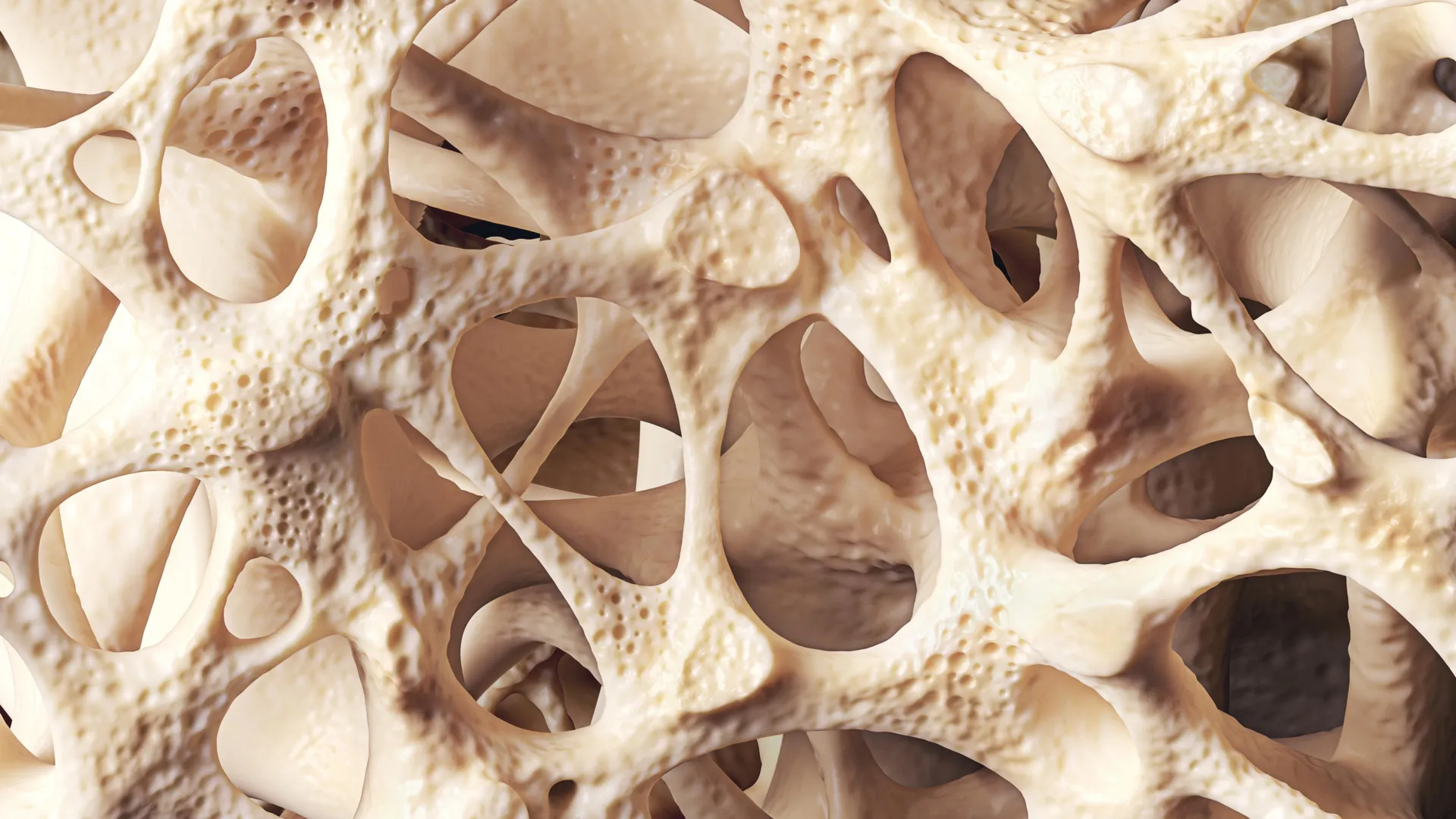
For decades, bone marrow has remained one of the most difficult tissues to study in situ. Described by researchers as a "gelatinous substance encased in a rigid mineral shell," the marrow is the body’s primary site for hematopoiesis—the process by which all blood and immune cells are formed. It serves as a sanctuary for hematopoietic stem cells (HSCs), which are responsible for the lifelong production of red blood cells, white blood cells, and platelets.
Traditional diagnostic and research tools, while effective for general quantification, have significant limitations when applied to this complex environment. Flow cytometry, the gold standard for many years, requires the mechanical or enzymatic dissociation of the bone marrow into a single-cell suspension. While this allows scientists to count and categorize cells with high precision, it completely destroys the spatial context—the "neighborhood" in which these cells reside. Understanding how a stem cell interacts with a neighboring blood vessel, a nerve fiber, or a malignant cancer cell is impossible once the tissue has been homogenized.
Conversely, standard fluorescence imaging techniques, which preserve tissue structure, have historically been limited by "spectral overlap." Most conventional microscopes can only distinguish between three to five different colors (markers) at once. Given that the bone marrow contains dozens of distinct cell types and signaling states, a five-color limit provides only a narrow, often insufficient, window into the tissue’s true complexity.
Technological Innovation: The Phenocycler 2.0 Breakthrough
The IU research team, led by co-lead author Sonali Karnik, PhD, and co-senior author Reuben Kapur, PhD, overcame these barriers by adapting the Phenocycler 2.0 (formerly known as CODEX) for use in mouse bone marrow. This multiplex imaging tool utilizes a sophisticated fluidics system and DNA-barcoded antibodies to perform iterative cycles of staining and imaging.
In this process, antibodies are tagged with unique synthetic DNA "barcodes." The tissue is exposed to a large panel of these antibodies simultaneously. However, instead of using permanent fluorescent dyes, the system uses "reporters"—fluorescently labeled DNA strands that bind only to specific barcodes. The machine images three markers at a time, washes away the reporters, and then applies the next set of reporters for the next three markers. This cycle repeats until all 25 markers are recorded. A computer then overlays these images to create a comprehensive, high-dimensional map of the tissue.
"Because mouse models are widely used to study human diseases, this technique offers a promising new method for investigating a range of conditions like autoimmune diseases, leukemia, and other disorders involving bone marrow," said Dr. Kapur, who serves as the director of the IU School of Medicine’s Herman B Wells Center for Pediatric Research.
A Chronology of Development and Discovery
The journey to this breakthrough began with the recognition that existing spatial biology tools were being successfully applied to soft tissues—such as the spleen, liver, and kidneys—but were failing when applied to the calcified environment of the bone.
- Phase I: Protocol Optimization (2021-2022): The IU team spent months refining the preparation of mouse femurs and tibias. The challenge was to decalcify the bone enough for sectioning while maintaining the delicate protein structures required for antibody binding.
- Phase II: Panel Design (2022-2023): Researchers curated a 25-marker antibody panel. This panel was designed to identify various stages of hematopoietic lineage, from primitive stem cells to mature immune cells, alongside structural markers for blood vessels and bone-forming cells (osteoblasts).
- Phase III: Validation and Imaging (Late 2023): The team successfully imaged intact marrow sections, proving that the Phenocycler could penetrate the complex marrow matrix without significant background noise or tissue degradation.
- Phase IV: Publication and Patenting (2024): After validating the reproducibility of the method, the findings were published in Leukemia. Simultaneously, the IU Innovation and Commercialization Office filed a provisional patent to protect the proprietary methodologies developed during the study.
Supporting Data: Quantifying the Impact
The shift from 3-5 markers to 25 markers represents a 500% to 800% increase in data density per tissue section. This allows for "neighborhood analysis," a computational approach where researchers can calculate the exact distance between different cell types.
In their published study, the researchers demonstrated the ability to distinguish between different "niches" within the marrow. For example, they could pinpoint the location of "quiescent" (dormant) stem cells versus those that had been activated to divide. In the context of cancer research, this data is invaluable. It allows scientists to see how leukemia cells "remodel" their environment to protect themselves from chemotherapy, often creating "sanctuary sites" where drugs cannot easily reach.
By maintaining the 25-marker panel, the researchers were able to identify:
- Hematopoietic Stem Cells (HSCs): The progenitors of all blood cells.
- Vascular Niches: Areas rich in endothelial cells that regulate cell trafficking.
- Endosteal Niches: Areas near the bone surface where specialized bone cells influence stem cell health.
- Immune Infiltrates: The presence and location of T-cells, B-cells, and macrophages.
Institutional Collaboration and Official Responses
The success of this project is attributed to the highly collaborative environment at the IU School of Medicine. The IU Cooperative Center of Excellence in Hematology (CCEH) provided the specialized infrastructure necessary for high-end hematologic research, while the Herman B Wells Center contributed expertise in pediatric oncology and stem cell biology.
Dr. Sonali Karnik, assistant research professor of orthopedic surgery, emphasized the versatility of the tool. "Bone marrow is difficult to study because it is gelatinous and encased in hard bone," she noted. "Since bone marrow plays an important role in blood and immune cell formation and houses valuable stem cells, our unique imaging approach offers a useful tool for a variety of research applications."
The research was supported by the National Institutes of Health (NIH), reflecting the federal government’s interest in advancing spatial biology to combat chronic and terminal illnesses. The broader scientific community has reacted with optimism, as this technique is expected to be adopted by laboratories worldwide that utilize mouse models for preclinical drug testing.
Broader Implications for Medicine and Drug Development
The implications of this imaging breakthrough extend far beyond the laboratory. In the realm of Leukemia and Blood Cancers, the ability to see the "spatial architecture" of a tumor within the marrow could lead to more targeted therapies. Current treatments often fail because they do not account for the protective microenvironment the marrow provides to cancer cells. With this new imaging capability, drug developers can observe exactly how a new compound affects both the cancer cells and the surrounding healthy tissue in real-time.
In Autoimmune Disease Research, the tool could help clarify why the bone marrow sometimes produces "rogue" immune cells that attack the body’s own tissues. By mapping the maturation process of these cells in the marrow, researchers may identify new "checkpoints" that can be targeted to prevent the onset of diseases like lupus or rheumatoid arthritis.
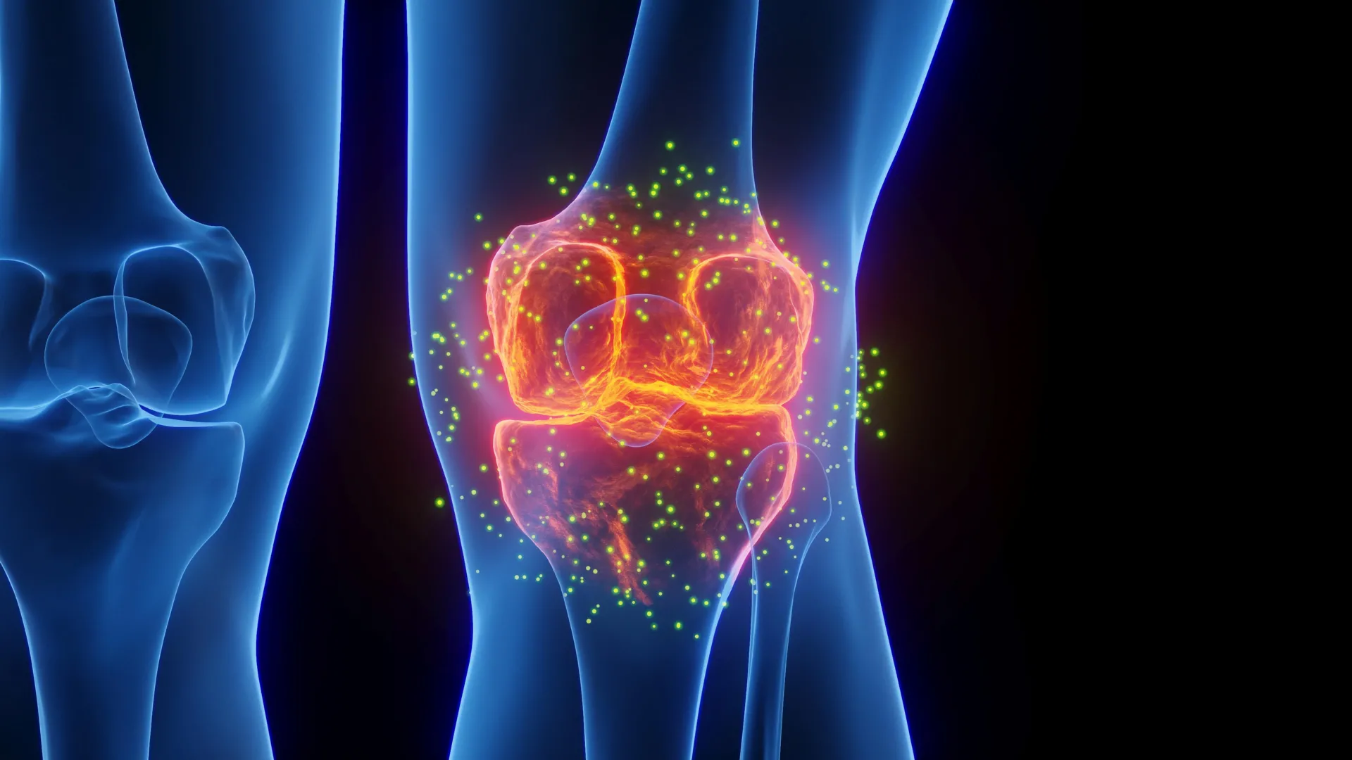
Furthermore, the technique holds promise for Musculoskeletal Disorders and aging research. As humans age, bone marrow often undergoes "adipogenic drift," where healthy blood-producing tissue is replaced by fat cells. This shift contributes to frailty and weakened immune systems in the elderly. The IU team’s methodology allows for a detailed study of this transition, potentially leading to interventions that can preserve bone marrow health into old age.
Future Horizons: Expanding the Map
The IU School of Medicine team is not resting on its current success. Work is already underway to expand the marker panel from 25 to 50 or more markers. This expansion will allow for even more granular detail, including the identification of specific signaling proteins and metabolic states of cells.
The team also plans to integrate other tissue types into their imaging protocols. "We are now working to expand the marker panel to include additional features such as bone, nerves, muscle and more immune and signaling cell types," the researchers stated. By including nerves and muscle, the team hopes to study the "neuro-vascular-bone" axis, a complex communication network that governs how the body responds to physical stress, injury, and infection.
The filing of a provisional patent by the IU Innovation and Commercialization Office suggests that this methodology could soon be standardized for commercial use, potentially through partnerships with biotechnology firms specializing in spatial proteomics. As the medical community moves toward "precision medicine," tools like this high-dimensional imaging technique will be essential for tailoring treatments to the specific cellular landscape of individual patients.
By unlocking the "black box" of the bone marrow, the researchers at Indiana University have not only solved a technical puzzle but have also opened a new frontier in the fight against some of the most challenging diseases of our time.