A groundbreaking study led by researchers at the Montefiore Einstein Comprehensive Cancer Center (MECCC) and the Albert Einstein College of Medicine has fundamentally altered the scientific understanding of glioblastoma, the most aggressive and lethal form of primary brain cancer. For decades, glioblastoma has been treated as a localized malignancy confined within the blood-brain barrier. However, the findings published on October 3 in the journal Nature Neuroscience reveal that the tumor’s influence extends far beyond brain tissue, actively dismantling the structural integrity of the skull and hijacking the body’s immune-cell production centers within the bone marrow.
The discovery that glioblastoma interacts with the skull marrow through physical and molecular channels suggests that the disease is systemic rather than local. This paradigm shift offers a compelling explanation for why traditional therapies, which focus exclusively on the brain, have consistently failed to provide long-term remission for the thousands of patients diagnosed each year. By identifying the skull as a key player in tumor progression, the research team has opened a new frontier for therapeutic intervention that could eventually lead to more effective treatments.
The Clinical Challenge of Glioblastoma
Glioblastoma multiforme (GBM) remains one of the most daunting challenges in modern oncology. According to data from the National Cancer Institute (NCI), approximately 15,000 individuals in the United States are diagnosed with this malignancy annually. Despite the implementation of the "Stupp Protocol"—the current standard of care involving maximal surgical resection followed by concurrent radiation and temozolomide chemotherapy—the prognosis remains grim. The median survival time for patients is roughly 15 months, a statistic that has seen little improvement over the last two decades.
The primary difficulty in treating GBM lies in its highly invasive nature and its ability to create a "cold" immune environment, where the body’s natural defenses are suppressed or recruited to aid tumor growth. Until now, the skull was largely viewed as a passive protective barrier. The Einstein study demonstrates that the skull is, in fact, a dynamic participant in the disease process, serving as a reservoir for immune cells that the tumor manipulates to its advantage.
Unveiling the Mechanism: Skull Erosion and Calvarial Channels
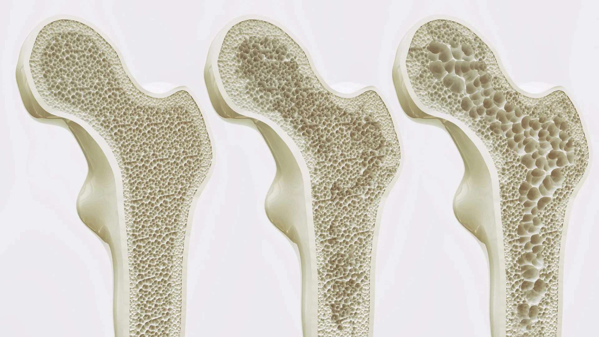
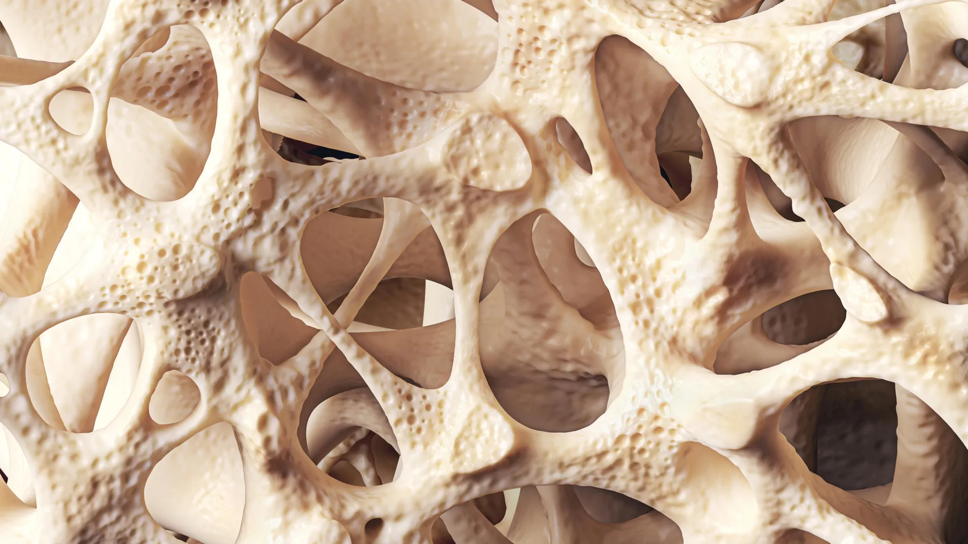
The research team, led by Jinan Behnan, Ph.D., an assistant professor at Einstein and a member of the MECCC, utilized advanced imaging and molecular tools to observe how glioblastoma affects the surrounding bone. Using mouse models of the disease, the scientists discovered significant erosion of the skull bone, particularly concentrated along the sutures—the fibrous joints where the various bones of the skull fuse together.
This bone loss was found to be highly specific to glioblastoma and other aggressive brain tumors. In control groups where mice were subjected to strokes, traumatic brain injuries, or non-CNS cancers, no such skull erosion occurred. To confirm the clinical relevance of these findings, the team conducted CT scans on human patients diagnosed with glioblastoma. The results mirrored the animal models, showing a marked reduction in skull thickness in the regions adjacent to the tumor.
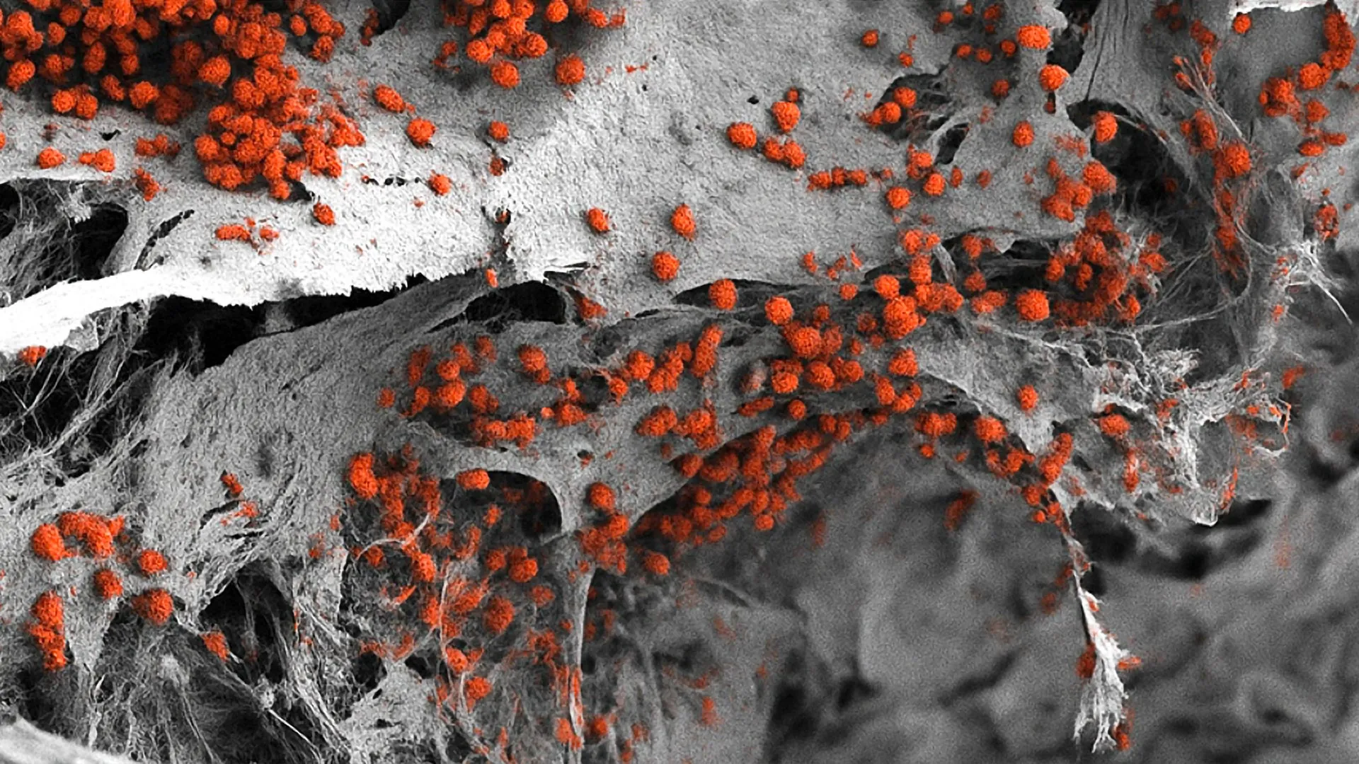
The mechanism behind this erosion involves the expansion of "calvarial channels." These are microscopic, hair-thin conduits that connect the brain’s surface to the bone marrow inside the skull. The researchers found that glioblastoma significantly increases both the number and the diameter of these channels. This structural change effectively creates a "superhighway" for molecular signaling, allowing the tumor to send instructions directly to the skull marrow.
The Immune Landscape: A Shift Toward Inflammation
The most significant finding of the study involves how these channels facilitate a shift in the immune landscape of the skull marrow. Using single-cell RNA sequencing, a technology that allows scientists to examine the genetic activity of individual cells, the researchers mapped the immune population within the marrow of mice with glioblastoma.
The results showed a dramatic imbalance. The tumor signaled the marrow to nearly double its production of pro-inflammatory myeloid cells, specifically neutrophils. Simultaneously, the production of B cells—which are responsible for producing antibodies—and other critical immune-fighting cells was nearly eliminated.
"The skull-to-brain channels allow an influx of these numerous pro-inflammatory cells from the skull marrow to the tumor, rendering the glioblastoma increasingly aggressive and, all too often, untreatable," explained study co-author E. Richard Stanley, Ph.D., a professor of developmental and molecular biology at Einstein.
This influx of inflammatory cells creates a microenvironment that shields the tumor from the immune system and promotes its rapid growth. Interestingly, the study found that this reaction was localized to the skull marrow. When the researchers examined the marrow in the femur (thigh bone), they found a completely different reaction: the tumor actually suppressed genes necessary for immune cell production there. This divergence underscores the unique, localized-yet-systemic relationship between the brain and the skull.
The Paradox of Osteoporosis Medications
In an attempt to mitigate the damage to the skull, the researchers experimented with FDA-approved drugs typically used to treat osteoporosis: zoledronic acid and denosumab. These medications are designed to prevent bone loss by inhibiting the cells that break down bone tissue.
The results were unexpected and serve as a cautionary tale for clinical practice. While both drugs were successful in halting the erosion of the skull bone, they had detrimental effects on cancer progression. In one type of glioblastoma model, zoledronic acid actually accelerated tumor growth.
Furthermore, both drugs were found to interfere with the efficacy of immunotherapy. When combined with anti-PD-L1—a checkpoint inhibitor designed to help T cells attack the tumor—the osteoporosis drugs blocked the beneficial immune response. This suggests that the structural changes in the bone and the immune shifts in the marrow are so deeply intertwined that standard bone-preserving treatments could inadvertently protect the tumor from modern immunotherapy.
Chronology of the Research and Global Collaboration
The study was a multi-year effort involving a diverse array of scientific disciplines, including neurosurgery, immunology, and molecular biology. The timeline of the research began with the observation of bone thinning in clinical glioblastoma cases, which led the team to investigate the biological "why" behind the phenomenon.
- Initial Observation: Researchers noticed localized skull thinning in GBM patients during routine imaging.
- Animal Modeling: The team developed mouse models to replicate the erosion and identified the role of calvarial channels.
- Molecular Mapping: Single-cell RNA sequencing was employed to identify the specific immune cells being recruited from the marrow.
- Comparative Analysis: The team compared skull marrow to femur marrow to determine if the effect was systemic or specific to the brain-skull axis.
- Pharmacological Testing: FDA-approved bone drugs were introduced to see if preventing bone loss could slow the disease.
- Publication: The findings were finalized and published in Nature Neuroscience in October 2024.
The project involved international collaboration, with contributors from Osaka University in Japan, Karolinska Hospital in Sweden, Duke University, the University of California, San Francisco, and the German Rheumatism Research Center in Berlin. This global effort highlights the perceived importance of the findings within the international oncology community.
Broader Implications and Future Treatment Strategies
The implications of this research for the future of glioblastoma treatment are profound. For decades, the failure of glioblastoma therapies was attributed largely to the blood-brain barrier’s ability to keep drugs out. However, this study suggests that the failure may also be due to the "immune-supply chain" coming from the skull.
Dr. Behnan and her colleagues argue that future treatment strategies must address the skull marrow’s immune imbalance. "Our discovery that this notoriously hard-to-treat brain cancer interacts with the body’s immune system may help explain why current therapies—all of them dealing with glioblastoma as a local disease—have failed," Dr. Behnan stated.
Potential future interventions might include:
- Targeted Marrow Modulation: Developing drugs that specifically prevent the production of pro-inflammatory neutrophils in the skull marrow while boosting B and T cell production.
- Channel Blocking: Investigating ways to close or narrow the calvarial channels to prevent the tumor from communicating with the bone marrow.
- Refined Immunotherapy: Redesigning immunotherapy protocols to account for the marrow’s inflammatory output, ensuring that drugs like anti-PD-L1 are not neutralized by the bone’s reaction.
Conclusion
The study by MECCC and Albert Einstein College of Medicine marks a turning point in neuro-oncology. By demonstrating that glioblastoma is a disease that reshapes the skeletal and immune architecture of the head, researchers have identified a previously invisible obstacle to successful treatment. While the findings regarding osteoporosis drugs suggest that the path forward is complex, the identification of the skull-brain axis provides a new map for scientists to follow in their quest to conquer one of the most devastating forms of cancer. As the medical community digests these findings, the focus will likely shift from purely intracranial treatments to a more holistic, systemic approach that views the brain, the skull, and the immune system as an integrated battlefield.