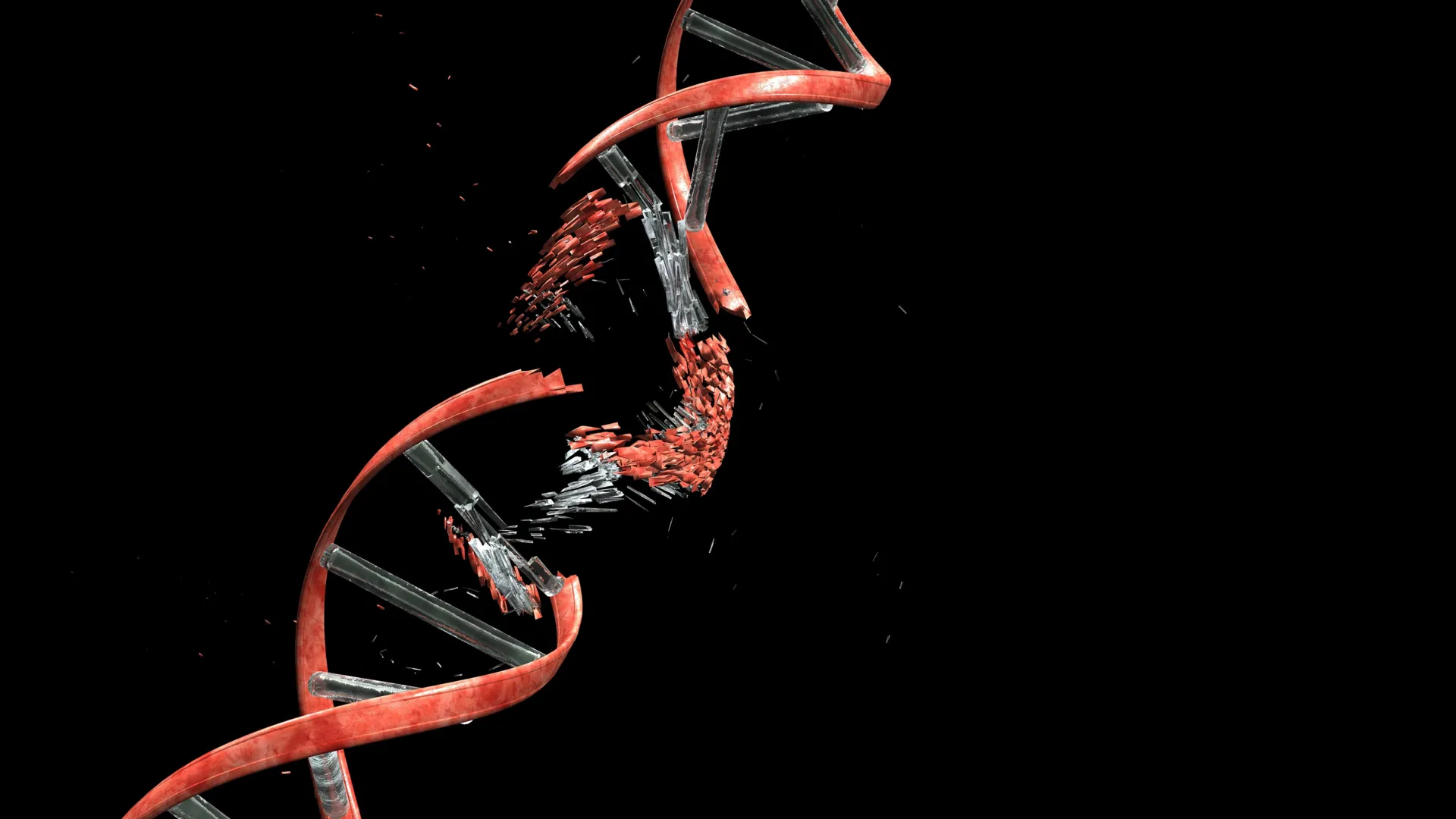
The formation of the human brain is a feat of biological engineering, requiring billions of newly born neurons to navigate a labyrinthine landscape to reach their ultimate destinations. This migration, essential for the assembly of the cerebral cortex, was long thought to be a purely structural process. However, a groundbreaking study published in the journal Nature has unveiled a startling biological cost to this journey. Researchers from Kyoto University’s Institute for Integrated Cell-Material Sciences (WPI-iCeMS), in collaboration with several international institutions, have discovered that the mechanical stress of migration causes widespread double-strand breaks in the DNA of developing neurons. While double-strand breaks are typically associated with cell death or the onset of cancer, the study reveals that in the context of the developing brain, this damage is a routine, manageable, and perhaps even foundational aspect of neurodevelopment.
The Mechanical Gauntlet of the Cerebral Cortex
The cerebral cortex is the seat of higher-order functions, including perception, memory, and voluntary movement. Its complex six-layer structure is built through a process called radial migration. During embryonic development, progenitor cells located near the center of the brain produce young neurons that must travel outward. To reach the cortical plate, these neurons must squeeze through an incredibly dense thicket of extracellular matrix, radial glial fibers, and other migrating cells.
This journey is not a stroll through an open field but a struggle through a microscopic gauntlet. As the neuron moves, its nucleus—the largest and stiffest organelle—must deform significantly to pass through gaps that are often much narrower than the nucleus itself. The Kyoto University study demonstrates that this physical constriction exerts immense pressure on the cell’s internal architecture, leading to unexpected consequences at the molecular level.
The Role of Topoisomerase IIβ and Mechanical Entrapment
At the heart of this discovery is an enzyme known as Topoisomerase IIβ (Top2β). Under normal physiological conditions, Top2β is a vital "janitor" for the genome. As DNA is replicated or transcribed, the double helix can become overwound and tangled, much like a telephone cord that has been twisted too many times. Top2β resolves this tension by intentionally cutting both strands of the DNA, allowing the helix to untwist, and then immediately re-ligating (reconnecting) the ends.
The research team found that the mechanical stress of migration disrupts this delicate "cut-and-paste" operation. When a neuron is squeezed through tight interstitial spaces, the physical pressure appears to trap Top2β in its intermediate state—after it has cut the DNA but before it can repair it. This results in the accumulation of double-strand breaks (DSBs), which are considered the most severe form of genomic damage because they sever both structural backbones of the DNA molecule.
"The process can be compared to cutting a tangled cable to remove twists and then reconnecting it," the researchers noted. "However, when neurons are subjected to mechanical stress while squeezing through tight spaces, the enzyme can become trapped midway through the process, leaving sections of DNA broken."
Replicating the Migration in Microchannels
To confirm that the damage was indeed caused by physical constriction, the scientific team utilized advanced microfluidic technology. They engineered synthetic microchannels that mimicked the narrow, confined spaces of the developing embryonic brain. By guiding living neurons through these channels, they were able to observe the cellular response in real-time.
Using fluorescent markers designed to highlight γH2AX—a protein that clusters at the site of DNA breaks—the team watched as the neurons’ nuclei deformed to enter the channels. As the cells navigated the constriction, the fluorescent signals flared, indicating a surge in double-strand breaks. Remarkably, once the neurons emerged from the other side of the microchannel and the mechanical pressure was relieved, the cells began an immediate recovery process. Within 24 hours, the majority of the DNA damage had been repaired, and the neurons continued to mature and integrate as if no trauma had occurred.
A Comparative Analysis: Neurons vs. Cancer Cells
One of the most intriguing aspects of the study is the comparison between migrating neurons and migrating cancer cells. It is well-documented that metastatic cancer cells also experience DNA damage as they squeeze through tissues to invade new parts of the body. However, the outcome for cancer cells is often chaotic. In many malignancies, the resulting DNA damage is random, leading to further mutations that can drive the progression of the disease or trigger "mitotic catastrophe" and cell death.
Neurons, however, appear to have evolved a more sophisticated "damage control" strategy. The study found that the DSBs in migrating neurons are not randomly distributed. Instead, they are concentrated in specific, non-coding regions of the genome—areas that do not contain the primary instructions for building essential proteins. By localizing the damage to these "safe zones," the neurons ensure that their critical functions remain intact even while their DNA is temporarily fractured. This suggests a highly specialized evolutionary adaptation that allows the brain to grow despite the inherent physical risks of its construction.
The Necessity of Ligase 4 and the Risks of Failed Repair
While the brain is designed to tolerate and repair this routine damage, the study highlights what happens when the repair machinery fails. The researchers focused on a mechanism known as non-homologous end joining (NHEJ), the primary pathway for fixing double-strand breaks. A key component of this pathway is the enzyme Ligase 4.
To test the long-term implications of unrepaired migration-induced damage, the team engineered a mouse model where newly formed neurons in the cerebellum lacked the Ligase 4 enzyme. Initially, these mice appeared healthy and reached adulthood without obvious defects. However, as they aged, they began to exhibit progressive motor dysfunction. The mice developed balance issues and a lack of coordination, symptoms that mirror human neurological conditions such as cerebellar ataxia.
This finding provides a critical link between the mechanical "bruising" of the genome during development and the onset of neurodegenerative symptoms later in life. It suggests that if the "history" of a neuron’s journey is not properly mended, the accumulated genomic instability may eventually compromise the nervous system’s integrity.
Implications for Human Health and Genomic Diversity
The implications of this research extend far beyond basic biology. It offers a new lens through which to view neurodevelopmental and neurodegenerative disorders. Conditions like microcephaly, lissencephaly, and various forms of intellectual disability are often tied to migration defects. This study suggests that in some cases, the primary issue may not just be that the neurons fail to reach their destination, but that the DNA damage incurred during the attempt remains unaddressed.
Furthermore, the study introduces the provocative concept of "somatic mosaicism" driven by mechanical stress. If every neuron undergoes a unique journey with varying levels of DNA breakage and repair, then every neuron may end up with a slightly different genetic "signature."
"It shifts how we think about the neuronal genome," says Professor Mineko Kengaku of WPI-iCeMS, the lead author of the study. "All neurons originate from the same DNA, but DNA damage and repair can introduce small genetic differences between individual neurons through a small mechanical journey. Some of that history may be written into the genome itself."
This "genomic history" could explain the immense diversity of neuronal responses in the human brain, contributing to why individuals respond differently to stimuli or why certain people are more resilient to brain diseases than others.
Collaborative Efforts and Future Directions
This study was a massive undertaking involving a multidisciplinary collaboration between Kyoto University, the University of Tokyo, the University of Osaka, the National University of Singapore, and the Tokyo Metropolitan Institute of Medical Science. By combining expertise in cell biology, micro-engineering, and genetics, the team has opened a new frontier in neuroscience.
Moving forward, the researchers aim to investigate whether environmental factors—such as prenatal stress or exposure to toxins—might exacerbate the DNA damage experienced by migrating neurons. They are also interested in whether the efficiency of the DNA repair process declines with age, potentially making the brain more susceptible to the "echoes" of its own developmental trauma as it enters the later stages of life.
Ultimately, the study transforms our understanding of the developing brain from a purely biological program into a physical struggle. It portrays the neuron not just as a messenger, but as a resilient survivor of a grueling mechanical journey, carrying the scars and repairs of its birth into the complex network of the adult mind. The discovery that "normal" development involves such high-stakes genomic maintenance provides a new framework for modern medicine to explore the roots of human cognition and the vulnerabilities of the human spirit.