

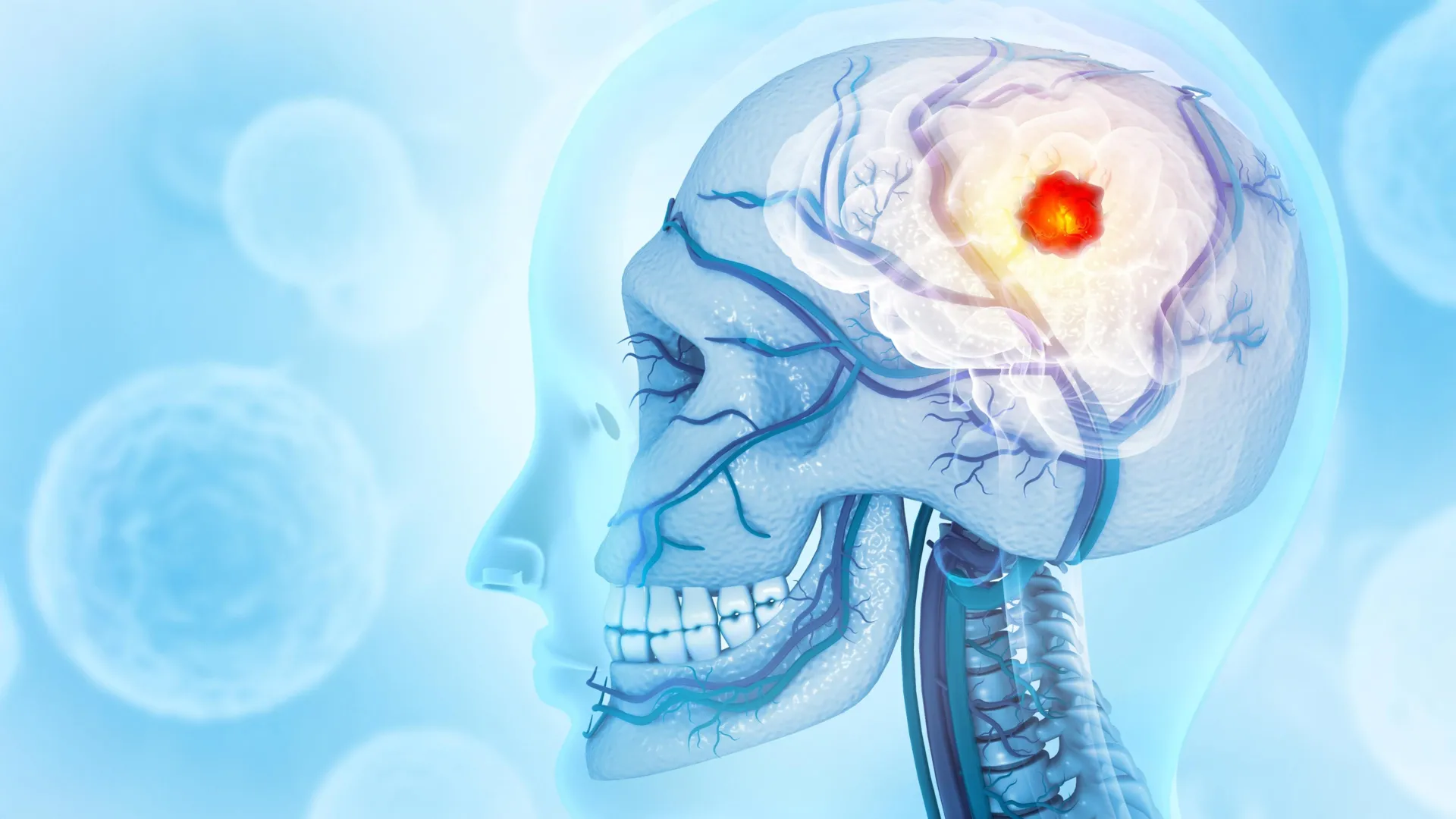
The understanding of glioblastoma, the most aggressive and lethal form of primary brain cancer, has undergone a fundamental shift following a landmark study by researchers at the Montefiore Einstein Comprehensive Cancer Center (MECCC) and the Albert Einstein College of Medicine. For decades, the medical community has approached glioblastoma as a localized neurological disease, confined within the blood-brain barrier. However, new evidence published on October 3 in the journal Nature Neuroscience reveals that the malignancy is a systemic predator that actively degrades the skull, manipulates the bone marrow, and hijacks the body’s immune system to facilitate its own growth.
This discovery provides a potential explanation for why traditional therapies—ranging from surgical resection to localized radiation and chemotherapy—frequently fail to prevent recurrence. By demonstrating that the tumor’s influence extends into the calvarial bone (the skullcap), the research team has opened a new frontier in neuro-oncology that may lead to more effective, multi-pronged treatment strategies.
The Clinical Challenge of Glioblastoma
Glioblastoma multiforme (GBM) remains one of the most daunting challenges in modern medicine. According to the National Cancer Institute (NCI), approximately 15,000 individuals in the United States are diagnosed with the disease annually. Despite advancements in neurosurgery and precision medicine, the prognosis remains grim. The current standard of care—the "Stupp Protocol"—which involves maximal safe surgical resection followed by a combination of radiotherapy and the chemotherapy drug temozolomide, yields a median survival time of only 15 months.
The primary difficulty in treating GBM lies in its highly invasive nature and the presence of the blood-brain barrier, which prevents many systemic drugs from reaching the tumor. However, the Einstein study suggests that the tumor has found its own "backdoor" to the rest of the body, utilizing the skull as a staging ground to manipulate the immune system.
Unveiling the Skull-Brain Connection
The research, led by Dr. Jinan Behnan, an assistant professor in the Leo M. Davidoff Department of Neurological Surgery at Einstein, was built upon the recent anatomical discovery of microscopic channels connecting the brain to the skull marrow. Historically, the skull was viewed as a static, protective casing. Recent biological insights have corrected this view, showing that the skull marrow is a dynamic site of hematopoiesis—the production of blood and immune cells—that communicates directly with the brain via these vascular channels.
Using advanced high-resolution imaging and three-dimensional reconstruction in mouse models, Dr. Behnan’s team observed that glioblastoma triggers a significant erosion of the skull bone. This degradation was most pronounced along the sutures, the fibrous joints where the various bones of the skull fuse together. This bone loss was not a general symptom of brain distress; the researchers compared the GBM models to mice with strokes or other types of traumatic brain injury and found that those conditions did not cause similar skull erosion. Furthermore, cancers originating in other parts of the body did not produce this specific effect, identifying the bone-thinning phenomenon as a unique signature of aggressive primary brain tumors.
To validate these findings in humans, the team analyzed CT scans of patients diagnosed with glioblastoma. The results mirrored the animal models: patients showed a measurable reduction in skull thickness in regions adjacent to the tumor, confirming that the laboratory observations have direct clinical relevance.
The Mechanism of Immune Hijacking
The study proposes that as the tumor erodes the skull, it increases both the diameter and the number of the channels linking the brain to the marrow. These expanded "highways" allow for a two-way exchange of molecular signals. The tumor sends signals out to the skull marrow, effectively "reprogramming" the production of immune cells.
Using single-cell RNA sequencing—a technology that allows researchers to see the genetic activity of individual cells—the team mapped the immune landscape of the skull marrow. They discovered a profound shift in the cellular population. In the presence of glioblastoma, the skull marrow nearly doubled its production of pro-inflammatory myeloid cells, particularly neutrophils. Simultaneously, the marrow saw a near-total depletion of B cells, which are responsible for producing antibodies, and other essential immune-regulating cells.
Dr. E. Richard Stanley, a co-author of the study and professor of developmental and molecular biology at Einstein, noted that this influx of pro-inflammatory cells creates an environment where the tumor can thrive. "The skull-to-brain channels allow an influx of these numerous pro-inflammatory cells from the skull marrow to the tumor, rendering the glioblastoma increasingly aggressive and, all too often, untreatable," Dr. Stanley explained.
A Systemic Disruption: Skull vs. Long Bones
One of the most striking findings of the research was the localized specificity of the immune disruption. While the skull marrow became a factory for pro-inflammatory cells, the researchers examined the marrow in the femur (the thigh bone) to see if the effect was uniform across the body.
The results showed a stark contrast. While the glioblastoma activated genes in the skull marrow to boost inflammatory cell production, it had the opposite effect on the femur marrow, where it suppressed genes necessary for the production of various immune cells. This divergence reinforces the idea that glioblastoma strategically manipulates its immediate surroundings—the skull—to create a protective, pro-tumor niche, while potentially weakening the broader systemic immune response.
The Osteoporosis Drug Paradox
Given the observed bone erosion, the researchers investigated whether existing medications designed to prevent bone loss could mitigate the tumor’s impact. They tested two FDA-approved drugs commonly used to treat osteoporosis: zoledronic acid (a bisphosphonate) and denosumab (a monoclonal antibody).
While both drugs successfully halted the erosion of the skull bone, the results regarding tumor progression were alarming. In one type of glioblastoma, zoledronic acid actually accelerated the growth of the tumor. Perhaps more significantly, both drugs were found to interfere with the efficacy of anti-PD-L1 immunotherapy. Immunotherapy is designed to "unmask" cancer cells so that T cells can attack them; however, the bone-preserving drugs appeared to block the beneficial influx of these tumor-fighting T cells.
This finding carries significant implications for clinical practice. Many cancer patients are prescribed bone-strengthening medications to prevent fractures or treat bone metastases. The Einstein study suggests that in the case of glioblastoma, these drugs might inadvertently neutralize the body’s remaining defenses and interfere with emerging immunotherapeutic treatments.
Chronology of the Discovery and Future Implications
The timeline of this discovery reflects a decade of evolving neuro-immunology. For years, the brain was considered an "immune-privileged" site, largely disconnected from the peripheral immune system. The discovery of lymphatic vessels in the brain in 2015 and the subsequent identification of skull-brain channels in 2018 set the stage for Dr. Behnan’s team.
The publication of this study in Nature Neuroscience on October 3, 2024, marks a pivotal moment in connecting these anatomical dots to a specific, deadly disease. The research team included a diverse array of international collaborators from institutions such as Osaka University in Japan, the Karolinska Institute in Sweden, and Duke University, highlighting the global importance of the work.
The implications for future glioblastoma treatment are profound. Dr. Behnan suggests that the failure of current therapies is due to their "local" focus. "Our discovery… may help explain why current therapies—all of them dealing with glioblastoma as a local disease—have failed," she stated.
The research points toward a new therapeutic paradigm:
- Marrow Regulation: Future treatments may need to target the skull marrow directly to prevent the overproduction of pro-inflammatory myeloid cells.
- Immune Restoration: Instead of just attacking the tumor, doctors may need to find ways to restore the balance of B and T cells within the skull’s internal environment.
- Combination Therapy Screening: The study highlights the urgent need to screen common medications (like those for osteoporosis) for their specific interactions with the unique microenvironment of the brain.
Conclusion
The Montefiore Einstein Comprehensive Cancer Center study redefines glioblastoma not as a solitary mass within the brain, but as a systemic architect that rebuilds the surrounding bone and bone marrow to suit its needs. By identifying the skull as a critical player in the progression of brain cancer, the researchers have provided a new roadmap for drug development.
As the medical community moves toward more personalized and systemic approaches to oncology, the focus may shift from the brain alone to the complex "crosstalk" between the brain, the skull, and the immune system. While the median survival for glioblastoma has remained stagnant for years, this new understanding of the "skull-marrow-tumor axis" offers a glimmer of hope for more durable and effective interventions in the fight against this devastating disease.

















