

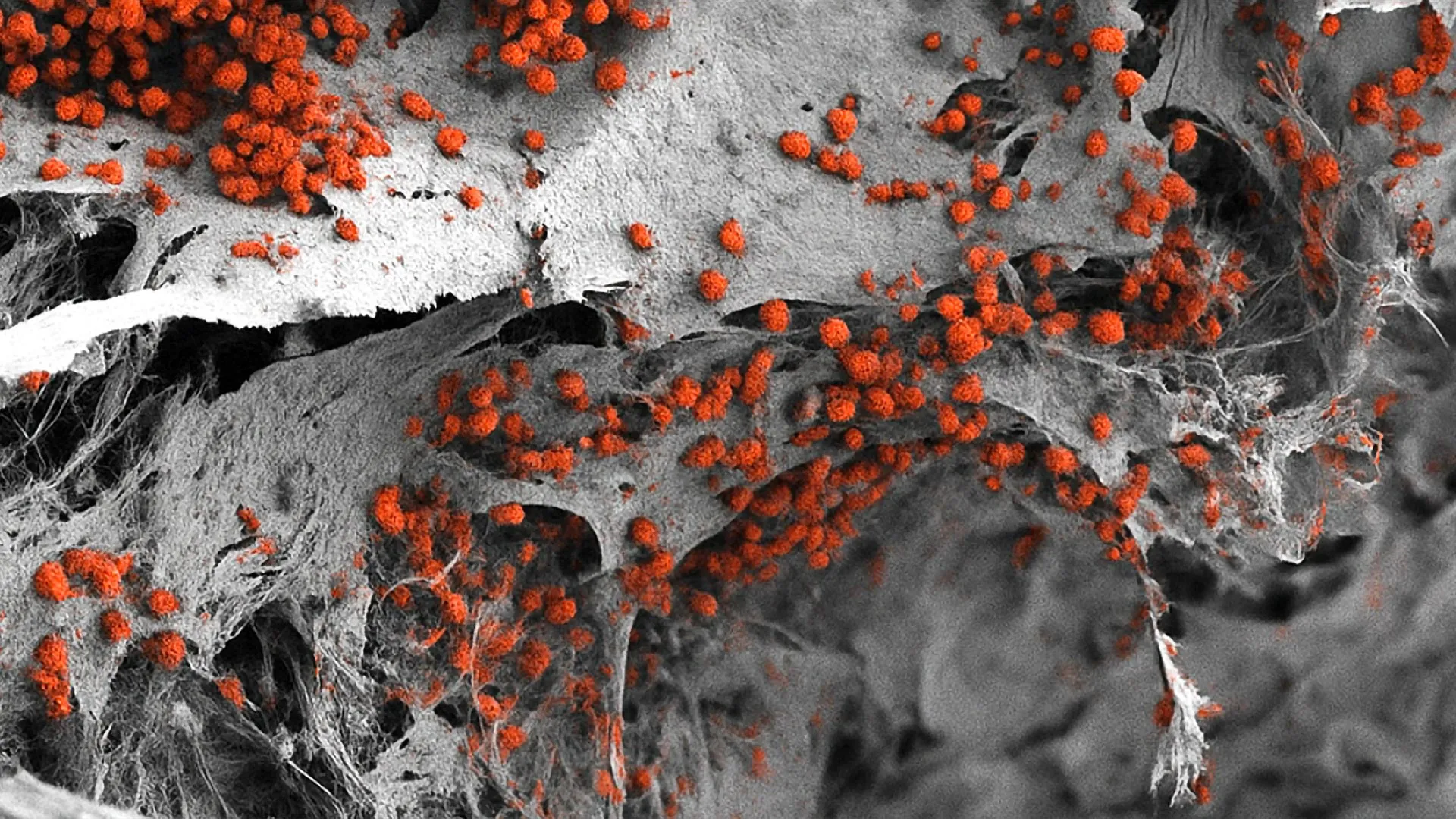
The human bone marrow is often described as the body’s primary "blood factory," a highly sophisticated and specialized tissue responsible for the continuous production of billions of new blood cells every day. This complex biological engine is comprised of an intricate network of bone cells, nerves, blood vessels, and a variety of supporting cell types that work in perfect synchronicity. For decades, reproducing this environment outside the human body has been one of the most significant challenges in regenerative medicine. However, a groundbreaking study from the University of Basel and University Hospital Basel has achieved a world first: the recreation of this complex network in a laboratory setting using exclusively human cells. This milestone, published in the prestigious journal Cell Stem Cell, represents a paradigm shift in how scientists study blood formation and disease, offering a more accurate alternative to traditional animal-based research models.
Under normal physiological conditions, the bone marrow operates with silent efficiency, largely ignored until a pathological state arises. It is only when this vital factory falters—most notably in cases of leukemia, multiple myeloma, or other hematological malignancies—that its function becomes the central focus of clinical intervention. Understanding the delicate balance of how blood is produced, and more importantly, how that process is disrupted by cancer, is essential for the development of effective therapies. The Basel-based research team, led by Professor Ivan Martin and Dr. Andrés García-García, has provided the scientific community with a new lens through which to view these processes, moving beyond the limitations of cross-species experimentation.
The Architecture of the Endosteal Niche
To appreciate the magnitude of this achievement, one must understand the specific microenvironments, or "niches," within the bone marrow. The bone marrow is not a uniform mass; it is a heterogeneous landscape where different zones support different stages of cell development. One of the most critical zones is the endosteal niche. Located near the inner surface of the bone, this niche is a hub of activity where hematopoietic stem cells (HSCs)—the "mother cells" of all blood—interact with bone-forming cells, blood vessels, and the nervous system.
The endosteal niche is of particular interest to oncologists because it is frequently implicated in the resilience of blood cancers. Research has shown that cancer cells can "hide" within this niche, utilizing its protective environment to evade the effects of chemotherapy and radiation. Until now, scientists lacked a human-centric model that included all the constituent parts of this niche—vessels, immune cells, nerves, and bone—in a single, unified 3D system. Previous models were either too simplified, utilizing only one or two cell types, or relied on animal tissue, which often fails to replicate the nuances of human molecular signaling.
Engineering the Human Bone Marrow Model
The development of this 3D model began with the construction of a synthetic framework designed to mimic the physical properties of human bone. The researchers utilized hydroxyapatite, a naturally occurring mineral form of calcium apatite that constitutes the primary inorganic component of human teeth and bones. This scaffold provided the necessary structural integrity and biochemical cues to support cell growth.
The most innovative aspect of the project involved the use of induced pluripotent stem cells (iPSCs). These are adult cells that have been genetically reprogrammed to an embryonic-like state, granting them the ability to differentiate into almost any cell type in the human body. By introducing these stem cells into the hydroxyapatite scaffold and applying a precise sequence of molecular signals and growth factors, the researchers guided the cells through a controlled developmental process.
Over a period of several weeks, the stem cells transformed into a diverse array of specialized cells, including osteoblasts (bone-forming cells), endothelial cells (which form blood vessels), and various stromal and neural components. The result was a functional, three-dimensional structure that measured approximately eight millimeters in diameter and four millimeters in thickness. This size is significantly larger than previous organoid models, providing a more robust platform for observation and experimentation.
Data and Validation: A Mirror to Human Biology
The Basel team conducted extensive analysis to verify that their laboratory-grown tissue accurately reflected the biological reality of a human bone marrow niche. Using advanced imaging and genetic sequencing, they compared the model’s cellular composition and gene expression profiles to actual human bone marrow samples. The data confirmed a high degree of similarity, particularly in the way the different cell types organized themselves spatially and communicated with one another.
Crucially, the model demonstrated the ability to support hematopoiesis—the process of blood cell formation—for several weeks. This longevity is vital for studying chronic diseases or the long-term effects of drugs. In traditional 2D cultures, blood stem cells often lose their functionality or die off quickly because they lack the physical and chemical support of their natural environment. The 3D human-only model overcomes this hurdle, maintaining the "stemness" of hematopoietic cells while allowing them to mature into red blood cells, white blood cells, and platelets.
A Chronology of Research Progress
The journey to this discovery has been decades in the making. The history of bone marrow research can be traced back to the mid-20th century, with the first successful bone marrow transplants performed by Dr. E. Donnall Thomas in the late 1950s. Since then, the field has evolved through several key phases:
- The Animal Model Era (1960s–Present): For over sixty years, the mouse has been the workhorse of hematology. While mouse studies provided the foundational knowledge of how stem cells differentiate, they often failed to predict human responses to drugs due to fundamental differences in immune system regulation and cell signaling.
- The Rise of In Vitro Cultures (1980s–2000s): Researchers began growing human marrow cells in plastic dishes (2D cultures). While useful for basic observations, these "flat" models could not replicate the mechanical stresses or the complex 3D architecture of living tissue.
- The Stem Cell Revolution (2006–Present): The discovery of iPSCs by Shinya Yamanaka in 2006 opened the door to creating patient-specific tissues. This technology became the cornerstone of the Basel team’s current work.
- The Bioengineering Milestone (2024): The successful integration of multiple cell lineages into a single, large-scale 3D human-only scaffold marks the current frontier of the field.
Implications for Animal Welfare and the 3Rs Principle
One of the most significant impacts of this research is its potential to drastically reduce the reliance on animal experimentation. The scientific community has long been guided by the "3Rs" principle: Replacement, Reduction, and Refinement of animal use in research. Professor Ivan Martin emphasized that while mouse studies have been invaluable, they are ultimately a proxy for human biology.
"Our model brings us closer to the biology of the human organism," Martin stated. By providing a platform that more accurately reflects human responses, researchers can conduct preliminary drug screenings and toxicity tests on the synthetic marrow before—or instead of—using animals. This not only aligns with ethical advancements but also improves the efficiency of the drug development pipeline, as many drugs that appear successful in mice ultimately fail in human clinical trials.
Future Prospects: Personalized Medicine and Drug Screening
The implications of this breakthrough extend far into the future of clinical oncology. One of the most promising applications is the development of personalized treatment plans. In the future, a patient diagnosed with leukemia could have their own cells used to create a "personalized bone marrow model" in the lab. Doctors could then test various combinations of chemotherapy or targeted biological therapies on that specific patient’s tissue to see which is most effective at eradicating the cancer cells while sparing the healthy ones.
However, challenges remain before this becomes a routine clinical tool. Dr. Andrés García-García noted that the current size of the model—while a feat of engineering—presents a hurdle for high-throughput drug testing. To test hundreds of different drug concentrations simultaneously, the system would need to be miniaturized into a "marrow-on-a-chip" format.
"For the specific purpose of drug development, the size of our bone marrow model might be too large," García-García explained. "To test many drugs or doses at the same time, the platform would need to be made smaller."
Analyzing the Broader Impact on the Pharmaceutical Industry
The pharmaceutical industry stands to benefit significantly from this technology. Currently, the cost of bringing a new drug to market exceeds $2 billion, with a high percentage of that cost attributed to failures in late-stage clinical trials. A human-only bone marrow model provides a "high-fidelity" environment to catch potential issues early. If a drug shows toxicity to the human endosteal niche in the lab, companies can pivot or abandon the compound before investing millions in human trials.
Furthermore, this model allows for the study of rare hematological diseases that may not have an equivalent animal model. By manipulating the genetic makeup of the iPSCs used to create the marrow, researchers can "program" the model to exhibit specific genetic disorders, providing a first-of-its-kind look at the progression of rare blood conditions in a human context.
The study from the University of Basel represents more than just a successful laboratory experiment; it is a foundational step toward a future where human disease is studied in human systems. As the team continues to refine the model—miniaturizing it for drug screening and expanding its complexity to include more immune components—the "blood factory" in the lab may soon become the gold standard for hematological research worldwide. By bridging the gap between synthetic engineering and biological reality, this research offers new hope for more effective, personalized, and ethical treatments for some of the most challenging diseases known to medicine.

















