The global healthcare landscape is currently facing a silent but escalating crisis as recent epidemiological data reveals that approximately 40% of adults worldwide are living with osteopenia. Characterized by a decrease in bone mineral density (BMD) that is lower than normal but not yet classified as osteoporosis, osteopenia serves as a critical clinical precursor to more severe skeletal fragility. This condition is particularly prevalent among postmenopausal women and the elderly, creating a significant burden on public health systems. In the United Kingdom alone, health authorities estimate that more than 500,000 fractures occur annually due to low bone density, many of which are directly attributable to the under-diagnosis and under-management of osteopenia. Because the condition develops without overt symptoms, it is frequently labeled a "silent" ailment, often only coming to light after a patient sustains a fragility fracture or undergoes routine screening due to age-related risk factors.
The Biological Mechanism of Bone Remodeling
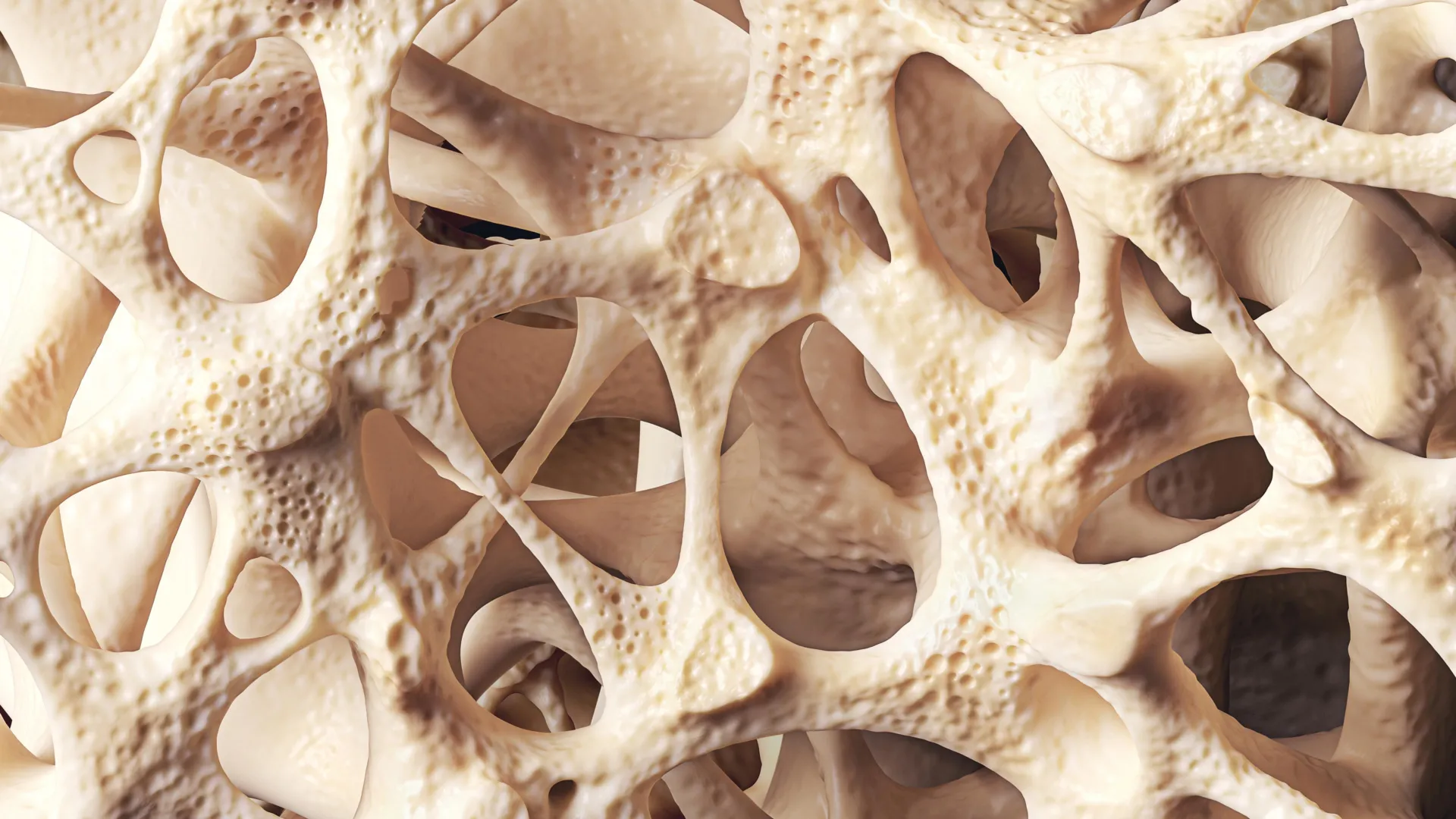
To understand the progression of osteopenia, it is essential to view bone not as a static structure, but as a dynamic, living tissue that is constantly being broken down and rebuilt. This physiological cycle, known as bone remodeling, involves two primary processes: resorption and formation. During resorption, specialized cells called osteoclasts break down old or damaged bone tissue, releasing minerals into the bloodstream. Subsequently, cells known as osteoblasts deposit new bone mineral to maintain skeletal integrity.
In a healthy young adult, these two processes are meticulously balanced. However, as the body ages, the equilibrium shifts. Clinical studies indicate that bone mass typically reaches its peak between the mid-20s and early 30s. This "peak bone mass" is a vital determinant of future skeletal health; the higher the peak, the more "bone bank" a person has to draw from as they age. Once this peak is passed, the rate of bone resorption begins to gradually outpace the rate of bone formation. Over decades, this net loss of mineral density results in the thinning of the bone’s internal architecture, leading first to osteopenia and potentially progressing to the more porous and brittle state of osteoporosis.
A Chronology of Bone Health Across the Lifespan
The trajectory of bone density follows a predictable chronological path, though individual rates of decline vary based on genetics and lifestyle.
- Growth Phase (Birth to Age 20): This is the period of rapid accumulation. Approximately 90% of peak bone mass is acquired by age 18 in girls and age 20 in boys. Adequate nutrition and physical activity during these formative years are the primary defenses against late-life osteopenia.
- Peak Stability (Age 25 to 35): Bone density remains relatively stable. The body focuses on maintaining the existing matrix through balanced remodeling.
- The Decline Phase (Age 40+): For both genders, a slow decline of roughly 0.5% per year begins.
- The Menopausal Transition (Age 50 to 60): In women, the decline accelerates sharply due to the loss of estrogen, sometimes reaching a loss of 2% to 3% of total bone mass per year during the first five years after menopause.
- Senior Maintenance (Age 70+): Bone loss continues for both men and women, often exacerbated by reduced physical activity and diminished nutrient absorption.
Primary Risk Factors and the Role of Hormonal Health
While aging is the universal driver of bone loss, several secondary factors can accelerate the transition to osteopenia. The most significant of these is hormonal fluctuation. Estrogen plays a protective role in the skeletal system by inhibiting the activity of osteoclasts. When estrogen levels plummet during menopause, the protective "brake" on bone resorption is removed, leading to a rapid thinning of the bone. Statistics show that approximately one in two women over the age of 50 will experience a fragility fracture in their remaining lifetime, a figure that underscores the vulnerability of the postmenopausal population.
Beyond hormones, lifestyle choices exert a cumulative impact on bone strength. Smoking has been clinically linked to reduced blood flow to the bones and interference with calcium absorption. Excessive alcohol consumption can disrupt the body’s nutrient balance and increase the risk of falls. Furthermore, physical inactivity—specifically a lack of weight-bearing movement—signals to the body that the skeleton does not need to maintain high density, leading to further atrophy.
Medical history also plays a decisive role. Patients with malabsorption syndromes, such as Crohn’s disease or coeliac disease, often struggle to absorb the calcium and Vitamin D necessary for bone formation. Additionally, the long-term use of certain medications, particularly glucocorticoids (steroids) used for asthma or rheumatoid arthritis, is a well-documented cause of secondary osteopenia.
Diagnostic Standards and the T-Score System
The primary tool for identifying osteopenia is the Dual-energy X-ray Absorptiometry (DXA) scan. This non-invasive, low-dose X-ray measures the mineral content in specific areas of the skeleton, usually the hip and the lumbar spine. The results are expressed as a T-score, which represents the number of standard deviations a patient’s BMD is above or below the mean for a healthy 30-year-old adult of the same sex.
- Normal: T-score of -1.0 or higher.
- Osteopenia: T-score between -1.0 and -2.5.
- Osteoporosis: T-score of -2.5 or lower.
While the DXA scan is the gold standard, clinicians also utilize the Fracture Risk Assessment Tool (FRAX). Developed by the World Health Organization, FRAX calculates a patient’s 10-year probability of suffering a major osteoporotic fracture. This tool is essential because it integrates BMD data with other clinical risk factors—such as age, BMI, smoking status, and family history—to determine whether pharmacological intervention is necessary even if a patient has not yet reached the threshold for osteoporosis.
Strategic Management and Lifestyle Interventions
Managing osteopenia is a multi-faceted endeavor aimed at stabilizing bone density and preventing the "fracture cascade," where one break leads to a loss of mobility and subsequent further bone loss.
The Role of Physical Activity
Not all exercise is created equal when it comes to bone health. To stimulate bone formation, the skeleton must be subjected to mechanical "loading" or strain. Weight-bearing aerobic exercises, such as brisk walking, dancing, and jogging, force the body to work against gravity, triggering osteoblasts to lay down new mineral. Resistance training (weightlifting) is equally vital, as the pull of muscles on the bone during contraction further stimulates density. Emerging research also highlights the benefits of balance-focused exercises like Tai Chi; while they may not significantly increase BMD, they drastically reduce the risk of falls, which are the immediate cause of most fractures.
Nutritional Optimization
Calcium and Vitamin D are the cornerstones of bone nutrition. Calcium provides the structural "bricks" for the bone matrix, while Vitamin D acts as the "gatekeeper" that allows the intestines to absorb that calcium. In regions like the UK, where sunlight exposure is limited for much of the year, Vitamin D deficiency is a widespread public health issue, making supplementation a common recommendation for those diagnosed with osteopenia. Dietary sources such as dairy, leafy greens (kale, collards), and fortified cereals remain the preferred method of intake, though many clinical guidelines suggest 1,000 to 1,200 mg of calcium daily for older adults.
Pharmacological Considerations
While not every patient with osteopenia requires medication, those at high risk (as determined by FRAX scores or previous fractures) may be prescribed antiresorptive drugs. Bisphosphonates are the most common class of medication used; they work by slowing the action of osteoclasts, thereby preserving existing bone mass. While more frequently associated with osteoporosis treatment, recent clinical trials suggest that early use in high-risk osteopenic patients can significantly lower the incidence of initial fractures.
Broader Economic and Public Health Implications
The implications of widespread osteopenia extend far beyond individual health. Fragility fractures represent a massive economic burden on healthcare systems. In the UK, the cost of treating hip fractures alone runs into billions of pounds annually, accounting for hospital stays, rehabilitation, and long-term social care. As the global population ages, these costs are projected to rise exponentially unless preventative measures are prioritized.
Public health experts are increasingly advocating for a "life-course approach" to bone health. This involves viewing osteopenia not as an inevitable part of aging, but as a preventable condition. There is a growing movement to implement Fracture Liaison Services (FLS) in hospitals—systems designed to ensure that any patient over 50 who suffers a fracture is automatically screened for low bone density and started on a management plan.
Conclusion: The Path Forward
Osteopenia should serve as a critical clinical "warning light." It represents a window of opportunity where lifestyle modifications and targeted medical interventions can effectively halt the progression toward osteoporosis. The evidence is clear: bone density can be maintained, and in some cases improved, through a dedicated regimen of nutrition, weight-bearing exercise, and risk management.
Ultimately, protecting the skeleton requires a long-term perspective. The habits formed in early adulthood—and the interventions staged in middle age—determine the quality of life in later years. By addressing osteopenia with the same urgency as hypertension or high cholesterol, society can reduce the global fracture burden and ensure that an aging population remains mobile, independent, and resilient.