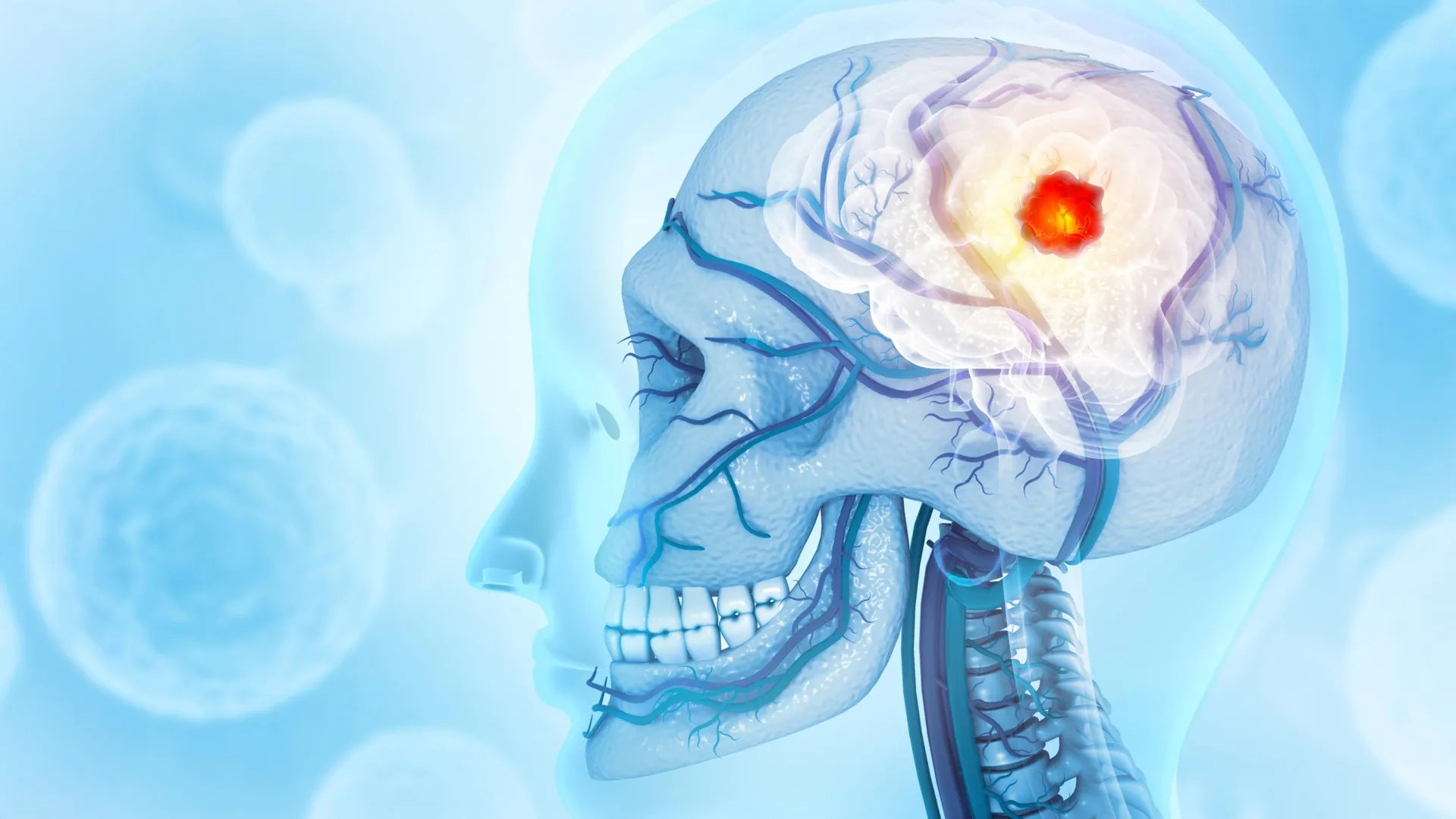
Researchers from the Montefiore Einstein Comprehensive Cancer Center (MECCC) and the Albert Einstein College of Medicine have fundamentally challenged the long-standing medical understanding of glioblastoma, the most aggressive and lethal form of primary brain cancer. Traditionally viewed as a localized malignancy confined within the brain’s protective barriers, glioblastoma has now been shown to exert a systemic influence that extends into the skeletal structure of the head. According to a study published on October 3 in the journal Nature Neuroscience, this cancer actively erodes the skull, fundamentally alters the biological composition of the bone marrow within it, and effectively "reprograms" the body’s immune response to favor tumor progression rather than suppression.
The findings provide a potential explanation for the persistent failure of current therapeutic interventions. While modern medicine has made significant strides in treating other forms of cancer, the prognosis for glioblastoma remains grim. By demonstrating that the disease is not merely a "brain tumor" but a condition that manipulates the surrounding bone and the immune cells produced within it, the research team, led by Jinan Behnan, Ph.D., has opened a new frontier for developing more effective treatment strategies.
The Stagnant Landscape of Glioblastoma Treatment
For decades, glioblastoma multiforme (GBM) has remained one of the most difficult challenges in oncology. According to data from the National Cancer Institute (NCI), approximately 15,000 individuals in the United States are diagnosed with this malignancy annually. Despite the implementation of a rigorous "standard of care" that typically includes surgical resection, high-dose chemotherapy (often temozolomide), and targeted radiation, the median survival time for patients remains a sobering 15 months.
The difficulty in treating glioblastoma stems from its highly invasive nature and the presence of the blood-brain barrier, which prevents many systemic drugs from reaching the tumor. However, the MECCC and Einstein study suggests that the problem may also lie in our narrow focus on the brain itself. Dr. Behnan, an assistant professor in the Leo M. Davidoff Department of Neurological Surgery and the department of microbiology & immunology at Einstein, noted that current therapies fail largely because they treat glioblastoma as a local disease. The new data suggests that the tumor’s reach is far more expansive, utilizing the skull as a secondary site for immune manipulation.
Mapping the Skull-Brain Connection
The foundation of this research lies in a relatively recent anatomical discovery: the existence of microscopic, thin channels that physically connect the brain to the skull’s bone marrow. Historically, the skull was viewed as a static, inert container for the brain. We now know it is a dynamic biological environment. Like the femur or the pelvis, the skull contains marrow that serves as a factory for immune cells.
Using advanced high-resolution imaging and mouse models of glioblastoma, Dr. Behnan’s team observed that as the tumor grows, it triggers a physical degradation of the skull. This erosion is not random; it is particularly concentrated along the sutures—the fibrous joints where the different plates of the skull fuse together. This phenomenon appears to be a unique hallmark of glioblastoma and other highly aggressive brain tumors. When the researchers examined mice with other conditions, such as strokes, traumatic brain injuries, or cancers originating in other parts of the body, they did not observe this specific skull erosion.
To confirm the clinical relevance of these findings, the team conducted CT scans on human patients diagnosed with glioblastoma. The results mirrored the animal models: patients exhibited a significant reduction in skull thickness in the exact regions identified in the laboratory. This structural decay facilitates the expansion of the pre-existing channels between the brain and the bone marrow, creating a larger "highway" for molecular signaling.
Immune Reprogramming and the Pro-Inflammatory Shift
The most profound discovery of the study involves how glioblastoma exploits these widened channels to alter the immune landscape. Through the use of single-cell RNA sequencing—a technology that allows scientists to examine the genetic expression of individual cells—the researchers analyzed the marrow within the skull.
They found that glioblastoma sends signals through the skull-brain channels that radically shift the production of immune cells. In a healthy state, bone marrow maintains a balance of various white blood cells, including B cells (which produce antibodies) and T cells (which attack infected or cancerous cells). However, in the presence of glioblastoma, the skull marrow nearly doubles its production of pro-inflammatory myeloid cells, specifically neutrophils. Simultaneously, the production of several types of B cells is nearly eliminated.
"The skull-to-brain channels allow an influx of these numerous pro-inflammatory cells from the skull marrow to the tumor, rendering the glioblastoma increasingly aggressive," explained study co-author E. Richard Stanley, Ph.D., a professor of developmental and molecular biology at Einstein.
This pro-inflammatory environment acts as a shield for the tumor. Rather than attacking the cancer, these myeloid cells contribute to an environment that promotes tumor growth and suppresses the body’s natural anti-cancer defenses. This suggests that glioblastoma is effectively "farming" the skull marrow to produce a steady supply of cells that aid its own survival.
A Systemic Contrast: Skull vs. Femur
One of the more surprising elements of the study was the discovery that glioblastoma does not affect all bone marrow equally. While the marrow in the skull was pushed into a hyper-inflammatory state, the marrow in the femur (thigh bone) reacted in the opposite manner. In the femur, the cancer appeared to suppress the genes necessary for producing various immune cells.
This distinction is critical for understanding the systemic nature of the disease. It indicates that the tumor has a localized, direct impact on the bones of the head via the skull-brain channels, while also exerting a different, perhaps hormone- or cytokine-driven, inhibitory effect on distant bones. This dual-action strategy further weakens the patient’s overall immune system, leaving them less capable of fighting the primary tumor.
The Pharmacological Paradox: Risks of Osteoporosis Drugs
Given the discovery that glioblastoma causes bone loss in the skull, the researchers investigated whether existing drugs used to treat bone density loss, such as those for osteoporosis, could mitigate the damage. They tested two FDA-approved medications: zoledronic acid and denosumab.
While both drugs were successful in halting the physical erosion of the skull, the biological consequences were unexpected and concerning. In certain types of glioblastoma, zoledronic acid was found to actually accelerate the progression of the tumor. Furthermore, both drugs interfered with the efficacy of immunotherapy.
Specifically, the researchers tested the drugs in combination with anti-PD-L1 therapy, a prominent class of immunotherapy designed to "unmask" cancer cells so that T cells can attack them. The bone-preserving drugs blocked the beneficial effects of the immunotherapy, preventing the recruitment of tumor-fighting T cells. This finding serves as a cautionary tale for clinicians, suggesting that common medications for bone health might inadvertently compromise the treatment of brain cancer patients.
Implications for Future Oncology
The research published in Nature Neuroscience represents a paradigm shift in neuro-oncology. By identifying the skull marrow as a key player in the progression of glioblastoma, the study provides a new set of targets for drug development.
The implications for future treatment include:
- Targeted Immune Restoration: Rather than just attacking the tumor cells, future therapies may need to focus on restoring the healthy balance of immune cells within the skull marrow—specifically by suppressing the overproduction of neutrophils while boosting T and B cell populations.
- Rethinking Clinical Trials: Future clinical trials for glioblastoma must account for the systemic immune state of the patient, including the health of their bone marrow and the potential interference of non-cancer medications.
- Diagnostic Imaging: The observation of skull erosion along sutures could potentially serve as a diagnostic marker or a method for monitoring the aggressiveness of a tumor through routine CT or MRI scans.
The study was a collaborative effort involving a wide range of international and domestic institutions, including Osaka University in Japan, Karolinska Hospital in Sweden, Duke University, and the University of California, San Francisco. This global cooperation underscores the urgency and complexity of the glioblastoma challenge.
As researchers move forward, the focus will likely shift toward finding ways to "close" or regulate the channels between the brain and the skull, or alternatively, finding ways to prevent the tumor from sending its manipulative signals to the marrow. While a cure remains elusive, understanding that glioblastoma is a multi-systemic predator rather than a isolated brain lesion is a vital step toward extending the lives of the thousands of patients diagnosed each year.