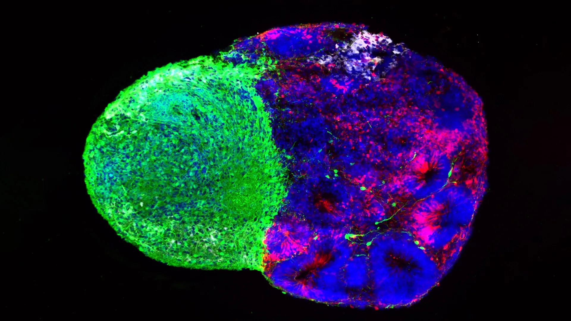
The intricate architecture of the human brain relies on a delicate balance of nutrient absorption and waste management, a process known as endocytosis. For decades, the mechanisms governing how neurons regulate the intake of extracellular material remained partially obscured, often categorized as a series of localized chemical reactions. However, researchers at Penn State University have recently unveiled a transformative discovery that shifts the paradigm of cellular biology: a hidden protein lattice known as the membrane-associated periodic skeleton (MPS) acts as a primary physical gatekeeper for the neuron. This structure, previously thought to be a mere scaffold for maintaining cellular shape, has been identified as a critical regulator of endocytosis, with profound implications for the development and progression of neurodegenerative conditions such as Alzheimer’s and Parkinson’s disease.
The study, led by Ruobo Zhou, an assistant professor of chemistry, biochemistry, and molecular biology at Penn State, and published in the journal Science Advances, provides the first comprehensive evidence that the MPS actively controls the rate and location of material uptake. By utilizing cutting-edge super-resolution microscopy, the team demonstrated that when this lattice is compromised, the uncontrolled influx of proteins—specifically those associated with plaque formation—accelerates cellular death. This discovery offers a new biological target for therapeutic interventions aimed at stabilizing the brain’s internal structure before irreversible cognitive decline occurs.
The Evolution of the Membrane-Associated Periodic Skeleton
The journey to this discovery began over a decade ago. In 2013, Ruobo Zhou was part of a research team at Harvard University that first identified the MPS. Using then-emerging imaging techniques, the team observed a highly organized, periodic structure composed of actin, spectrin, and associated proteins forming rings around the axons and dendrites of neurons. At the time, the scientific community largely viewed the MPS as a "passive" skeleton—a structural framework similar to the beams of a building, designed to prevent the long, thin protrusions of neurons from collapsing or snapping under mechanical stress.
However, the regularity of the MPS—spaced at approximately 180 to 190 nanometers apart—suggested a level of organization that hinted at a more complex functional role. In the years following the initial discovery, researchers began to wonder if this lattice influenced the movement of proteins within the cell membrane. The Penn State study represents the culmination of this inquiry, proving that the MPS is not just a support system but a dynamic "traffic controller" that determines which molecules are allowed to enter the neuron and at what speed.
Mapping the Nanoscale: Methodology and Super-Resolution Imaging
To observe the MPS in action, the Penn State team had to overcome the physical limitations of traditional light microscopy. Standard microscopes are limited by the diffraction of light, meaning they cannot clearly resolve structures smaller than 200 nanometers. Given that the MPS operates at the nanoscale—roughly 10,000 times thinner than a human hair—the researchers employed super-resolution fluorescence microscopy. This technology allows scientists to bypass the diffraction limit, providing a clear view of individual protein rings and their interactions with the cellular environment.
The researchers cultivated primary neurons in a laboratory setting and introduced fluorescent markers to specific proteins. By tracking these proteins in real-time, they could observe the process of endocytosis as it occurred. The experiment involved exposing the neurons to various external molecules, including nutrients and signaling proteins, and monitoring how the MPS reacted.
A critical phase of the study involved the deliberate manipulation of the MPS. The team used chemical agents and genetic tools to either strengthen or disrupt the lattice. They found that when the MPS was intact, endocytosis occurred at a steady, regulated pace. However, when the lattice was pharmacologically disrupted, the rate of material uptake skyrocketed. This suggested that the MPS acts as a physical barrier, a mesh-like "gate" that prevents the cell from being overwhelmed by the fluid and fragments surrounding it.
The Feedback Loop of Cellular Decay
One of the most significant findings of the study is the discovery of a "positive feedback loop" that contributes to cellular degradation. The researchers observed that the MPS is not only a regulator of endocytosis but is also a victim of its own regulatory failure.
When the rate of endocytosis increases—whether due to aging, environmental stress, or genetic factors—the process itself begins to weaken the MPS. As the cell pulls in more material, molecular signals are triggered that direct internal enzymes to cleave the proteins making up the lattice. This structural breakdown creates more gaps in the "gate," which in turn allows for even faster and more chaotic endocytosis.
"We discovered that this membrane skeleton is actively regulating the nutrient uptake process of neurons," Zhou stated. "You can think of it as a gatekeeper, guarding this physical barrier to not allow nutrient uptake to happen. When a neuron needs to take in a specific nutrient, this gatekeeper will open the gates and let it in." However, in a state of disease, the gatekeeper effectively loses control, leading to a cascade of cellular instability.
Linking the MPS to Alzheimer’s Pathogenesis
The implications of this feedback loop are particularly stark in the context of Alzheimer’s disease. A hallmark of Alzheimer’s is the accumulation of amyloid-beta plaques in the brain. These plaques are formed from the breakdown of amyloid precursor protein (APP).
In their cellular models, the Penn State team increased the levels of APP to mimic the early stages of neurodegeneration. They found that neurons with a weakened or damaged MPS took in APP at a significantly higher rate than healthy neurons. Once inside the cell, this APP was processed into amyloid-B42, a toxic fragment that is highly prone to aggregation.
The study demonstrated that the MPS acts as a first line of defense against the internal accumulation of these toxins. In neurons where the MPS was stabilized, the uptake of APP was limited, and the subsequent production of toxic amyloid-B42 was reduced. Conversely, neurons with a compromised MPS showed rapid accumulation of toxic fragments and displayed clear markers of apoptosis, or programmed cell death.
Jinyu Fei, a graduate student in the chemistry department and lead author of the study, noted the importance of this finding: "We created a model which is very much like Alzheimer’s disease and found that in some aging neurons, or neurons under pathologic conditions, the endocytosis of toxic proteins was enhanced, which caused stressing conditions, ultimately leading to neuron deaths."
Supporting Data and Broader Biological Context
The data provided by the Penn State study adds a crucial layer to our understanding of "proteostasis"—the process by which cells maintain the health of their protein population. In neurodegenerative diseases, proteostasis fails, leading to the "clogging" of neurons with misfolded proteins.
Statistical analysis from the study showed a direct correlation between the density of the MPS lattice and the frequency of clathrin-mediated endocytosis (the most common form of nutrient uptake). In regions of the neuron where the MPS was densest, endocytic events were rare. In regions where the MPS was sparse or damaged, endocytic pits formed with nearly triple the frequency.
Furthermore, the study highlighted that this mechanism is not limited to a single type of uptake. The MPS appeared to regulate nearly every major pathway of endocytosis, including macropinocytosis and fast endophilin-mediated endocytosis. This suggests that the MPS is a universal regulator of the neuron’s interaction with its external environment.
Scientific Reactions and the Shift in Neurobiology
The broader scientific community has reacted to these findings with cautious optimism. For years, Alzheimer’s research has focused heavily on clearing plaques after they have already formed—a strategy that has seen limited success in clinical trials. The Penn State research suggests a "pre-emptive" strategy: focusing on the structural integrity of the neuron itself to prevent the toxic uptake from happening in the first place.
Independent experts in neurocytology have noted that this research bridges the gap between structural biology and pathology. While it was known that the cytoskeleton changes during aging, the specific functional consequence of those changes on nutrient uptake was a "missing link" in the literature. By identifying the MPS as the regulator, Zhou and his team have provided a concrete physical mechanism for why aging brains become more susceptible to protein toxicity.
Implications for Future Therapies
The discovery of the MPS’s role as a gatekeeper opens several new avenues for drug development. Current Alzheimer’s treatments, such as monoclonal antibodies like lecanemab, work by targeting and removing amyloid-beta from the brain tissue. While effective at clearing plaques, these treatments often come late in the disease’s progression.
A therapy based on the Penn State findings would likely focus on "stabilizing the gate." This could involve:
- Small Molecule Stabilizers: Developing drugs that reinforce the bonds between actin and spectrin in the MPS, making the lattice more resistant to enzymatic cleavage.
- Enzyme Inhibitors: Identifying the specific "molecular scissors" that cut the MPS during high-stress endocytosis and developing inhibitors to block their activity.
- Early Diagnostic Tools: Using the state of the MPS as a biomarker for early-stage neurodegeneration, potentially allowing for intervention years before cognitive symptoms appear.
"We think this could open the door for future therapies such as a protein target for neurodegenerative disease treatment," Fei said. "Preserving or stabilizing the MPS might offer a way to slow the early, hidden cellular changes that precede Alzheimer’s symptoms."
Conclusion
The identification of the membrane-associated periodic skeleton as a functional gatekeeper represents a landmark shift in neurobiological research. By moving beyond the view of the cellular skeleton as a passive structure, the Penn State team has uncovered a dynamic system that sits at the heart of how neurons live, age, and die.
As the global population ages, the prevalence of neurodegenerative diseases is expected to rise sharply, with Alzheimer’s cases projected to reach 13.8 million in the United States alone by 2050. The discovery of the MPS’s regulatory role provides a vital new map for scientists racing to understand these conditions. While further research is needed to translate these laboratory findings into clinical treatments, the "gatekeeper" within our neurons may hold the key to preserving cognitive health in the face of aging and disease.