Scientists at the Johns Hopkins Kimmel Cancer Center and the Johns Hopkins Bloomberg School of Public Health have unlocked a fundamental mechanism by which a rare and aggressive form of kidney cancer progresses. In a study published April 22 in Cell Reports, researchers detailed how abnormal combinations of rearranged genes create proteins that form microscopic liquid droplets within cellular nuclei. These droplets act as hyper-efficient command centers that dysregulate gene expression, driving the rapid growth and metastasis of tumors. Supported by funding from the National Institutes of Health (NIH), the breakthrough identifies a novel vulnerability in cancer biology that could pave the way for targeted therapies where none currently exist.
The findings focus primarily on translocation renal cell carcinoma (tRCC), an uncommon malignancy driven by chromosomal aberrations. Unlike more prevalent forms of kidney cancer, tRCC typically affects younger populations and lacks standard, effective treatment protocols outside of surgical resection in early stages. By discovering that these disease-driving fusion proteins rely on physical phase separation—the formation of liquid condensates—to exert their malignant influence, researchers have illuminated a tangible target for future pharmaceutical intervention.
Background Context and the Mechanics of Translocation Renal Cell Carcinoma
Translocation renal cell carcinoma arises from a structural breakdown and incorrect repair of chromosomes. During this genetic accident, a segment of DNA is swapped, fusing the tail end of the TFE3 gene with the beginning of one of roughly 20 other genes. The most frequent fusion partners identified in clinical pathology are PRCC, NONO, and SFPQ, with NONO and SFPQ alone accounting for approximately 40 percent of all TFE3 gene fusions.
These genetic alterations produce chimeric proteins—molecules known as TFE3 fusion proteins—that do not exist in healthy human physiology. While oncologists and molecular biologists have long recognized that these aberrant proteins trigger tRCC, the precise biochemical pathways by which they initiate and sustain tumor formation remained elusive.
Historically, studying rare cancers like tRCC presents significant hurdles due to small patient cohorts and the complexity of targeting transcription factors, which are notoriously difficult to inhibit using traditional small-molecule drugs. The Johns Hopkins team circumvented this barrier by analyzing the physical properties of the fusion proteins themselves rather than merely attempting to block their chemical interactions.
Chronology and Methodology of the Discovery
The research journey leading to the April 2022 publication involved a multidisciplinary collaboration between cell biologists, oncologists, and computational geneticists.
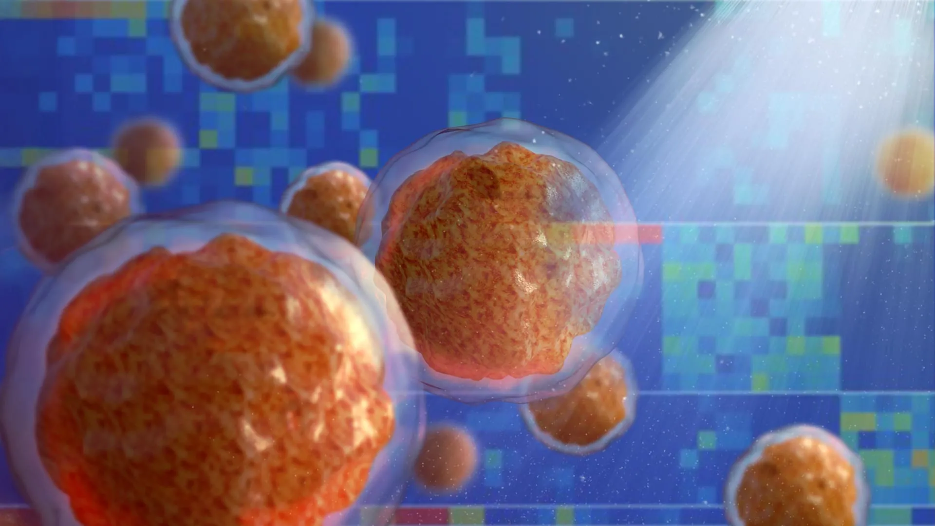
Initially, senior author Danfeng "Dani" Cai, Ph.D., assistant professor of biochemistry and molecular biology at the Johns Hopkins Bloomberg School of Public Health, and her laboratory team engineered patient-derived kidney cancer cells by attaching fluorescent glowing tags to the TFE3 fusion proteins. Utilizing advanced live-cell microscopy, the researchers observed a striking phenomenon: rather than distributing evenly throughout the cell, the fusion proteins coalesced into distinct, glowing puncta—dots—within the nucleus, the compartment where cellular DNA is housed.
Recognizing these structures as liquid-liquid phase condensates—a specialized area of research in the Cai laboratory—the team investigated their function. They discovered that these droplets were not inert aggregations; instead, they actively concentrated marker proteins typically found on active genes alongside transcription-activating proteins.
To map out the exact interaction between these droplets and the cellular genome, Dr. Cai partnered with Eneda Toska, Ph.D., an assistant professor of oncology at the Johns Hopkins Kimmel Cancer Center. Utilizing genomic mapping techniques, the investigators analyzed how the TFE3 fusion proteins interface with chromatin, the complex of DNA and protein that makes up chromosomes.
The chromatin inside a healthy cell is dynamically packaged, resembling beads on a string. Where the string is tightly wound around the histone beads, genes are effectively silenced. Where the string is open and accessible, transcription machinery can bind to switch genes on. Dr. Toska’s analysis revealed that TFE3 fusion proteins actively remodel this chromosomal landscape. By executing chemical modifications, the fusion proteins open and close chromatin sites with precision, upregulating target genes that govern cell proliferation and cellular migration—two hallmarks required for tumor expansion and metastasis.
In the final phase of the experimental timeline, the researchers performed targeted deletions of specific segments within the TFE3 fusion proteins to isolate the structural domains responsible for condensate formation. When they removed a tiny segment that normally forms a coiled-coil structure—a structural motif where alpha-helices wind around each other—the functional consequences were immediate. The modified fusion proteins lost their ability to form liquid droplets, and correspondingly, they failed to activate the cancer-promoting genes.
Expert Statements and Official Responses
The discovery has generated considerable interest within the broader oncological research community due to its conceptual overlap with other difficult-to-treat malignancies.
"Other cancers, such as Ewing sarcoma and leukemia, are caused by fusion genes as well," notes Dr. Danfeng Cai. "It is entirely possible that these fusion genes form similar droplets, or condensates, that regulate genes in these cancers and could react to similar treatment strategies."
Dr. Eneda Toska emphasized the structural remodeling capacity of the chimeric proteins discovered during the study. "We found that these fusion proteins open and close different sites on the chromatin by making chemical modifications," Dr. Toska explained. "They bind, regulate, and redesign the chromosome landscape, interacting with target genes that promote cell proliferation and movement—functions that cancer needs to grow and spread."
Dr. Cai also highlighted the evolutionary hijack mechanism at play. "Individually, all the protein components found in the TFE3 fusions, including full-length TFE3, NONO, and SFPQ, are typically involved in the cell machinery that turns on genes to make proteins," she stated. "However, we found when in the form of these fusion proteins, they acquire an even stronger ability to control what genes get tuned on."
Supporting Data and Funding Sources
The rigorous scope of the study required substantial infrastructural and financial backing from federal agencies and specialized research foundations. The work was supported by grants from the National Institutes of Health (NIH), specifically the National Institute of General Medical Sciences (grant R35GM142837); the National Cancer Institute (grants K22CA245487, R01CA276187, and K01CA245124); and the National Human Genome Research Institute (grants R01HG013409 and R01HG010889).
Additional financial contributions included a Department of Defense Kidney Cancer Idea Development Award (grant W81XWH2210900), a Jayne Koskinas Ted Giovanis grant, and a Johns Hopkins Provost Catalyst Award.
The extensive author roster reflects the cross-disciplinary nature of the investigation, featuring contributions from Choon Leng So, Ye Jin Lee, Wanlu Chen, Binglin Huang, Emily De Sousa, Yangzhengyu Gao, Marie Elena Portuallo, Sumaiya Begum, Kasturee Jagirdar, Vito Rebecca, and Hongkai Ji of the Johns Hopkins Bloomberg School of Public Health; Bujamin Vokshi of the Johns Hopkins University School of Medicine; and W. Marston Linehan of the National Cancer Institute. In disclosures accompanying the study, Dr. Toska reported receiving research grants and consulting fees from AstraZeneca, as well as consulting fees from Menarini.
Broader Impact, Therapeutic Implications, and Future Directions
The implications of this study extend well beyond the immediate clinical pathology of translocation renal cell carcinoma. By demonstrating that oncogenic fusion proteins rely on physical phase separation to execute their transcriptional programs, the Johns Hopkins team has added momentum to an emerging paradigm in molecular biology: the targeting of biomolecular condensates for therapeutic intervention.
For decades, drug discovery has focused on blocking active enzymatic pockets on proteins using small molecules. However, transcription factors and fusion proteins often lack well-defined enzymatic pockets, rendering them "undruggable" by traditional pharmacological standards. Liquid condensates present an entirely different structural target. Because these droplets depend on weak, multi-valent interactions between specific protein domains—such as the coiled-coil regions identified in the TFE3 fusions—pharmaceutical chemists can theoretically design small molecules or drugs designed to dissolve or prevent the formation of these condensates.
Looking ahead, the Johns Hopkins research team aims to systematically identify all additional molecular components residing within the TFE3-driven liquid droplets. Pinpointing these co-factors will enable high-throughput drug screening assays. By testing chemical libraries for compounds capable of disrupting these specific condensates, scientists hope to develop the first targeted, non-surgical therapies for patients suffering from tRCC and potentially translate these findings to other fusion-gene-driven cancers like leukemias and sarcomas.