Glioblastoma has long been understood by neuro-oncologists as one of the most formidable adversaries in modern medicine. As the most common and lethal form of primary malignant brain cancer in adults, it is characterized by relentless growth, invasive infiltration into healthy neural architecture, and a near-inevitable recurrence profile. For decades, the medical community has approached this devastating malignancy strictly as a localized affliction of the central nervous system. Treatment paradigms have historically centered on surgical resection to debulk the main tumor mass, followed by aggressive courses of fractionated radiation therapy and systemic chemotherapy, typically utilizing temozolomide. Yet, despite these grueling multi-modality interventions, the prognosis remains grim; the median survival time for diagnosed individuals hovers stubbornly at approximately 15 months.
This clinical intransigence has prompted researchers to look beyond the immediate boundaries of the tumor bed to understand why conventional therapies consistently falter. A landmark study published on October 3 in the journal Nature Neuroscience has fundamentally shifted this perspective. Conducted by a multidisciplinary team of investigators from the Montefiore Einstein Comprehensive Cancer Center (MECCC) and the Albert Einstein College of Medicine, the research reveals that glioblastoma is not merely a localized brain disease. Instead, it operates as a systemic disruptor that actively erodes the skull, fundamentally alters the composition of bone marrow housed within the cranial vault, and reshapes the body’s immune landscape. These profound systemic effects not only explain the failure of localized therapies but also issue a critical warning regarding the use of common bone-density medications in this patient population.
Unveiling the Cranial-Neural Axis
To understand the magnitude of this discovery, one must examine the anatomical and physiological relationship between the brain and the skull. Traditionally, the cranium was viewed simply as a protective, inert casing designed to shield the delicate neural tissues of the central nervous system from physical trauma. However, recent scientific breakthroughs have painted a far more dynamic picture, demonstrating that the skull and the brain are intimately connected through an intricate network of microscopic channels. These vascular and structural pathways facilitate a constant, bi-directional exchange of signaling molecules, fluids, and immune cells between the cranial bone marrow and the intracranial environment.
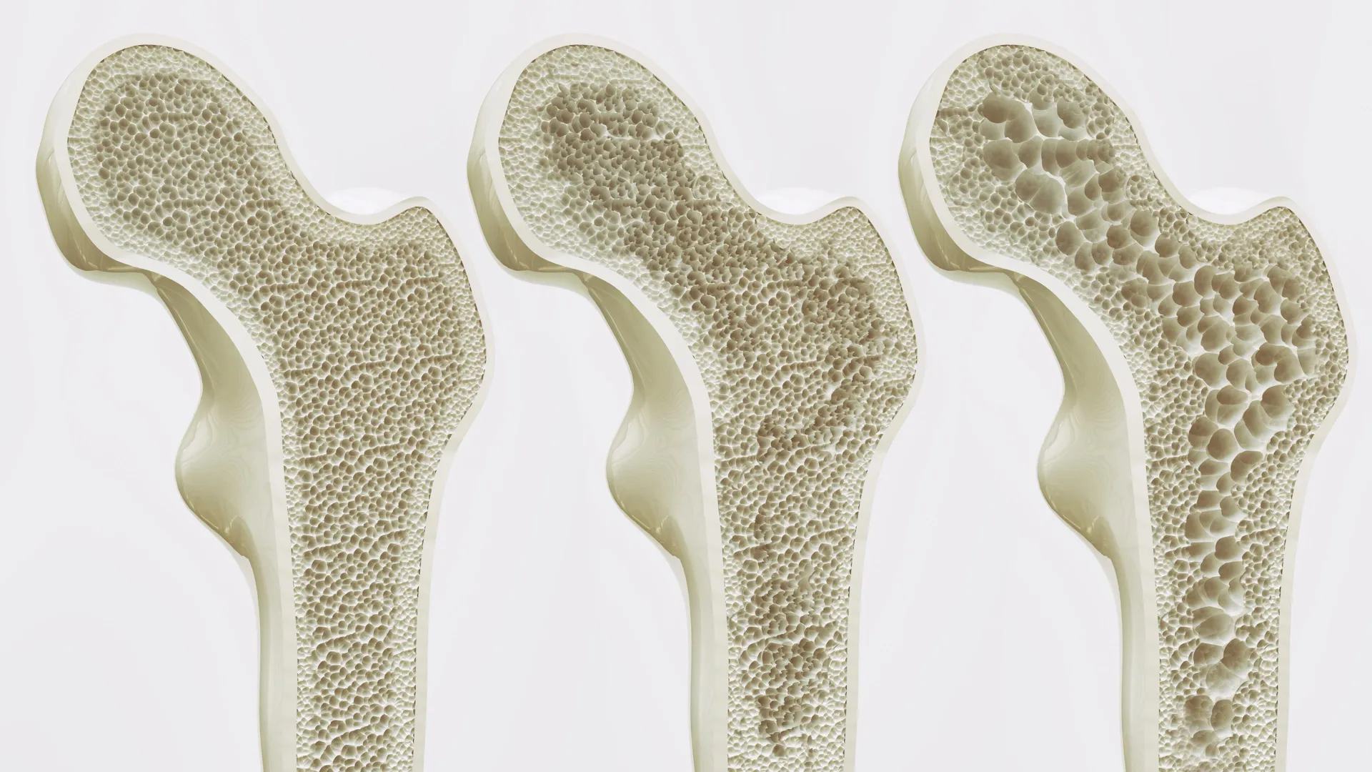
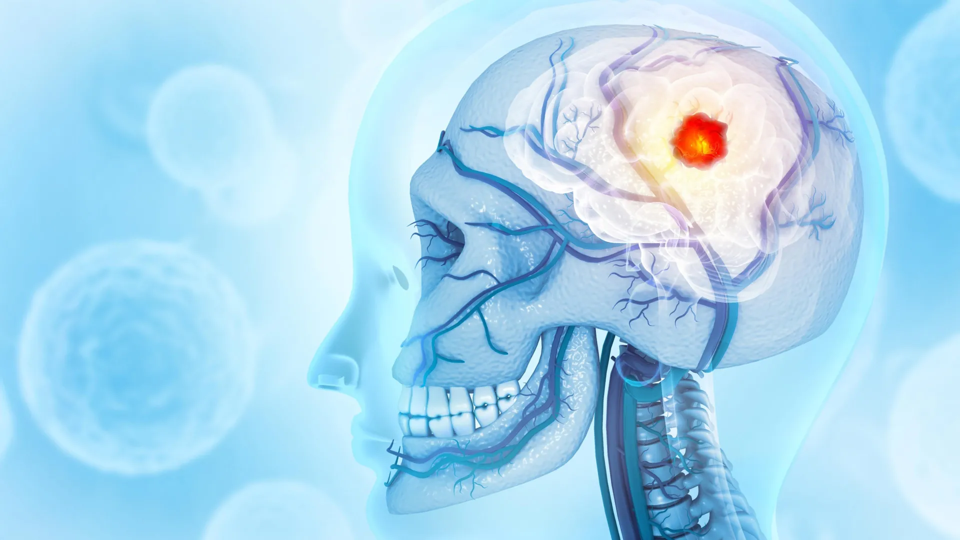
The research team, led by corresponding author Dr. Jinan Behnan—an assistant professor in the Leo M. Davidoff Department of Neurological Surgery and the Department of Microbiology & Immunology at Einstein, alongside his colleagues at the NCI-designated MECCC—set out to investigate how glioblastoma exploits this cranial-neural axis. Utilizing advanced, high-resolution imaging modalities, the investigators observed murine models engineered to develop two distinct subtypes of glioblastoma. The results of these imaging analyses were striking: the presence of the brain tumors induced significant structural degradation of the skull bones.
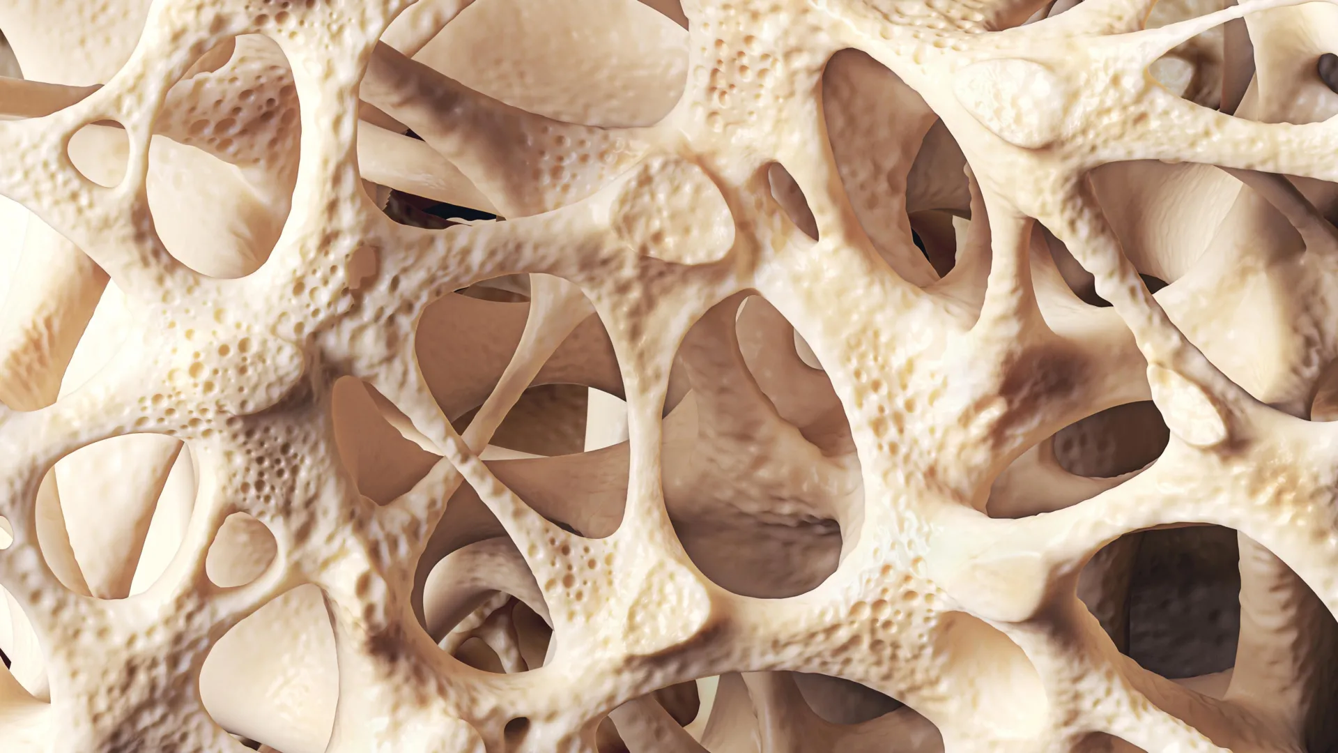
This pathological erosion was most pronounced along the cranial sutures—the fibrous joints where adjacent plates of the skull fuse together. Crucially, this bone loss appeared to be a unique hallmark of glioblastoma and other highly aggressive intracranial malignancies. Control experiments involving mice subjected to ischemic strokes, traumatic brain injuries, or metastatic cancers originating outside the central nervous system displayed no comparable cranial degradation. To validate these animal models against human pathology, the researchers analyzed computed tomography (CT) scans from human glioblastoma patients. The clinical imaging corroborated the murine findings, revealing localized reductions in skull thickness in precisely the same anatomical regions observed in the laboratory models.
As the skull bones eroded in the mice, the microscopic channels linking the cranium to the brain underwent significant physical remodeling, increasing in both number and cross-sectional area. The researchers hypothesized that these dilated channels serve as superhighways for pathological communication, allowing the tumor to project molecular distress signals directly into the cranial bone marrow and, conversely, permitting altered immune cells to migrate backward into the brain.
A Radical Shift in Bone Marrow Immunity
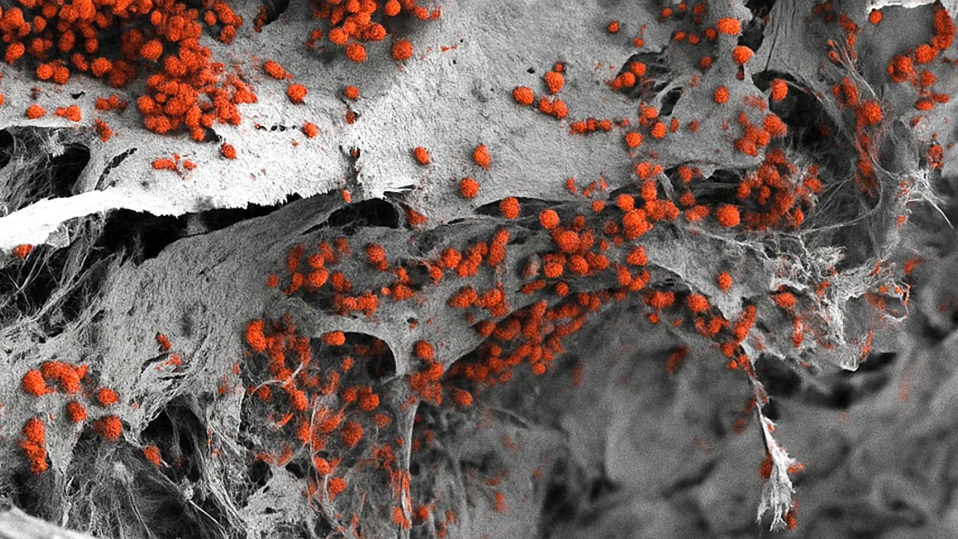
Like other flat and irregular bones throughout the skeletal system, the human skull contains specialized red bone marrow that serves as a primary factory for the continuous production of hematopoiesis—the generation of red blood cells, platelets, and a diverse array of immune cells. When Dr. Behnan’s team performed single-cell RNA sequencing to analyze the cellular composition of the skull marrow in tumor-bearing mice, they uncovered a radical reprogramming of the immune microenvironment.
Glioblastoma profoundly skewed the developmental trajectory of immune cells within the cranial vault, triggering a massive surge in pro-inflammatory myeloid cells. Specifically, the levels of inflammatory neutrophils within the skull marrow nearly doubled compared to healthy controls. Concurrently, the disease nearly eradicated several crucial populations of B cells, including those responsible for antibody production, alongside other essential adaptive immune lineages.
Co-author Dr. E. Richard Stanley, a professor of developmental and molecular biology at Einstein, emphasized the clinical implications of this cellular imbalance. The structural widening of the skull-to-brain channels acts as a conduit, driving a massive influx of these rogue pro-inflammatory cells directly from the cranial marrow into the tumor microenvironment. Rather than mounting an effective defense against the malignancy, these recruited cells are hijacked by the tumor, rendering the glioblastoma increasingly aggressive, treatment-resistant, and ultimately lethal.
Intriguingly, the study revealed a striking dichotomy in how different skeletal sites respond to the systemic presence of brain cancer. While the skull marrow was activated to churn out pro-inflammatory immune cells, the bone marrow of the femur—located far from the cranium in the lower extremity—exhibited the exact opposite response. In the femur, glioblastoma suppressed the genes required for normal immune cell production. This regional disparity underscores the complex, systemic nature of the disease, proving that glioblastoma exerts highly localized effects on the adjacent cranial bones that are entirely distinct from its systemic footprints elsewhere in the body.
The Unintended Danger of Osteoporosis Therapies
Armed with the revelation that glioblastoma actively degrades skull bone and manipulates cranial marrow, the research team pivoted to investigate whether existing pharmaceutical interventions designed to prevent bone loss could alter the course of the disease. Millions of individuals worldwide, particularly aging populations, rely on anti-osteoporosis medications to preserve bone mineral density and prevent fractures. Among the most common and effective treatments are bisphosphonates, such as zoledronic acid, and monoclonal antibodies, such as denosumab, both of which are approved by the U.S. Food and Drug Administration (FDA).
To test their efficacy in this novel context, the researchers administered these two anti-osteoporosis drugs to mouse models bearing glioblastoma tumors. Both agents successfully halted the pathological erosion of the skull bones, achieving their primary pharmacological objective of preserving bone density. However, the downstream consequences for the brain tumors were alarming and divergent.
One of the tested medications, zoledronic acid, actively fueled tumor progression, accelerating the growth of specific glioblastoma subtypes. Furthermore, when the researchers attempted to combine these bone-loss therapies with immunotherapy—specifically anti-PD-L1 checkpoint inhibitors, which are designed to unleash the body’s T cells against cancer—both zoledronic acid and denosumab completely neutralized the therapeutic benefits of the immunotherapy. By blocking the bone-remodeling pathways, the drugs inadvertently hindered the mechanisms required for tumor-fighting T cells to mount an effective assault on the malignancy.
This unexpected pharmacological interaction carries profound implications for clinical practice. Many glioblastoma patients, particularly elderly individuals or those undergoing corticosteroid treatments that induce bone loss, are at high risk for osteoporosis and skeletal fractures. The findings from the Nature Neuroscience study suggest that clinicians must exercise extreme caution when prescribing standard anti-resorptive medications to patients diagnosed with brain cancer, as certain agents may inadvertently promote tumor growth and compromise the efficacy of emerging immunotherapies.
Broader Clinical Implications and Future Therapeutic Strategies
The publication of this study marks a conceptual turning point in neuro-oncology. For decades, clinical trials targeting glioblastoma have focused almost exclusively on optimizing surgical margins, delivering higher doses of focal radiation, or developing targeted small-molecule inhibitors that cross the blood-brain barrier to attack tumor cells directly. The repeated failure of these trials to significantly extend overall survival has long frustrated researchers and clinicians alike.
By reframing glioblastoma as a systemic disease that engages in active crosstalk with the cranial skeleton and the immune system, this research opens entirely new avenues for therapeutic intervention. The data strongly indicate that future treatment strategies must look beyond the brain parenchyma and incorporate multi-systemic approaches.
Specifically, therapeutic protocols will need to focus on restoring the normal immunological balance within the skull bone marrow. Rather than merely halting bone loss with traditional anti-osteoporosis agents—which carry the risk of exacerbating tumor growth—future therapies will likely need to selectively suppress the hyper-production of pro-inflammatory neutrophils and monocytes while actively rescuing the synthesis of protective T and B cells. Designing pharmacological agents that can selectively modulate cranial bone marrow immunity without triggering systemic toxicity will be a primary objective for translational researchers in the coming years.
The Collaborative Effort and Global Scope
The breadth of this discovery reflects a massive, highly collaborative international research effort. The published paper, titled Brain Tumors Induce Widespread disruption of Calvarial Bone and Alteration of Skull Marrow Immune Landscape, features contributions from a vast consortium of scientists spanning multiple prestigious institutions across the United States, Japan, and Sweden.
Alongside Dr. Behnan and Dr. Stanley, the Montefiore Einstein Comprehensive Cancer Center and Albert Einstein College of Medicine contingent included Abhishek Dubey, Biljana Stangeland, Imane Abbas, Dr. David Fooksman, Wade R. Koba, Dr. Jinghang Zhang, Dr. Benjamin T. Himes, Dr. Derek Huffman, Zhiping Wu, Rachel Welch, David Reynolds, Dr. Kostantin Dobrenis, Qinge Ye, Kevin Fisher, and Dr. Emad Eskandar.
International collaborators brought specialized expertise to the project, including Erika Yamashita, Yutaka Uchida, and Professor Masaru Ishii from Osaka University in Osaka, Japan; Robert A. Harris from the Karolinska Hospital Solna in Stockholm, Sweden; Gregory M. Palmer from Duke University Medical Center in Durham, North Carolina; Olivia R. Lu and Professor Winson S. Ho from the University of California, San Francisco; and Alexander F. Fiedler from the German Rheumatism Research Center (DRFZ) and Freie Universität Berlin in Germany.
As approximately 15,000 individuals in the United States continue to receive a glioblastoma diagnosis each year—facing a formidable disease that has resisted meaningful improvements in long-term survival for decades—this collaborative milestone offers a renewed sense of direction. By dismantling the long-held dogma that glioblastoma is merely a localized brain tumor, researchers have charted a new map for therapeutic innovation, bringing the medical community one step closer to turning the tide against one of cancer’s most relentless frontiers.