Why do some new skills seem to click almost instantly while others remain frustratingly difficult even after repeated practice? For decades, neuroscientists have attributed the disparity in learning outcomes primarily to innate cognitive talent, the intensity of focus, or the frequency of repetition. However, recent evidence suggests that the physical architecture of the brain—and its constant, rhythmic dialogue with the rest of the body—may be the missing variable in the equation. A groundbreaking study published in the journal iScience on August 25, 2026, by researchers at Tohoku University, suggests that the secret to permanent mastery may lie in a previously overlooked mechanism: the physiological state of the brain following a training session, modulated by the vagus nerve.
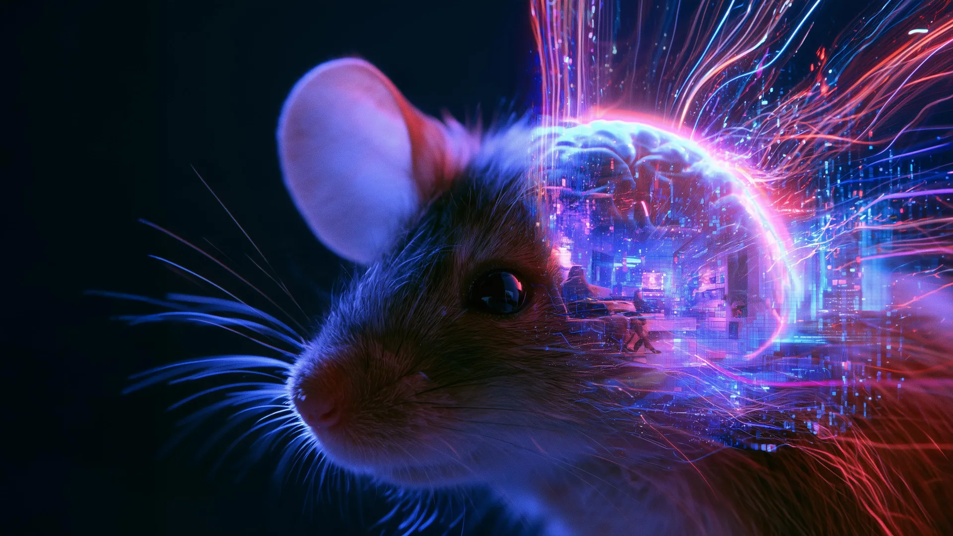
The study, which utilized mouse models to observe the long-term consolidation of motor skills, indicates that stimulating the vagus nerve after a period of learning can act as a catalyst for memory retention. By manipulating the blood volume oscillations within the cerebellum, researchers have uncovered a potential "hidden window" for synaptic plasticity that could, in the future, be harnessed to accelerate learning in humans.
The Anatomy of the Vagus Nerve and Neuromodulation
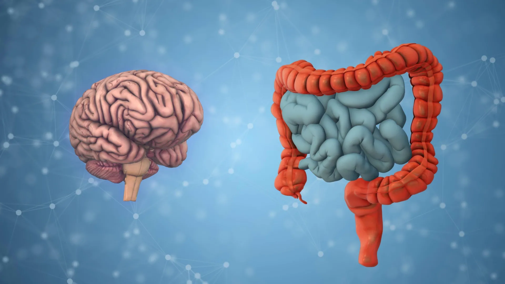
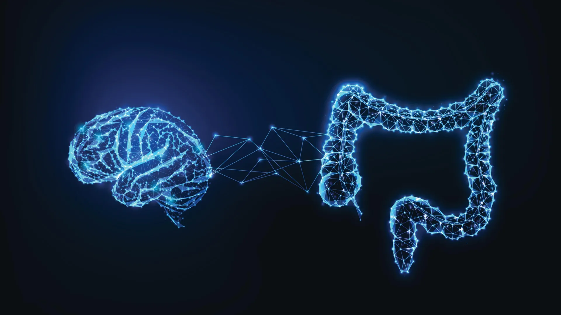
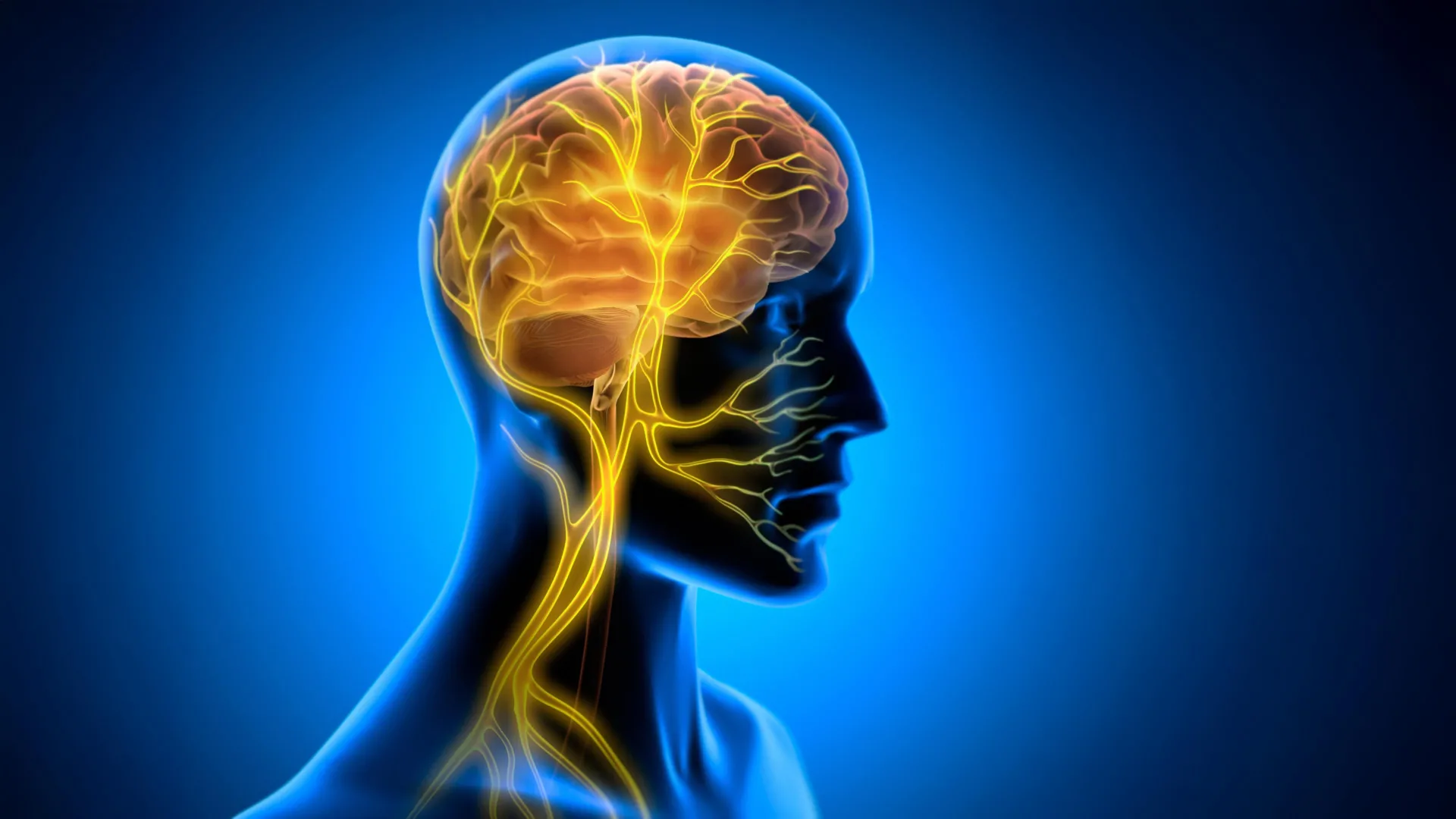
The vagus nerve is the longest cranial nerve in the human body, serving as the primary superhighway for the autonomic nervous system. It creates a continuous loop of bidirectional communication, relaying sensory feedback from the heart, lungs, and digestive tract to the brainstem, while simultaneously carrying regulatory signals from the brain back to the visceral organs. Because of its expansive reach, the vagus nerve has long been a target for medical intervention.
Vagus Nerve Stimulation (VNS) involves the use of an implanted or transcutaneous device to deliver mild electrical pulses to the nerve. Since the late 1990s, VNS has been clinically approved for the treatment of refractory epilepsy and treatment-resistant depression. Its mechanism of action has historically been understood through the lens of neurotransmitter modulation—specifically, its ability to influence the release of norepinephrine and acetylcholine, chemicals critical for arousal and focus.
However, the Tohoku University research team, led by Professor Ko Matsui, hypothesized that VNS might affect the brain through a more physical, mechanical pathway: the regulation of cerebral blood flow. By modulating the vascular environment of the brain, VNS may be creating a metabolic "sweet spot" that allows the neural connections formed during practice to solidify into long-term memories.
Chronology of the Tohoku University Experiment
The research project followed a rigorous experimental timeline to isolate the effects of VNS on motor learning. The team focused on a task known as the horizontal optokinetic response (HOKR). This task requires mice to coordinate eye movements in response to moving visual stripes—an automatic reflex similar to the human ability to stabilize vision while observing a train passing by.
- Baseline Training (Days 1–3): Mice were subjected to HOKR training sessions. During this phase, baseline performance levels were established to ensure all subjects began from an equitable position.
- Intervention Phase (Post-Training): Unlike previous studies that applied VNS during the learning task to enhance focus, the Tohoku team applied VNS exclusively after the training sessions concluded.
- Observation and Measurement (Days 4–7): Researchers utilized fiber photometry to track blood volume activity in the cerebellar flocculus—the specific region of the brain responsible for HOKR motor learning.
- Data Correlation: By the fifth day, the researchers cross-referenced the intensity of blood volume oscillations with the performance improvements of the mice.
The results were statistically significant. Mice that received VNS post-training demonstrated a marked improvement in long-term retention of the eye-movement task compared to control groups that received no stimulation. Crucially, the stimulation had no immediate impact on performance during the training itself, suggesting that the intervention specifically targets the consolidation phase—the period when the brain transitions information from short-term to long-term memory.
The Role of Vascular Rhythms in Learning
One of the most striking findings of the study was the discovery of a two-phase vascular response triggered by VNS. Upon stimulation, local blood volume in the cerebellar flocculus initially decreased before experiencing a sustained rise. When the stimulation was repeated, these fluctuations formed rhythmic oscillations.
"Our findings suggest that VNS may open a hidden window of opportunity for enhanced learning by making the brain environment more receptive to long-lasting change," explains Professor Matsui. The team’s data indicated a direct correlation between the amplitude of these blood volume oscillations and the success rate of the learning task. Those mice that exhibited more pronounced rhythmic vascular changes showed significantly higher proficiency in their motor skills by the end of the study period.
This discovery moves beyond traditional neurochemical explanations of learning. It suggests that the brain is not just a computational organ, but a dynamic, metabolic environment. By rhythmically pulsing blood into specific regions, the body may be providing the necessary nutrients and waste-clearance signals required to stabilize new synaptic connections. Lead author Junyu Chen noted, "Our brains may be more strongly influenced by the body than we imagine. By tuning the brain’s metabolic environment, including rhythmic vascular movements, we may eventually unlock capacities that would otherwise remain latent."
Broader Implications for Neuro-Rehabilitation and Education
The implications of this research are vast, spanning the fields of clinical neurology, cognitive psychology, and educational technology. If VNS can be refined to optimize the brain’s "receptivity" to information, it could fundamentally change the approach to neuro-rehabilitation.
For patients recovering from strokes or traumatic brain injuries, the process of relearning basic motor functions—such as grasping objects or walking—is often exhausting and slow. If a post-training VNS protocol can enhance the consolidation of these functions, recovery timelines could be drastically shortened. Furthermore, the ability to "tune" the brain’s vascular environment could eventually be used to address learning disabilities, where the standard synaptic consolidation process is impaired.
However, researchers caution that the leap from mice to humans is significant. The complexity of the human brain, combined with the nuances of VNS delivery, means that any clinical application is still years away. The current study focused on a relatively simple motor task; whether these findings translate to complex intellectual learning, such as language acquisition or mathematical reasoning, remains to be seen.
Expert Perspectives and Future Directions
The scientific community has reacted with cautious optimism. Experts in neuroplasticity emphasize that the study provides a compelling, evidence-based argument for the "body-to-brain" communication loop. By shifting the focus from the brain as an isolated entity to the brain as part of an integrated system, the Tohoku University team has opened a new frontier in neuroscience.
Future research, according to the team, will focus on two key areas:
- Protocol Optimization: Determining the ideal frequency, duration, and timing of VNS to maximize results while minimizing side effects.
- Mechanism Mapping: Identifying the exact pathways through which the vagus nerve communicates with the cerebellar vasculature to trigger these oscillations.
As the scientific community continues to explore the "hidden window" of opportunity discovered by the Tohoku researchers, the traditional understanding of how we acquire skills is undergoing a necessary transformation. We are moving toward an era where learning may no longer be viewed as a purely mental exertion, but as a physiological process that can be managed, supported, and ultimately enhanced through the sophisticated interplay of the body’s major communication pathways.
As we continue to map these interactions, the boundary between the body’s internal health and our cognitive potential will continue to blur, offering the potential for a new generation of bio-technological interventions designed to make learning more efficient, more durable, and more accessible than ever before.