The revelation that these structures, termed "non-synaptic varicosities," are a standard feature of healthy neurons—rather than mere hallmarks of pathology—has profound implications for our understanding of neurodegenerative diseases, neural plasticity, and the fundamental mechanics of communication within the central nervous system.
A Paradigm Shift in Axonal Morphology
Axons are the long, slender projections of nerve cells that transmit electrochemical impulses to distant targets. Traditional histology and electron microscopy have historically relied on chemical fixation and dehydration—processes that involve treating tissue with solvents and heat to preserve it. While effective for observing gross anatomy, these methods often shrink or distort the delicate, water-rich structures of the brain.
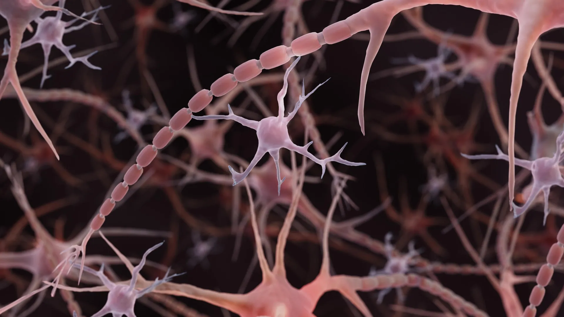
The Johns Hopkins team, led by Dr. Shigeki Watanabe, an associate professor of cell biology and neuroscience, employed a sophisticated alternative known as high-pressure freezing electron microscopy. By flash-freezing samples, researchers were able to capture the neurons in a state remarkably close to their living condition, avoiding the “raisin-like” shrinkage caused by traditional chemical dehydration. Under these conditions, the axons revealed a distinct, beaded architecture. These swellings are not the well-known synaptic varicosities—the specialized sites where neurotransmitters are released—but rather smaller, repeating irregularities along the length of the axon itself.
The Chronology of Discovery
The journey toward this discovery began years prior, grounded in observations made by Dr. Watanabe during his work with the nematode Caenorhabditis elegans. When he noted recurring structural irregularities along the axons of these worms, he initiated a cross-disciplinary dialogue with collaborators, including Dr. Graham Knott, to determine if these features were incidental or functional.
Following the initial hypothesis that the internal protein skeleton might be responsible for the “pearling,” graduate student Jacqueline Griswold conducted rigorous tests to determine if disrupting the cytoskeleton would eliminate the swellings. The results were unexpected: the pearls remained, indicating that the structure was governed by forces outside of, or in addition to, the internal protein framework. This realization led the team to investigate membrane physics, collaborating with theoretical biophysicist Dr. Padmini Rangamani of the University of California San Diego.
The timeline of formal findings began in earnest in late 2024, when the study was published online. The significance of the work was immediately recognized by the National Institute of Mental Health (NIMH), which awarded a major grant to Watanabe and Rangamani to further investigate the nexus of membrane physics and neural signaling. This momentum continued into 2025 and 2026, as the researchers successfully identified these same pearled structures in human cortical tissue, confirming that the phenomenon was not limited to murine models but was a feature of human biology.
Mechanics of the Membrane: Physics Over Anatomy
The collaborative team utilized mathematical modeling to understand the physical forces at play. By treating the axonal membrane as a fluid structure subject to tension and surface area constraints, they demonstrated that “pearling” is an emergent property of the membrane’s physical environment. The model suggested that as the membrane’s tension fluctuates—often dictated by the concentration of cholesterol and the local chemical environment—the diameter of the axon naturally shifts.
Experimental data confirmed this theory. When the researchers introduced substances that altered the fluidity of the membrane—such as removing cholesterol—the structural integrity of the pearls changed, leading to a measurable decline in the velocity of electrical signals traveling down the axon. This suggests that the “beads” act as a regulatory mechanism. A wider axonal diameter, in certain configurations, may act as a highway for ions, allowing for more rapid signal transmission by reducing resistance and preventing the “traffic jams” that occur in narrower, more constricted channels.
The Role of Neural Activity and Plasticity
Perhaps the most compelling finding is that these pearls are not fixed in size. The Johns Hopkins study revealed that after high-frequency electrical stimulation, the pearl-like regions underwent significant morphological changes, becoming longer and wider. These changes persisted for at least 30 minutes, suggesting that the structure of an axon can be remodeled in response to its own activity.
This provides a potential physical basis for synaptic plasticity, the process by which connections between neurons strengthen or weaken over time—a cornerstone of learning and memory. If the axon itself changes its physical geometry based on the frequency of the signals it carries, the nervous system possesses an intrinsic mechanism to fine-tune its own speed and efficiency. This challenges the long-held view that neural communication is governed solely by the strength of the synapses themselves, suggesting that the "cables" are just as plastic as the connections they form.
Broader Implications and Future Directions
The implications of this discovery extend far beyond basic neuroanatomy. For decades, clinicians have identified beaded, swollen axons as clear indicators of neurodegeneration, often associated with conditions such as Parkinson’s disease, Alzheimer’s, or multiple sclerosis. In these pathological states, beading is frequently accompanied by the breakdown of the cell membrane and internal skeletal damage.
The distinction between "normal" pearling and "pathological" beading is now a critical area of investigation. If researchers can define the precise physical parameters that separate functional, healthy pearling from destructive, disease-driven swelling, it could open new doors for diagnostic medicine. Distinguishing between a neuron that is adapting its shape to optimize signaling and one that is undergoing terminal degradation could lead to earlier interventions in neurodegenerative disease.
Furthermore, the research has inspired a wider effort to catalog and control axonal dimensions. In July 2026, researchers at the University of Edinburgh published findings in PLOS Biology detailing a high-throughput screen of 880 compounds in zebrafish. By identifying 33 compounds that could modulate axon width, they provided a proof-of-concept that the physical diameter of neural wiring could, in theory, be pharmacologically influenced. While this work is in its infancy, it points toward a future where the physical structure of the brain’s wiring could be a target for therapeutic intervention, potentially offering ways to accelerate signal transmission in brains affected by injury or age-related decline.
Official Responses and Scientific Consensus
The scientific community has largely received these findings as a transformative contribution to the field. By moving away from the "static tube" model, the Johns Hopkins team has provided a new framework for researchers to interpret high-resolution imaging data. The involvement of the NIMH via substantial, long-term funding highlights the perceived weight of this research; the agency’s commitment to modeling how physical properties dictate neural behavior suggests a shift in focus toward the biophysics of the brain.
Despite the excitement, the researchers remain cautious, noting that their findings do not imply that all axons are identical or that the same pearling mechanisms operate uniformly across all regions of the brain. The human studies, specifically those involving tissue from epilepsy surgeries, demonstrate the presence of these structures but leave open the question of how variations in pearling might manifest in different clinical populations.
Conclusion
The discovery of pearled axons represents a shift from a structuralist view of the brain—where function is solely defined by connectivity—to a biophysical view, where the material properties of the neurons themselves are integral to the system’s performance. As the field continues to explore the mechanisms of membrane physics and rapid endocytosis, the textbook illustration of the neuron will likely need to be redrawn to reflect this nuanced, flexible, and highly responsive anatomy. By understanding how the brain "breathes" and reshapes its own architecture in real-time, science inches closer to deciphering the immense complexity of the human mind, moving past the static diagrams of the past toward a dynamic, living map of neural communication.