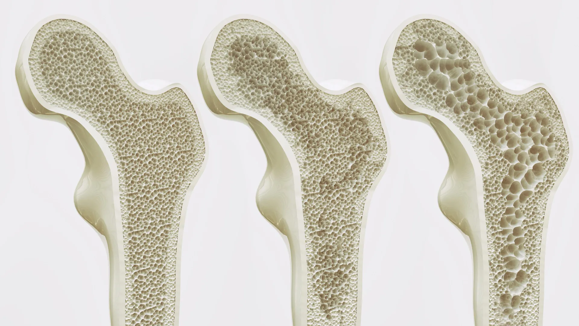
The human body relies upon a sophisticated, subterranean biological engine to sustain life: the bone marrow, an intricate and highly specialized tissue matrix that serves as our primary blood factory. Operating largely out of view within the rigid confines of our skeletal structure, this dynamic tissue orchestrates the continuous production of red blood cells, white blood cells, and platelets. It is a biological marvel composed of a dense network of bone cells, delicate nerve fibers, intricate blood vessels, and a myriad of specialized immune and stromal cells. Yet, despite its critical importance to human health, studying this complex ecosystem has long posed a formidable challenge to the scientific community. For decades, researchers striving to understand hematopoiesis—the formation of blood cellular components—and the devastating pathologies that disrupt it, such as leukemia and multiple myeloma, have been forced to rely on two imperfect proxies: animal models, predominantly mice, and heavily simplified, two-dimensional human cell cultures.
These traditional methodologies, while foundational to the history of biomedical science, carry inherent limitations. Murine bone marrow, though useful, diverges significantly from human physiology in terms of cellular composition, immune response kinetics, and microenvironmental architecture. Consequently, therapeutics that perform brilliantly in preclinical animal trials frequently falter when translated into human clinical trials. Simultaneously, standard in vitro cell cultures lack the complex three-dimensional architecture and cellular crosstalk characteristic of living human tissue.
A monumental paradigm shift has now arrived from the Department of Biomedicine at the University of Basel and the University Hospital Basel, where a multidisciplinary team of scientists has successfully engineered a fully functional, highly realistic human bone marrow model constructed entirely from human cells. Published in the peer-reviewed journal Cell Stem Cell, this breakthrough achievement marks the first time researchers have been able to recreate the intricate structural and functional complexity of the human bone marrow microenvironment in a laboratory setting without relying on animal substrates. Led by Professor Ivan Martin and Dr. Andrés García García, the research team has unveiled a biomimetic platform that not only mirrors native human biology with unprecedented fidelity but also heralds a new era for pharmacological testing, blood cancer research, and the ethical mandate to reduce, refine, and replace laboratory animal experiments.
Decoding the Endosteal Niche: Anatomy of a Breakthrough
To comprehend the significance of the Basel team’s achievement, one must examine the microarchitectural landscape of the bone marrow. The tissue is not a homogenous soup of cells; rather, it is divided into distinct, highly specialized microenvironments known as niches. Among these, the endosteal niche occupies a position of paramount importance. Situated in close proximity to the hard inner surface of the bone, the endosteal niche acts as a master regulator of hematopoietic stem cells, governing their quiescence, self-renewal, and differentiation into mature blood lineages. Furthermore, scientific literature increasingly points to the endosteal niche as a critical sanctuary where malignant cells, particularly in hematological malignancies like acute myeloid leukemia, can hide, evade conventional chemotherapy, and develop resistance to treatment.
Historically, replicating this specific niche in an artificial environment proved to be an insurmountable hurdle. The endosteal niche is a bustling metropolis of diverse cellular and structural players, requiring the simultaneous integration of mineralized bone matrix, vascular endothelial cells, peripheral nerves, and specialized immune components. Previous attempts to model this environment in vitro typically succeeded in incorporating only one or two of these elements, yielding systems that lacked the physiological relevance required for translational medicine.
The Chronology and Methodology of Innovation
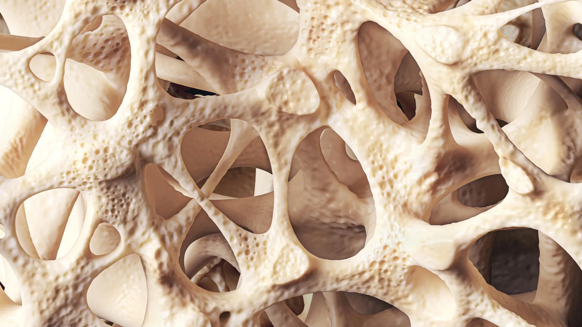
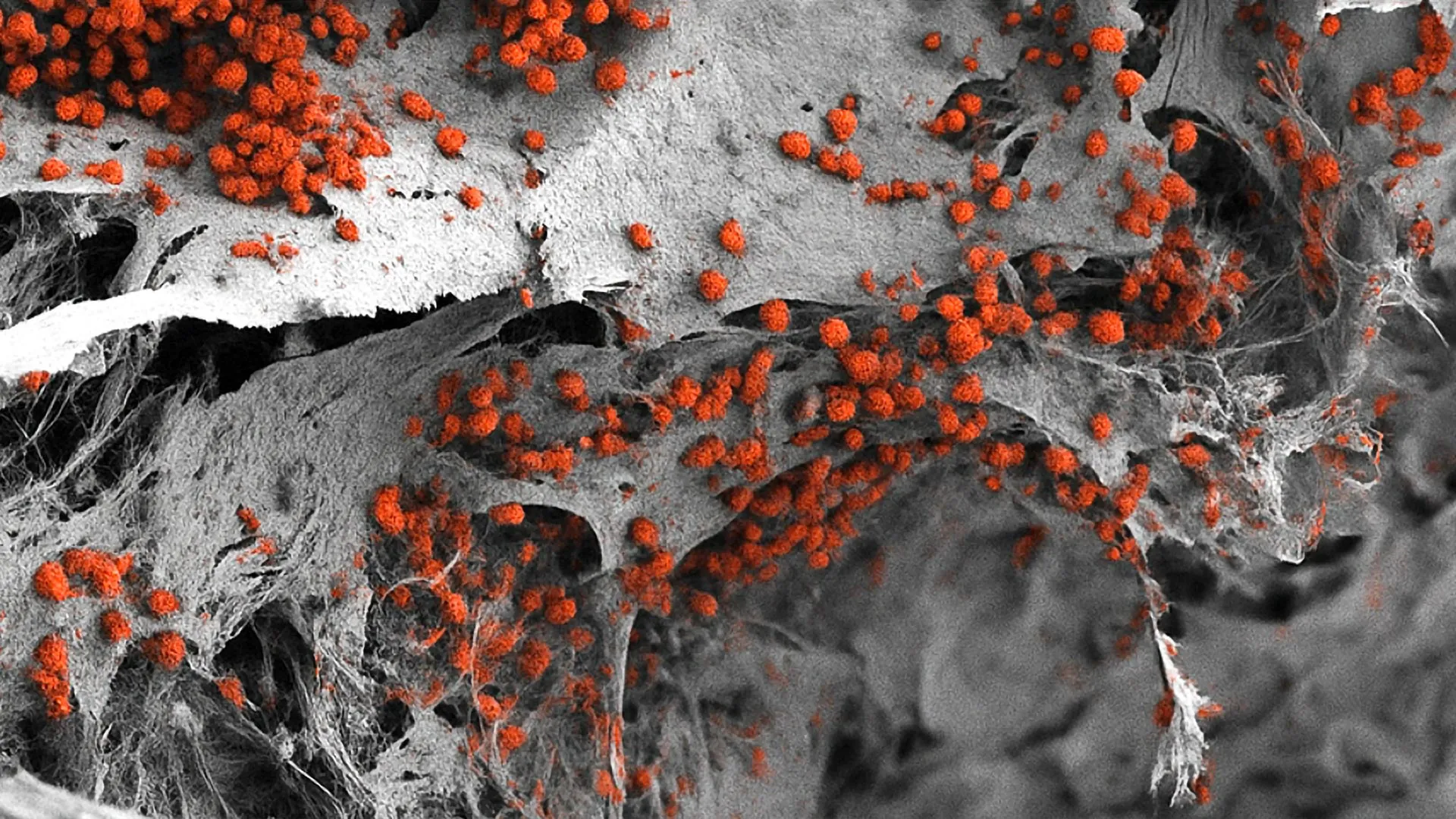
The genesis of this breakthrough spans years of meticulous bioengineering and molecular biology research at the University of Basel. The scientific journey began with the conceptualization of an advanced three-dimensional structural framework. Rather than utilizing organic bone matrices of animal origin, the researchers engineered an artificial bone scaffold fabricated from hydroxyapatite, a naturally occurring calcium phosphate mineral that constitutes the primary mineral component of human teeth and bones. This scaffold provided the necessary mechanical rigidity and biochemical cues to mimic the natural skeletal environment.
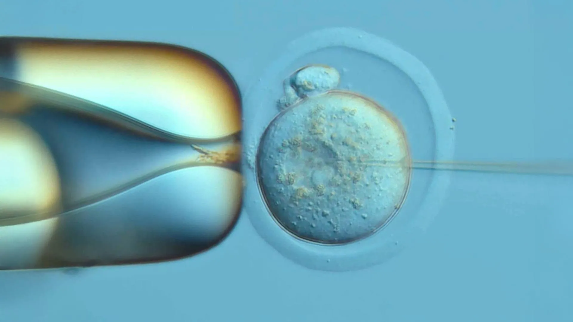
The next critical phase involved mastering cellular derivation. Utilizing cutting-edge molecular biology techniques, the team took human somatic cells and reprogrammed them into human induced pluripotent stem cells (hiPSCs). These versatile stem cells possess the remarkable capacity to differentiate into virtually any cell type in the human body when exposed to the appropriate biochemical and biophysical signals. By seeding these hiPSCs onto the hydroxyapatite scaffold and subjecting them to a carefully orchestrated sequence of biochemical developmental cues, the researchers guided the cells to differentiate in situ.
The resulting structure is a sophisticated, three-dimensional biological construct measuring a robust eight millimeters in diameter and four millimeters in thickness—dimensions significantly larger than any previously achieved human bone marrow models. Histological and molecular analyses confirmed that this multi-cellular construct successfully established the diverse cellular taxonomy characteristic of the native endosteal niche. Crucially, when maintained in specialized bioreactors, this engineered system demonstrated the remarkable ability to sustain human blood cell formation continuously in vitro for a period of several weeks, proving that the artificial microenvironment was not merely structurally similar to native marrow, but functionally active as well.
Quantifying the Impact: Data, Scalability, and Preclinical Promise
The implications of this study extend far beyond basic developmental biology, offering tangible solutions to some of the most persistent bottlenecks in pharmaceutical research and regulatory science. In the landscape of modern drug development, the attrition rate remains alarmingly high. Only a tiny fraction of candidate molecules that demonstrate efficacy in preclinical models ultimately successfully navigate clinical trials to achieve regulatory approval. A significant driver of this failure rate is the poor predictive validity of animal models for complex human systemic diseases.
By introducing a human-cell-derived bone marrow platform, the scientific community gains a powerful auxiliary tool that bridges the gap between traditional in vitro assays and human clinical reality. The University of Basel model allows researchers to observe human hematopoiesis in real time, tracking how healthy blood cell production responds to various biochemical stimuli, environmental stressors, and pathogenic agents. More importantly, it opens unprecedented avenues for oncology research. Because the endosteal niche plays a central role in tumor homing and drug resistance in blood cancers, scientists can now populate the scaffold with patient-derived malignant cells to study how leukemias interact with their surrounding stromal and vascular environments.
However, transitioning from a conceptual triumph to an industrial-scale high-throughput screening tool requires addressing practical engineering hurdles. Dr. Andrés García García has candidly addressed one of the primary physical limitations of the current prototype: its scale. While the current dimensions—eight millimeters by four millimeters—are ideal for maintaining complex cellular interactions and long-term blood cell formation, they are presently too large for high-throughput drug screening applications. In modern pharmaceutical laboratories, screening chemical libraries typically involves multi-well plates containing hundreds of miniaturized samples tested simultaneously against various drug candidates and concentration gradients. To adapt the Basel bone marrow model for this level of high-throughput pharmacological screening, the platform must be successfully downscaled without sacrificing its cellular complexity or structural integrity. Engineering teams are already looking toward microfluidic integration and advanced bio-printing techniques to achieve this miniaturization.
The Ethical Imperative: Reducing Reliance on Animal Experiments
At the heart of the University of Basel’s communication regarding this breakthrough is a steadfast commitment to the principles of Humane Experimental Technique—commonly known as the Three Rs: Replacement, Reduction, and Refinement. For decades, biomedical research involving hematological disorders and bone marrow physiology has been inextricably linked to the sacrifice of millions of laboratory mice. While murine models have undeniably expanded human understanding of biological systems, mounting ethical scrutiny and scientific recognition of interspecies physiological differences have accelerated the push for viable alternatives.
Professor Ivan Martin emphasized this philosophical and scientific shift in public statements following the publication of the study in Cell Stem Cell. "We have learned a great deal about how bone marrow works from mouse studies," Martin noted, acknowledging the foundational role that animal research has played in medical history. Nevertheless, he was quick to highlight the imperative of evolution: "However, our model brings us closer to the biology of the human organism. It could serve as a complement to many animal experiments in the study of blood formation in both healthy and diseased conditions."
This sentiment resonates deeply within the European research ecosystem, where institutional frameworks increasingly prioritize non-animal methodologies whenever scientifically feasible. The creation of a fully humanized bone marrow model does not instantly render animal testing obsolete, but it provides a rigorous, biologically faithful alternative that can significantly reduce the volume of murine subjects required for preliminary investigations, mechanistic studies, and initial toxicity screenings. By screening candidate molecules through human cell-based marrow platforms first, researchers can filter out ineffective or overtly toxic compounds before any animal subjects are ever introduced to the experimental pipeline.
Toward the Horizon of Personalized Medicine
Perhaps the most exciting long-term implication of the Basel research lies in the realm of personalized medicine. Traditional cancer treatments are largely designed around population averages—therapies that demonstrate statistical efficacy in broad clinical trials are administered to patients based on standardized protocols, despite the fact that individual tumor biology varies wildly from person to person. This "one-size-fits-all" approach frequently results in trial-and-error treatment regimens characterized by severe side effects and delayed therapeutic optimization.
Looking forward, the research team at the University of Basel envisions a future where the techniques developed in this study can be harnessed to construct patient-specific bone marrow models. By taking somatic cells from an individual diagnosed with a blood cancer, reprogramming them, and using them to construct a personalized three-dimensional endosteal niche in vitro, clinicians could theoretically recreate that specific patient’s bone marrow ecosystem in the laboratory.
Within this personalized avatar, oncologists could directly test a panel of diverse chemotherapeutic agents, immunotherapies, or novel targeted drug combinations to observe which specific regimen successfully eradicates the malignant cells while preserving healthy hematopoietic stem cell function. This empirical approach would allow medical teams to identify the most efficacious, patient-specific treatment strategy prior to administering the first dose of actual therapy, thereby maximizing therapeutic response rates, minimizing unnecessary toxicity, and fundamentally transforming the clinical management of hematological malignancies.
Conclusion and Future Outlook
The successful recreation of a fully functional, three-dimensional human bone marrow model using exclusively human cells marks a watershed moment in biomedical engineering and translational research. By bridging the chasm between simplified cellular monolayers and whole-organism animal testing, Professor Ivan Martin, Dr. Andrés García García, and their colleagues at the University of Basel have delivered a sophisticated, physiologically relevant platform that promises to accelerate blood cancer research, refine drug development pipelines, and significantly curb the reliance on laboratory animals.
While significant developmental work remains—particularly in miniaturizing the platform for high-throughput pharmacological screening and establishing clinical workflows for personalized medicine—the foundational architecture is now securely in place. As laboratories around the world begin to adopt and build upon this human cell-based paradigm, the scientific community moves one step closer to a future where medical research is increasingly defined by human relevance, precision, and ethical responsibility.