
The 2026 Nikon Small World competition has officially concluded, unveiling a collection of 86 images that bridge the gap between technical scientific observation and aesthetic fine art. Since its inception in 1975, the competition has served as a premier global platform for photographers and scientists to showcase the intricate beauty of the microscopic world. This year’s top honors reflect a significant evolution in imaging technology, as researchers and hobbyists alike employ increasingly sophisticated rigs to capture phenomena that occur at speeds and scales previously inaccessible to the human eye.
A Masterclass in High-Speed Precision
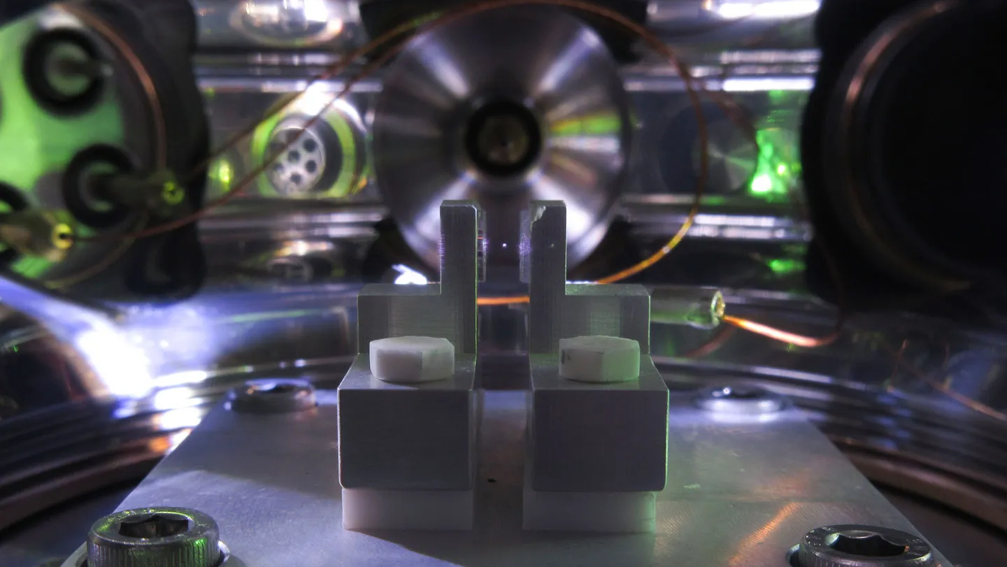
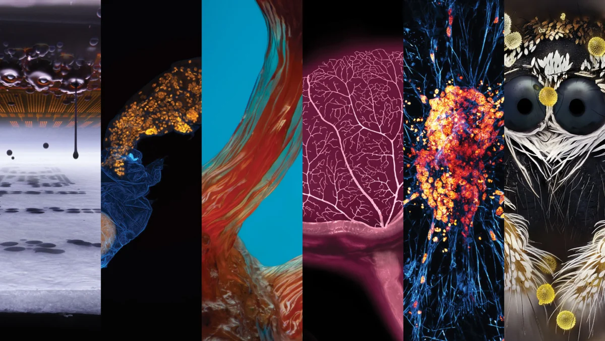
The first-place entry, captured by German design student Ole Bielfeldt, offers a profound look at the mechanics of everyday technology: the inkjet printer. Bielfeldt’s image captures individual ink droplets suspended in mid-air, a feat achieved by overcoming the physical limitations of standard consumer electronics. To secure the shot, Bielfeldt constructed a custom imaging apparatus. Recognizing that the standard vibration and movement of printer nozzles would blur any potential image, he modified a printer to keep the print head stationary while moving the paper substrate instead.

The technical challenge was compounded by the velocity of the ink. Traveling at approximately 20 meters per second, the droplets move far faster than the frame rate of standard high-speed cameras. Bielfeldt solved this by operating in total darkness, utilizing a strobe-like flash to freeze the motion at the exact millisecond of ejection. The result is a crisp, high-resolution portrait of fluid dynamics that turns a mundane office task into a revelation of engineering precision. Bielfeldt’s success serves as a reminder that even the most ubiquitous tools—items that have become "invisible" through constant use—harbor complex, artistic beauty when examined under a different lens.
The Evolution of the Nikon Small World Competition
The Nikon Small World competition was originally established to foster a greater appreciation for the work performed under the light microscope. Over five decades, it has grown from a niche academic gathering into a significant cultural event that draws entries from dozens of countries. The selection process is rigorous, involving a panel of experts from diverse scientific fields who evaluate entries based on content, artistic composition, and, crucially, technical proficiency.
The 2026 winners, announced on October 21, demonstrate a clear trend: the integration of DIY engineering with high-end optics. As digital camera sensors have become more sensitive and lighting technology has advanced, the barrier to entry for high-level photomicrography has lowered for those willing to invest the time in custom setups. This year’s field included contributions from professional cell biologists, independent researchers, and amateur photographers, highlighting the democratization of high-level scientific documentation.

Ecological and Biological Narratives
While the top prize focused on mechanical engineering, the runners-up provided a window into the biological complexities of our planet. John-Oliver Dum’s second-place photograph of a pollen-dusted jumping spider highlights the intersection of entomology and patience. Capturing such an image requires an intimate knowledge of seasonal rhythms; Dum’s process involved years of preparation, including growing specific plants to attract spiders during their peak activity windows. His work emphasizes a growing sentiment among science communicators: that the preservation of global biodiversity begins with an appreciation for the "unknown" small-scale interactions occurring in our own backyards.
Similarly, the work of Dr. Gonzalo Quiroga-Artigas, who captured a tardigrade emerging from its cuticle, offers a rare glimpse into the life cycle of one of Earth’s most resilient organisms. By documenting the exact moment a tardigrade leaves behind its molted skin—and a protective egg—Quiroga-Artigas provides more than just a striking image; he offers a data point for developmental biologists studying the synchronization of molting and reproduction in microscopic invertebrates.
Medical Imaging and the Future of Regenerative Medicine
The competition also underscored the practical applications of photomicrography in medical research. Satu Paavonsalo and Dr. Sinem Karaman’s 19th-place image of a mouse eye’s vascular network demonstrates the shift from two-dimensional slides to three-dimensional imaging. By moving away from flattened retinal samples, the researchers were able to visualize the fractal nature of blood vessels in a way that could eventually help in understanding the pathogenesis of obesity-related vascular diseases.

Likewise, the work of Faith Fernandes and Dr. James Glover at UC Berkeley regarding human induced pluripotent stem cells (iPSCs) illustrates the future of regenerative medicine. By reprogramming adult cells into neurons, the team was able to capture the vibrant, branching architecture of the human nervous system in development. This research is not merely for show; it is part of a broader, global effort to develop treatments for neurodegenerative conditions like Parkinson’s disease. The ability to visualize these cells in high detail allows researchers to assess the quality of stem cell differentiation before moving to more invasive clinical applications.
The Cosmic Connection: Slime Molds and Scientific Modeling
Perhaps the most surprising entry was Eric Epstein’s sixth-place photograph of a slime mold (Hemitrichia sp.) found in Brooklyn, New York. Beyond its visual appeal, the image serves as a reminder of the unexpected parallels between terrestrial biology and astrophysics. Slime molds have been used by researchers to model the distribution of dark matter in the cosmic web, as their growth patterns mirror the way galaxies form in the universe. Epstein’s work acts as a bridge, reminding the public that even a common forest fungus can hold the keys to understanding the structure of the cosmos.
Implications and Broader Impact
The 2026 Nikon Small World competition serves as more than just a showcase of visual talent. It acts as an unofficial census of the current state of scientific visualization technology. The reliance on "focus stacking"—the process of combining multiple images taken at different focal depths—was evident in nearly every top-tier entry, including Epstein’s 240-image stack. This technique, once reserved for high-end laboratories, is now standard practice for top-tier entries, indicating a widespread adoption of computational photography in science.

Furthermore, the competition highlights the vital role of "citizen science." Many of the most compelling images were not produced in multi-million dollar institutional facilities, but in home-built rigs in garages or local parks. This reflects a shift in how scientific discovery is perceived by the public. When a student or a local nature enthusiast can produce images of the same caliber as a university researcher, it invites broader public engagement with the scientific process.
The implications for education are also significant. By transforming complex biological or physical processes into high-art, these photographers are creating a powerful tool for STEM (Science, Technology, Engineering, and Mathematics) advocacy. When a student sees the "stars" within a neuron or the "gem-like" egg of a tardigrade, the abstract concepts of biology move from the textbook into the realm of the tangible and the awe-inspiring.
As the scientific community looks toward 2027 and beyond, the Nikon Small World competition will likely continue to evolve alongside the technology it celebrates. With the advent of AI-enhanced image processing and more accessible light-sheet microscopy, the next few years may see even deeper penetration into the biological and physical secrets of the world around us. For now, the 2026 collection stands as a testament to the fact that, regardless of how much we think we understand the world, there remains an infinite amount of beauty waiting to be seen just beneath the threshold of our perception. Whether through the lens of a repurposed printer or the eye of a jumping spider, the message is clear: the microscopic world is not just a subject of study, but a vast, untapped frontier of human wonder.