
A multidisciplinary team of researchers at Cornell University, in collaboration with international partners, has achieved a significant milestone in neurotechnology by developing a microscale neural implant roughly the size of a grain of salt. This device, known as a microscale optoelectronic tetherless electrode (MOTE), represents a leap forward in bio-integrated electronics, capable of wirelessly transmitting high-fidelity brain activity data from a living subject for over a year. The breakthrough, recently detailed in the journal Nature Electronics, suggests a future where brain-machine interfaces (BMIs) are less invasive, more durable, and capable of operating in environments previously considered off-limits to electronic sensors.
The development was spearheaded by Alyosha Molnar, a professor in the School of Electrical and Computer Engineering at Cornell, alongside Sunwoo Lee, an assistant professor at Nanyang Technological University (NTU) in Singapore. Lee’s involvement began during his tenure as a postdoctoral researcher in Molnar’s laboratory, illustrating a long-term commitment to solving one of the most persistent challenges in neuroscience: how to record internal biological signals without the use of bulky wires or large, battery-dependent hardware that can damage delicate tissue.
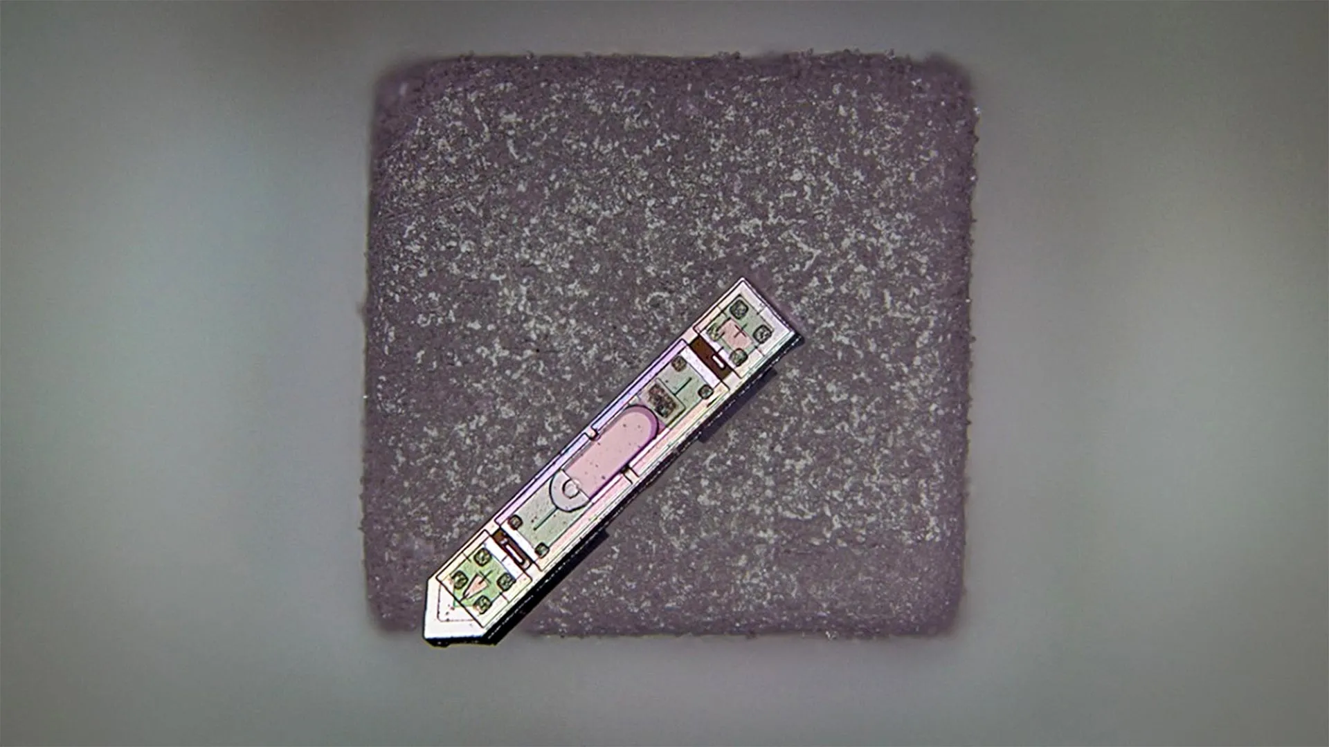
The Engineering of the MOTE Device
The MOTE device is a masterclass in miniaturization, measuring approximately 300 microns in length and 70 microns in width. To put this in perspective, a standard human hair is about 100 microns thick, meaning the implant is narrower than a few strands of hair bundled together. Despite its diminutive stature, the MOTE is a fully integrated system containing power harvesting, sensing, and communication components.
The core of the device is constructed from aluminum gallium arsenide (AlGaAs), a semiconductor material chosen for its specific optoelectronic properties. Unlike traditional silicon-based chips that rely on radio frequency (RF) waves for communication, the MOTE utilizes light. Specifically, the device is powered by red and infrared laser beams that can safely penetrate several millimeters of biological tissue. This light is captured by a semiconductor diode on the device, which converts the photons into electrical energy to power the internal circuitry.
To transmit data back to the researchers, the MOTE employs a method known as pulse position modulation (PPM). This technique involves emitting tiny pulses of infrared light that encode the electrical signals—the "spikes" of neuronal activity—detected by the electrode. By varying the timing of these light pulses, the device can relay complex information with extremely low power consumption. This approach is analogous to the optical communication systems used by satellites, adapted here for the microscopic landscape of the mammalian brain.
A Chronology of Micro-Neural Interface Development
The journey toward the MOTE began over a decade ago as the neuroscience community recognized the limitations of the "Utah Array" and other early-generation neural probes. While these devices provided groundbreaking insights into brain function, they were typically "tethered" by physical wires protruding through the skull, which posed significant risks of infection and restricted the natural movement of the subject.
Between 2015 and 2018, the Cornell team focused on the fundamental physics of optical power delivery. The challenge was twofold: ensuring the light could reach the device without causing thermal damage to the brain and designing a circuit small enough to fit on a microscopic footprint while remaining sensitive enough to detect microvolt-level neural signals.
By 2020, the team had transitioned from theoretical models to the first prototypes. The primary hurdle during this phase was the integration of the low-noise amplifier and the optical encoder. These components had to be fabricated using the same semiconductor processes used in commercial microchips but scaled down to a level that pushed the boundaries of existing lithography techniques.
The most recent phase of the project involved longitudinal testing in living animals. Unlike previous micro-implants that often failed within weeks due to the body’s immune response or "foreign body reaction," the MOTE demonstrated remarkable longevity. The results published in Nature Electronics confirm that the device remained functional and continued to transmit clear data for more than 12 months, a duration that sets a new benchmark for wireless micro-implants.
Supporting Data and Technical Specifications
The efficiency of the MOTE is largely attributed to its unconventional communication protocol. Most wireless implants use radio frequency identification (RFID) or Bluetooth-like protocols, which require relatively large antennas and significant power. The Cornell team’s data shows that by using optical links, the MOTE can operate on a power budget of only a few microwatts.
Data highlights from the research include:
- Dimensions: 300 µm x 70 µm x 30 µm.
- Longevity: 400+ days of continuous operation in vivo.
- Signal-to-Noise Ratio (SNR): Comparable to larger, wired electrodes, despite the massive reduction in scale.
- Power Source: External 800-900nm laser (near-infrared), which minimizes tissue heating and maximizes penetration depth.
The use of aluminum gallium arsenide is also a critical data point. AlGaAs is not only efficient at converting light to electricity but is also more biocompatible than many other high-performance semiconductors when properly encapsulated. The device’s ability to remain "dark" (non-transmitting) until triggered by an external light source further preserves its lifespan and prevents interference with other biological processes.
Official Responses and Academic Significance
The successful deployment of the MOTE has garnered significant attention from the global scientific community. Professor Alyosha Molnar emphasized the uniqueness of the achievement, noting that the device represents the smallest wireless neural implant currently capable of reporting electrical activity.
"By using pulse position modulation for the code, we can use very, very little power to communicate and still successfully get the data back out optically," Molnar stated. This efficiency is the key to the device’s size; without the need for large batteries or bulky antennas, the device can be reduced to the point where it is virtually imperceptible to the surrounding tissue.
Dr. Sunwoo Lee, now at NTU, highlighted the collaborative nature of the project and its implications for future medical devices. "The challenge was not just making it small, but making it robust enough to survive the harsh environment of the living body for a long period," Lee noted in discussions regarding the research. The stability of the MOTE over a year suggests that the encapsulation techniques and the choice of optical communication have effectively bypassed the degradation issues that plague other micro-electronics.
While not directly involved in the Cornell study, peers in the field of neuroprosthetics have noted that this technology addresses the "scalability problem." Current BMIs like those developed by Neuralink or Blackrock Neurotech are highly capable but involve larger footprints. The MOTE offers a path toward "distributed" sensing, where dozens or even hundreds of independent sensors could be scattered throughout the brain to provide a high-resolution map of activity without the trauma associated with a single large implant.
Analysis of Broader Impact and Future Applications
The implications of the MOTE extend far beyond basic neuroscience research. One of the most significant potential applications lies in the field of diagnostic imaging. Currently, patients with metallic neural implants are often barred from undergoing Magnetic Resonance Imaging (MRI) because the strong magnetic fields can cause the metal to heat up or move, potentially damaging the brain. However, the semiconductor materials used in the MOTE are largely non-metallic and non-ferromagnetic.
According to the research team, the MOTE could potentially allow for simultaneous neural recording during an MRI scan. This would provide clinicians with a "dual-view" of brain health: the high-resolution structural and blood-flow data from the MRI combined with the real-time electrical data from the MOTE. Such a capability would be invaluable for treating conditions like epilepsy, where identifying the exact origin of a seizure is critical for surgical intervention.
Furthermore, the technology is not limited to the brain. The researchers envision the MOTE being adapted for use in the spinal cord to monitor recovery after injury, or in peripheral nerves to help control advanced prosthetic limbs. The small size makes it ideal for integration into "smart" medical devices, such as artificial skull plates or orthopedic implants, where the electronics could be embedded directly into the structure of the replacement part.
The future of the MOTE may also involve "opto-electronic" integration at a systemic level. By combining these sensors with future innovations in transparent cranial windows or light-emitting skull caps, researchers could create a completely non-invasive way to "talk" to the brain’s internal circuitry.
Conclusion
The development of the microscale optoelectronic tetherless electrode marks a turning point in the evolution of bio-electronics. By solving the dual challenges of power delivery and wireless communication at a microscopic scale, the Cornell-led team has provided a tool that could fundamentally change how we observe and interact with the nervous system. As the technology moves toward human clinical trials, the focus will likely shift to further refining the surgical delivery methods and expanding the bandwidth of the optical data links. For now, the MOTE stands as a testament to the power of miniaturization and a harbinger of a new era in long-term, high-resolution biological monitoring.


Leave a Reply