

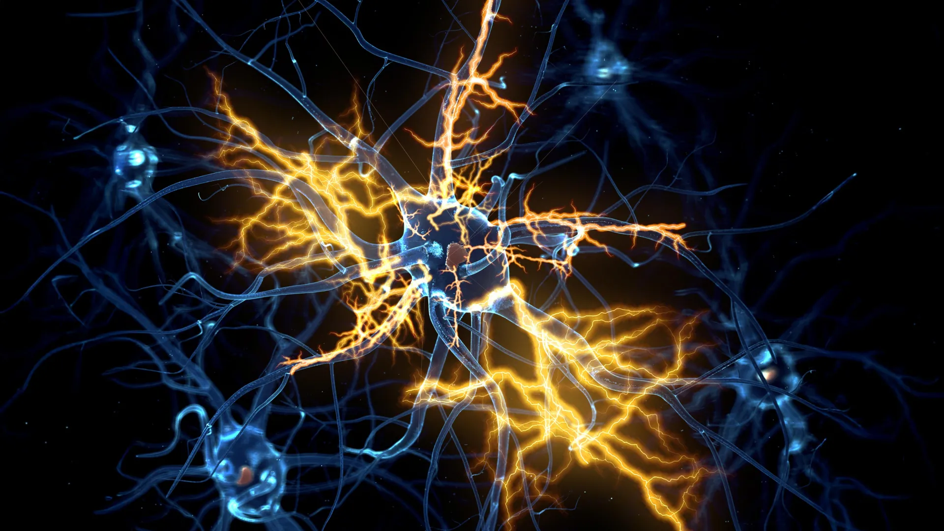
The traditional medical consensus that damage to the central nervous system is inherently permanent has been challenged by a landmark study from the University of Cambridge. Researchers have successfully developed sophisticated, miniature models of the human brain and spinal cord, using these "organoids" to pinpoint the exact developmental window when neurons lose their ability to regenerate. Most significantly, the team discovered that this regenerative decline is not an inevitable decay but is controlled by a specific genetic network that can be manipulated. By utilizing an existing pharmaceutical compound, the scientists demonstrated that it is possible to flip this biological switch, potentially paving the way for future treatments for paralysis, motor neurone disease, and multiple sclerosis.
The Challenge of Central Nervous System Repair
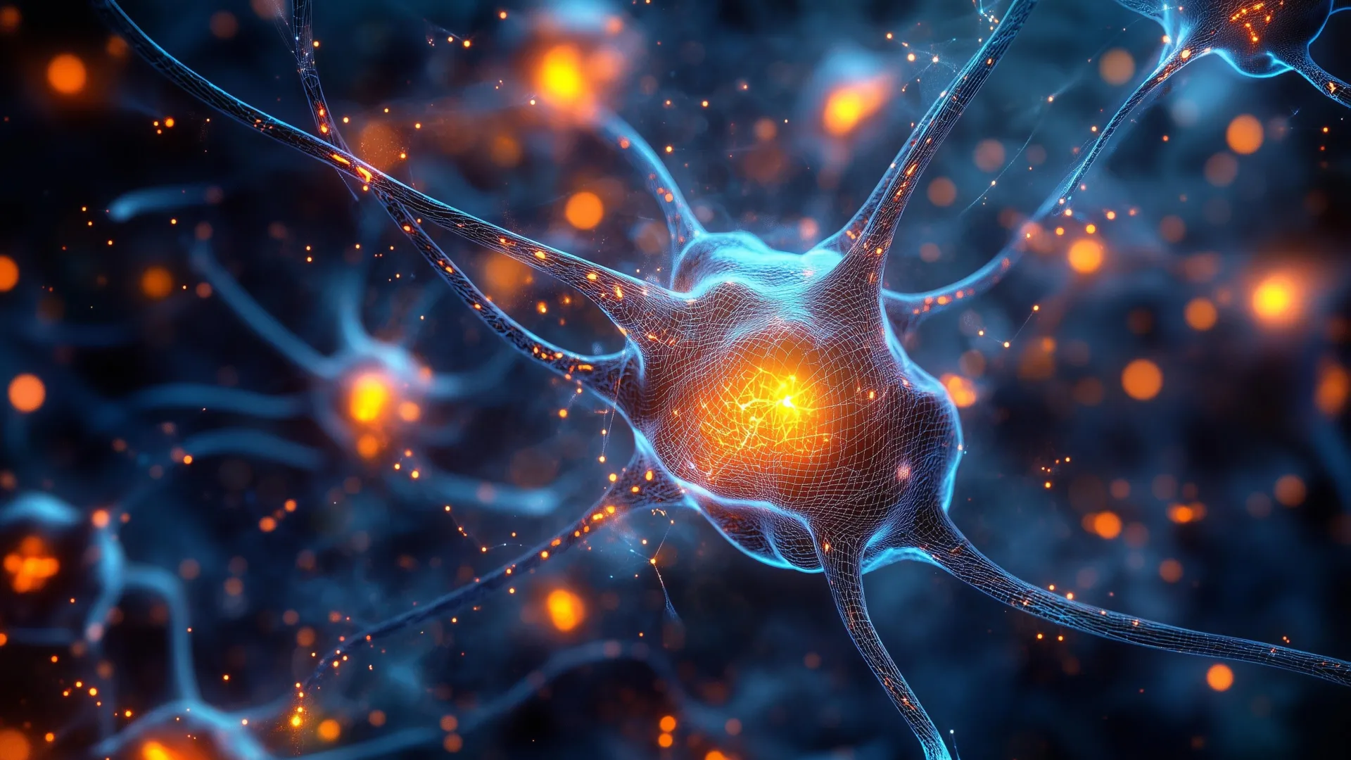
In the human body, the central nervous system (CNS)—comprising the brain and the spinal cord—serves as the command center for every movement and sensation. This system relies on neurons that extend long, slender fibers known as axons. These axons function like biological electrical wires, carrying signals across vast distances to facilitate communication between the cerebral cortex and the limbs. During the early stages of embryonic and fetal development, these axons possess a remarkable ability to grow, navigate through complex tissues, and form the intricate circuitry required for human life.
However, as the nervous system matures, this intrinsic growth capacity drastically diminishes. Unlike the peripheral nervous system, which can often repair itself after a minor injury, the CNS is notorious for its inability to heal. When the brain or spinal cord is damaged by trauma or neurodegenerative disease, the axons typically fail to regrow across the site of the injury. This failure is often compounded by the formation of scar tissue and inflammatory responses that create a hostile environment for repair. For decades, the medical community has viewed the resulting loss of function—whether it be paralysis, sensory loss, or cognitive decline—as a permanent condition.
Advancing the Frontier of Organoid Technology
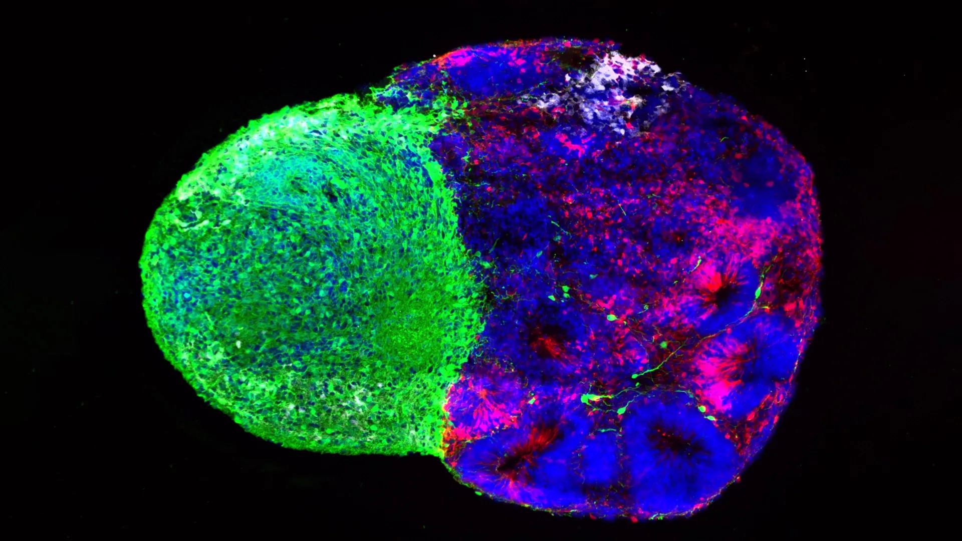
The breakthrough at the University of Cambridge, led by Dr. András Lakatos of the Department of Clinical Neurosciences, stems from years of refinement in stem cell research. In 2021, Dr. Lakatos and his team successfully grew "brain organoids"—pea-sized clusters of cells derived from human stem cells that mimic the architecture and molecular behavior of the human cerebral cortex. These models allowed researchers to observe the cellular malfunctions associated with conditions like Amyotrophic Lateral Sclerosis (ALS) in a controlled, human-specific environment.
In their latest study, published in the journal Cell Reports, the team took this technology a step further. Recognizing that the brain and spinal cord function as a single integrated circuit, they developed a system that includes both brain and spinal cord organoids. Because these structures are physically distinct in the human body, the researchers kept them in separate compartments within the laboratory. They then observed as axons from the brain organoids spontaneously grew across a physical gap to integrate with the spinal cord tissue. This created a functional neural circuit capable of sending signals that triggered contractions in nearby muscle cells, effectively recreating the human "movement loop" in a dish.
The 150-Day Threshold: A Chronology of Regenerative Decline
One of the most significant contributions of the study is the establishment of a clear timeline for when human neurons lose their regenerative potential. By maintaining these organoid systems for over a year, the Cambridge team was able to monitor the neurons at various stages of maturity, roughly corresponding to different periods of human gestation and infancy.
The data revealed a striking "turning point" in neural development. Up until approximately day 150 of development—which corresponds to the middle of the second trimester of pregnancy—the neurons exhibited a robust ability to regrow their axons after being severed. However, after this 150-day mark, the regenerative capacity plummeted.
George Gibbons, the study’s first author, noted that this decline is not a result of the cells becoming "tired" or "old," but is rather a programmed biological transition. "Neurons taken from less mature organoids regrew long fibers after injury, but those from more mature organoids showed a sharp drop in their ability to regrow," Gibbons explained. "In other words, poor regeneration is built into human neurons as they mature in the central nervous system."
This finding suggests that the loss of repair capacity is a trade-off: as the brain moves from a state of "growth and exploration" to a state of "stability and synapse formation," it intentionally shuts down the machinery required for long-distance axon growth to ensure that established connections remain secure.
Identifying the Genetic Switch and the Role of Lynestrenol
To understand the mechanics behind this decline, the researchers conducted an intensive analysis of gene activity within the connecting neurons. They identified a specific network of genes that acts as a regulatory switch. As the neurons reached the 150-day maturity threshold, this network would activate, effectively "locking" the cell’s growth potential.
The researchers hypothesized that if they could interfere with this network, they might be able to trick the mature neurons into returning to a more youthful, regenerative state. When they experimentally blocked key regulators within this genetic circuit, the results were remarkable: the mature neurons regained the ability to grow axons, bridging the gaps caused by injury.
To translate this finding into a potential clinical application, the team screened a vast database of existing pharmaceutical compounds to see if any known drugs could influence this genetic network. Their search identified lynestrenol, a synthetic progestogen currently used in various contraceptive pills and treatments for menstrual disorders. When applied to the damaged, mature organoids, lynestrenol significantly boosted axon regrowth.
While the researchers cautioned that lynestrenol itself may not be the final "cure" for spinal cord injury—given that it is a hormone with systemic effects—its success serves as a "proof of principle." It demonstrates that the regenerative block in human neurons is chemically reversible and that targeted drug therapies could potentially stimulate repair in the human CNS.
Supporting Data and Statistical Context
The implications of this research are underscored by the global burden of neurological injuries. According to the World Health Organization (WHO), between 250,000 and 500,000 people suffer a spinal cord injury every year, most of which result in permanent disability. Furthermore, neurodegenerative diseases like Motor Neurone Disease (MND) affect approximately 1 in 300 people across their lifetime, characterized by the progressive death of the very neurons the Cambridge team is studying.
The Cambridge study provides several key data points that offer hope for these populations:
- Regenerative Window: The identification of the 150-day developmental mark provides a specific biological benchmark for future research.
- Functional Recovery: The fact that the lab-grown circuits could trigger muscle contractions proves that the regrown nerves are not just structurally present but are electrophysiologically active.
- Genetic Targets: The identification of the specific gene network narrows the search for drug targets from thousands of possibilities to a manageable cluster of regulators.
Shifting Away from Animal Models
A critical aspect of this study is its reliance on human-derived organoids rather than traditional animal models. For decades, spinal cord research has relied heavily on rats and mice. However, there are fundamental differences between rodent and human neurobiology. Rodent neurons often possess a higher baseline for regeneration than human neurons, and many drugs that show promise in mice fail to produce results in human clinical trials.
"Much of what we know about nerve regeneration comes from rodents, whose neurons behave differently from human neurons," said Dr. Lakatos. "Our sophisticated organoid models help bridge the knowledge gap from animal models to what we see in patients."
By using human stem cells, the researchers can ensure that the biological pathways they are manipulating are the same ones present in a human patient. This "human-first" approach not only increases the likelihood of successful clinical translation but also contributes to the global scientific effort to reduce the reliance on animal testing in medical research.
Official Reactions and Broader Implications
The study has been met with cautious optimism from the scientific and medical communities. Organizations such as Spinal Research and the UK Research and Innovation Medical Research Council, which funded the study, have highlighted the importance of understanding the internal "roadblocks" to nerve repair.
Medical analysts suggest that this research could lead to a paradigm shift in how we approach spinal cord injuries. Currently, most treatments focus on stabilizing the injury site, reducing inflammation, and intensive physical therapy to make the most of remaining function. The Cambridge study suggests a future where "regenerative medicine" actually means what it says: physically regrowing the lost connections to restore original function.
However, challenges remain. In a living patient, regrowing an axon is only half the battle; that axon must then find its way to the correct destination and form a functional synapse without causing "wrong" connections that could lead to pain or involuntary movements.
Dr. Lakatos acknowledged these hurdles, stating, "Although we still need to show that this strategy will also help to re-establish appropriate connections between the brain and spinal cord cells, this gives us hope that one day we may be able to treat conditions previously thought untreatable."
Conclusion
The work at the University of Cambridge marks a significant milestone in the field of neuroscience. By creating a functional, human-specific model of the central nervous system’s communication lines, researchers have moved beyond observing damage to understanding the fundamental biological reasons why that damage persists. The discovery that the loss of nerve regeneration is a reversible genetic event, rather than an irreversible fact of nature, provides a new roadmap for the treatment of paralysis and neurodegeneration. While the journey from the laboratory dish to the hospital bedside is long, the "switch" for human nerve repair has finally been found.
















Leave a Reply