

In a significant advancement for regenerative medicine and hematology, researchers at Weill Cornell Medicine have identified a specific molecular mechanism that governs the transition of blood stem cells from a dormant state to an active, regenerative one. The study, published in the journal Nature Immunology, identifies the protein FLI-1 as the primary "switch" required for hematopoietic stem cells (HSCs) to multiply and produce the diverse array of cells that constitute the human blood and immune systems. This discovery addresses a long-standing challenge in clinical medicine: the difficulty of expanding and activating stem cells for use in bone marrow transplants and gene therapies.
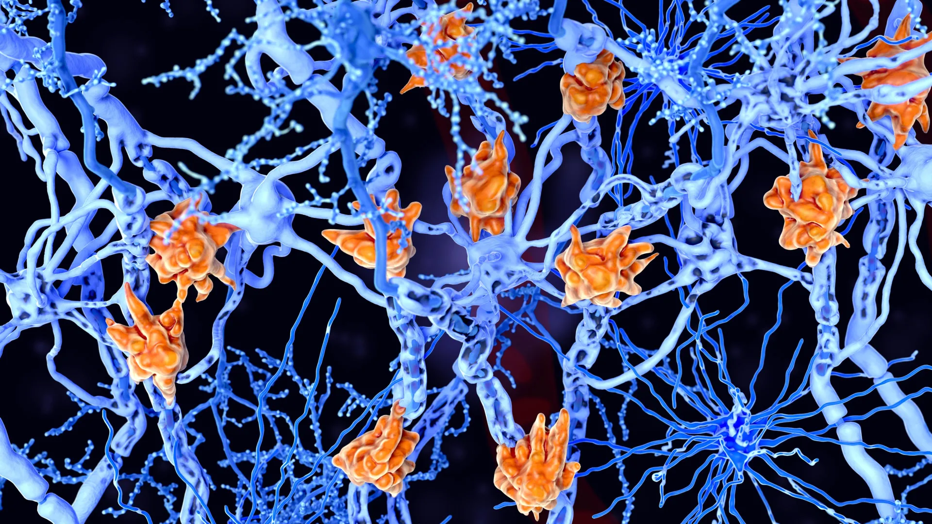
Hematopoietic stem cells are the foundation of the body’s blood production. Located primarily within the bone marrow, these cells are characterized by their "quiescence"—a state of metabolic hibernation where they divide very slowly. This dormancy protects the stem cell pool from exhaustion and genetic damage over a lifetime. However, when the body faces an injury, infection, or medical intervention like chemotherapy, these cells must "wake up" and enter an activated state to replenish the blood supply. The Weill Cornell study reveals that without the transcription factor FLI-1, this critical transition cannot occur effectively, leaving the body unable to repair its blood-forming tissues.
The Biological Mechanism of the FLI-1 Switch
The research team, led by Dr. Shahin Rafii, director of the Hartman Institute for Therapeutic Organ Regeneration and the Ansary Stem Cell Institute at Weill Cornell Medicine, utilized high-resolution single-cell profiling to map the genetic differences between dormant and active stem cells. By analyzing thousands of individual cells, the investigators identified that FLI-1, a DNA transcription-regulating protein, acts as a master controller.
Transcription factors like FLI-1 work by binding to specific sequences of DNA, essentially turning genes "on" or "off." The study demonstrated that FLI-1 controls a vast network of thousands of genes. Crucially, FLI-1 does more than just trigger cell division; it facilitates the "cross-talk" between blood stem cells and their immediate environment, known as the vascular niche.
The vascular niche is a specialized microenvironment within the bone marrow composed of endothelial cells that line the blood vessels. The study found that FLI-1 is responsible for restoring the physical and chemical connections between stem cells and these endothelial cells. When FLI-1 is absent, stem cells remain isolated and unresponsive to the signals that normally trigger regeneration. When FLI-1 is activated, the stem cells become "co-adaptive," meaning they synchronize their behavior with the surrounding blood vessels to facilitate rapid expansion and migration into the bloodstream.
Addressing the Challenges of Bone Marrow Transplants
Bone marrow transplantation is a cornerstone treatment for various forms of leukemia, lymphoma, and other blood disorders. The process involves replacing a patient’s diseased bone marrow with healthy blood stem cells. However, the procedure is often limited by the quantity and quality of the donor cells. In many cases, particularly when using adult donors or cells that have been harvested after chemotherapy, the stem cells are "exhausted" or stuck in a deep quiescent state, leading to poor "engraftment"—the process by which the new cells take root and begin producing blood.
"The approach we outlined in this study could substantially improve the efficiency of marrow transplants and marrow-cell-targeted gene therapies," said Dr. Rafii, who is also the chief of the division of regenerative medicine at Weill Cornell Medicine. He noted that the discovery is particularly vital for patients where the donor supply of viable stem cells is extremely limited. By transiently boosting FLI-1 levels, clinicians could theoretically "prime" these cells to be more aggressive in their regenerative efforts once infused into the patient.
Innovation in Gene Therapy and Blood Disorders
The implications of the FLI-1 discovery extend into the burgeoning field of gene therapy. For conditions such as beta-thalassemia and sickle cell anemia, doctors harvest a patient’s own stem cells, use viral vectors or CRISPR technology to insert or correct a gene, and then re-infuse the cells. A major bottleneck in this process is the "ex vivo" expansion phase—growing enough of these modified cells in a laboratory setting to ensure a successful treatment.
Currently, stem cells often lose their potency or die when researchers attempt to multiply them in the lab. By understanding that FLI-1 is the driver of the regenerative state, scientists can now develop protocols to temporarily activate this switch during the lab-growth phase. This would allow for a much higher yield of functional, therapeutic cells, potentially reducing the cost and increasing the safety of gene therapy procedures.
Solving the Umbilical Cord Blood Puzzle
One of the most intriguing aspects of the study is how it explains a well-known phenomenon in pediatrics and hematology: why umbilical cord blood is often superior to adult blood for certain transplants. It has long been observed that blood stem cells derived from human umbilical cords have a much higher regenerative potential than those found in adult bone marrow.
The Weill Cornell team addressed this puzzle by comparing the molecular profiles of cord blood cells and adult cells. They found that cord blood stem cells naturally possess higher levels of FLI-1 activity. This elevated baseline of FLI-1 allows infant stem cells to interact more dynamically with the vascular niche, explaining their high potency and superior ability to restore blood production in a new host. By identifying FLI-1 as the source of this "youthful" vigor, the researchers have provided a blueprint for potentially "rejuvenating" adult stem cells to match the performance of cord-derived cells.
Safety and the mRNA Delivery Method
While FLI-1 is essential for healthy regeneration, its overactivity is a known risk factor for certain types of leukemia. Permanent activation of FLI-1 could cause stem cells to divide uncontrollably, leading to cancer. To bypass this risk, the research team developed a sophisticated delivery method based on modified messenger RNA (mRNA)—the same technology utilized in the development of modern COVID-19 vaccines.
By using modified mRNA, the researchers can introduce FLI-1 into the stem cells for a very brief period—typically only a few days. This "transient" expression provides the necessary jump-start to wake the cells from hibernation and encourage initial expansion without altering the cells’ genome permanently.
"The stem cells we prime with FLI-1 modified mRNA in this way wake up from hibernation, expand and functionally and durably engraft in the recipient host, without any evidence of cancer," explained Dr. Tomer Itkin, study co-first author and current director of Tel Aviv University’s Neufeld Cardiovascular Research Institute. This temporal control ensures that once the cells have successfully integrated into the host’s bone marrow, the FLI-1 levels return to normal, allowing the cells to resume their natural regulatory cycles.
Computational Analysis and Collaborative Effort
The study’s success relied heavily on advanced bioinformatics. Co-first author Sean Houghton, a bioinformatics analyst, noted that the research required extensive computational modeling to decipher how FLI-1 integrates with other known signaling pathways, such as those involved in cell survival and self-renewal.
"We showed that stem cell activity is not autonomous but also is not fully determined by endothelial cell vascular niche signals—it depends instead on signaling and adaptability between the two," Houghton stated. This shift in perspective—from viewing the stem cell as an independent actor to viewing it as part of a communicative "duet" with the blood vessels—marks a significant change in how scientists approach tissue engineering.
The research was a multi-disciplinary effort supported by several branches of the National Institutes of Health (NIH), including the National Heart, Lung, and Blood Institute and the National Institute of Diabetes and Digestive and Kidney Diseases. Additional support came from the Hartman Institute for Therapeutic Organ Regeneration and the Selma and Lawrence Ruben Daedalus Fund for Innovation.
Future Outlook and Clinical Implementation
The next phase for the Weill Cornell team involves scaling up the mRNA-based method for preclinical testing in larger models. The ultimate goal is to transition into human clinical trials, where the FLI-1 activation technique could be used to treat patients with bone marrow failure, genetic blood diseases, and those recovering from aggressive cancer treatments.
If successful, this approach could set a new standard for precision medicine in hematology. By focusing on the molecular cues that govern cell behavior, doctors may soon be able to "program" stem cells to behave with maximum efficiency, ensuring that even the most vulnerable patients have access to life-saving regenerative therapies. The discovery of the FLI-1 switch not only clarifies a fundamental mystery of cell biology but also provides a practical tool for the next generation of medical interventions.


Leave a Reply