

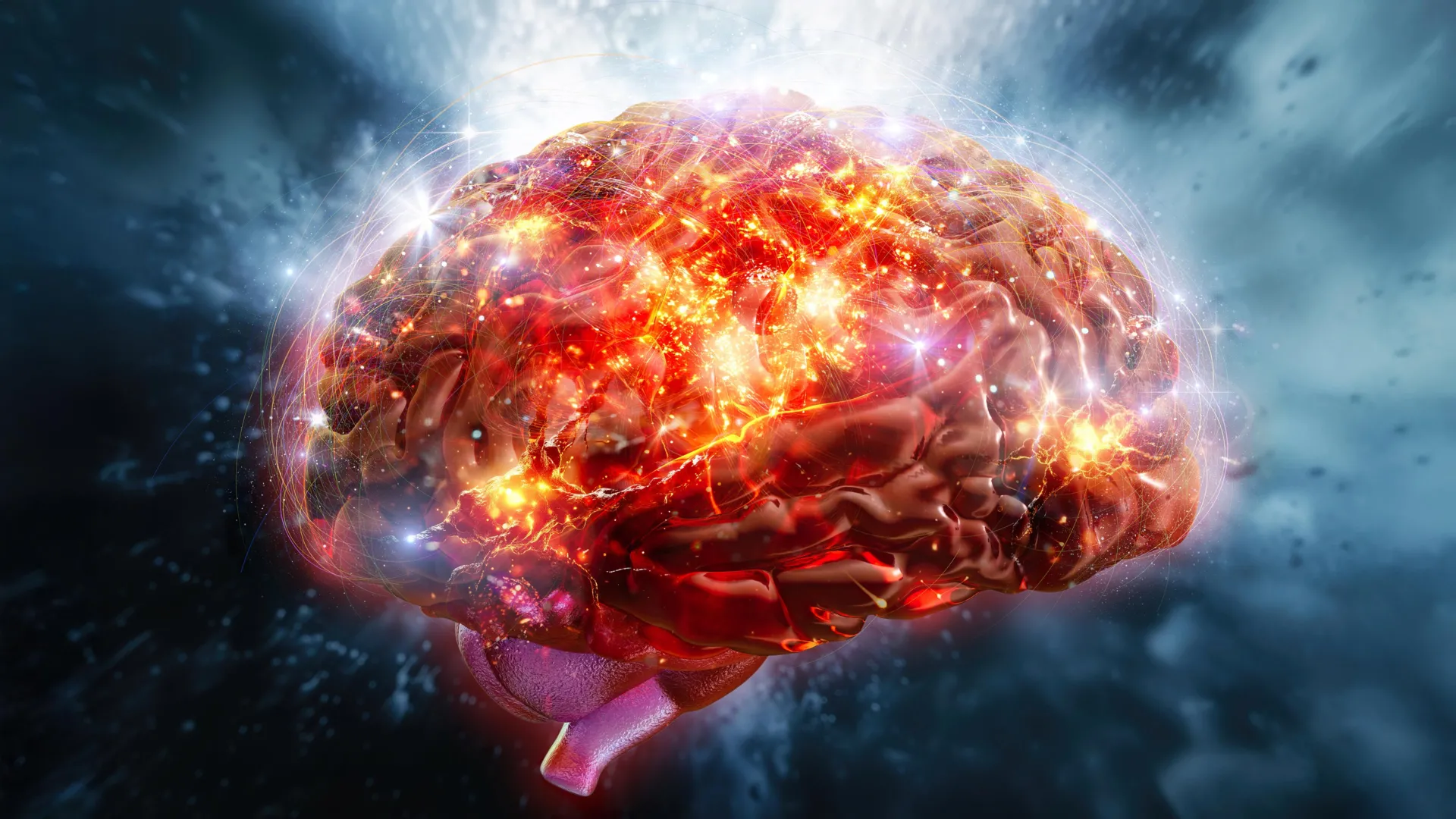
The traditional understanding of physical fitness has long been centered on the periphery of the human body, focusing on the hypertrophy of skeletal muscle fibers, the increased efficiency of the cardiovascular system, and the expansion of lung capacity. However, groundbreaking research published in the journal Neuron suggests that the true engine of athletic progression may reside within the folds of the brain. A study led by researchers at the University of Pennsylvania has identified a specific neural mechanism that orchestrates the body’s adaptation to exercise, revealing that the brain undergoes significant remodeling to facilitate improvements in endurance, speed, and metabolic efficiency.
This research shifts the paradigm of sports science from a muscle-centric view to a neuro-metabolic perspective. By tracking the activity of specific neurons during and after physical exertion, scientists have discovered that the brain does not merely signal the body to move; it actively processes the "stress" of exercise long after the workout has ended, creating a neurological blueprint that allows the heart and muscles to become stronger over time.
The Role of the Ventromedial Hypothalamus in Physical Adaptation
At the heart of this discovery is a region of the brain known as the ventromedial hypothalamus (VMH). Historically, the VMH has been recognized by neuroscientists as a critical hub for homeostatic regulation, controlling appetite, body weight, and blood glucose levels. However, the study led by J. Nicholas Betley, an associate professor of biology at the University of Pennsylvania, demonstrates that the VMH also serves as a command center for physical endurance.
Within the VMH, the research team focused on a distinct population of nerve cells known as steroidogenic factor-1 (SF1) neurons. Using advanced optogenetic and calcium imaging techniques, the researchers monitored these neurons in mice as they performed treadmill exercises of varying intensities. The data revealed that SF1 neurons are not only highly active during the bout of exercise but, crucially, they remain in a state of heightened firing for at least sixty minutes following the cessation of physical activity.
This prolonged neural "afterglow" suggests that the brain enters a period of consolidation after exercise, similar to how the brain processes memories during sleep. This post-exercise activity appears to be the mechanism by which the brain "teaches" the rest of the body how to handle future physical stress more effectively.
Experimental Methodology and the Chronology of Endurance Gains
To understand the long-term implications of this neural activity, the research team designed a two-week longitudinal study. Mice were subjected to daily treadmill sessions where researchers measured their baseline endurance, including maximum running speed and the time taken to reach the point of physical exhaustion.
During the first week, the mice showed standard physiological responses to training. However, by the end of the second week, the group showed a marked increase in performance. These mice were able to run significantly longer distances and maintain higher velocities than they could at the start of the trial. When the researchers conducted brain scans of these "trained" mice, they found a profound change: the number of active SF1 neurons had increased, and the intensity of their firing patterns had strengthened.
The chronology of these changes indicates a cumulative effect. Each exercise session served as a stimulus that triggered a neural response, which then persisted into the recovery phase. Over fourteen days, these repeated bouts of post-exercise neural firing resulted in a permanent shift in the brain’s regulatory set-points, allowing for superior energy mobilization and fatigue resistance.
The Critical Discovery: The Post-Exercise Window
The most significant revelation of the study came when the researchers intervened in the neural signaling process. Using sophisticated chemical tools to "silence" the SF1 neurons, the team tested whether the neurons were necessary for the physical gains observed.
In the first experimental variation, the researchers blocked the SF1 neurons during the actual exercise session. As expected, the mice showed reduced performance. However, the second variation provided the most startling data: when the researchers allowed the neurons to function normally during exercise but blocked them only during the one-hour recovery period immediately following the workout, the mice failed to gain any endurance over the two-week training period.
Despite having completed the same amount of physical work as the control group, the mice with silenced post-exercise neurons remained at their baseline fitness levels. This finding confirms that the "work" done by the brain during the recovery phase is just as essential for building fitness as the physical work done by the muscles during the workout. It suggests that the brain must "authorize" the physiological adaptations—such as increased mitochondrial density in muscles or improved stroke volume in the heart—that lead to increased fitness.
Supporting Data: Metabolism and Glucose Utilization
The study’s findings are bolstered by metabolic data collected during the trials. The SF1 neurons in the VMH are intrinsically linked to how the body manages its fuel sources. During high-intensity exercise, the body relies heavily on glucose stored in the liver and muscles. As these stores deplete, fatigue sets in.
The researchers observed that the continued firing of SF1 neurons after exercise correlated with a more efficient stabilization of blood sugar levels. This suggests that the brain is actively managing the replenishment of glycogen stores and optimizing the transition from carbohydrate burning to fat oxidation. By improving metabolic flexibility, the brain allows the body to recover faster and perform at a higher threshold in subsequent sessions.
Analysis of the data indicates that without the sustained activity of these neurons, the body’s recovery is fragmented. This lack of coordination between the central nervous system and the metabolic system results in a failure to adapt, explaining why the mice in the "blocked" group did not see improvements in their running capacity.
Expert Perspectives and Theoretical Implications
While the study was conducted on murine models, the implications for human physiology are profound. The VMH and SF1 neurons are highly conserved across mammalian species, meaning they function similarly in humans.
"We have traditionally viewed exercise as a bottom-up process—muscles get tired, they send signals to the brain, and the brain reacts," noted a theoretical analysis of the study. "This research suggests a top-down model where the brain determines the ceiling of our physical capabilities based on how it processes the recovery period."
This aligns with the "Central Governor Theory" proposed by some exercise physiologists, which posits that the brain limits physical performance to prevent catastrophic injury. The Neuron study provides a specific cellular mechanism for this theory, suggesting that training is actually the process of the brain "re-programming" its governor to allow for higher limits.
Clinical and Societal Applications
The ability to manipulate or support the activity of SF1 neurons opens new doors for clinical interventions. The researchers highlighted several key areas where these findings could be applied:
- Geriatric Health: As humans age, the brain’s plasticity decreases, and the body’s ability to adapt to exercise wanes. By understanding the neural triggers for endurance, medical professionals may develop therapies to help older adults maintain mobility and cardiovascular health even when their physical capacity for high-intensity training is limited.
- Stroke and Injury Recovery: For patients recovering from neurological damage, the "connection" between the brain and the body’s metabolic systems is often disrupted. This research could lead to rehabilitation protocols that specifically target neural recovery to accelerate physical healing.
- Athletic Optimization: Elite athletes are constantly seeking ways to shorten recovery times. If the post-exercise neural window can be optimized through specific nutritional or environmental interventions (such as temperature control or cognitive rest), athletes could potentially see faster gains in performance.
- Combating Sedentary Lifestyles: One of the primary reasons individuals abandon exercise programs is the "lag time" between starting a routine and seeing results. If science can find ways to "jumpstart" the neural adaptations described in the study, people might experience the benefits of exercise—such as increased energy and mental clarity—much sooner, leading to higher adherence rates.
Future Research Directions
While the study identifies the SF1 neurons as key players, many questions remain regarding the exact molecular signals that these neurons send to the rest of the body. Future research is expected to investigate the specific hormones or neurotransmitters involved in this brain-to-muscle communication.
Furthermore, J. Nicholas Betley and his team are interested in exploring whether different types of exercise—such as resistance training versus aerobic training—trigger different neural pathways within the hypothalamus. "When we lift weights, we think we are just building muscle," Betley noted. "It turns out we might be building up our brain when we exercise."
The study concludes by emphasizing that exercise should be viewed as a holistic event that engages the entire nervous system. The "clear-headedness" and "sharpness" reported by individuals after a workout may not just be a side effect of increased blood flow, but a visible sign of the brain performing the essential work of physiological remodeling.
This research was made possible through the support of several major institutions, including the National Institutes of Health (NIH), the National Science Foundation (NSF), and the National Research Foundation of Korea. As scientists continue to map the neural circuitry of fitness, the definition of "being in shape" will likely evolve to include not just the strength of one’s heart or limbs, but the efficiency and resilience of the brain’s metabolic command centers.














Leave a Reply