
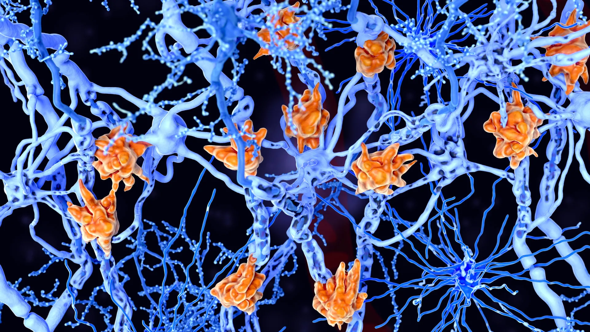
The long-standing paradigm of neuroscience, which positioned neurons as the sole architects of thought, emotion, and memory, is undergoing a fundamental shift following a landmark study published in the journal Nature. Researchers from the University of Arizona and the National Institutes of Health (NIH) have demonstrated that astrocytes—star-shaped non-neuronal cells once dismissed as mere "brain glue"—play a decisive role in the formation, storage, and extinction of fear memories. This discovery suggests that the brain’s "support staff" may actually be functioning as co-directors of neural activity, specifically within the amygdala, the brain’s primary emotional processing center.
For over a century, the scientific community categorized astrocytes as "glia," a term derived from the Greek word for glue. Their known functions were limited to "housekeeping" duties: maintaining the blood-brain barrier, providing nutrients to neurons, and cleaning up metabolic waste. However, the new research led by Lindsay Halladay, an assistant professor at the University of Arizona’s Department of Neuroscience, alongside Andrew Holmes and Olena Bukalo of the NIH’s Laboratory of Behavioral and Genomic Neuroscience, proves that astrocytes are integral to the complex signaling required for emotional learning.
The Evolution of the Tripartite Synapse
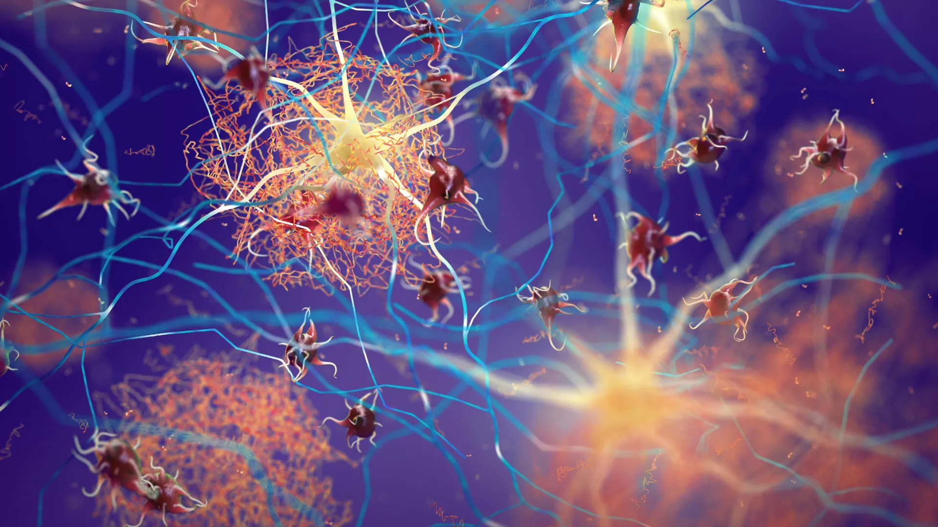
To understand the magnitude of this discovery, it is necessary to look at the evolving concept of the "tripartite synapse." Traditionally, a synapse was viewed as a two-way communication point where one neuron releases neurotransmitters to be received by another. In recent decades, the theory of the tripartite synapse has emerged, suggesting that a third player—the astrocyte—is present at the junction, actively monitoring and modulating the chemical exchange.
The study in Nature provides some of the most robust evidence to date for this theory in the context of complex emotional behavior. By utilizing advanced imaging and genetic manipulation, the research team showed that astrocytes do not just sit idly by while neurons fire; they encode information about fearful stimuli and maintain those signals over time. When a threat is perceived, astrocytes in the amygdala exhibit specific activity patterns that mirror the intensity of the fear.
"Astrocytes are interwoven among neurons in the brain, and it seemed unlikely they were there just for housekeeping," Halladay noted. "We wanted to understand what they’re actually doing—and how they’re shaping neural activity in the process."
Methodology: Observing Fear in Real Time
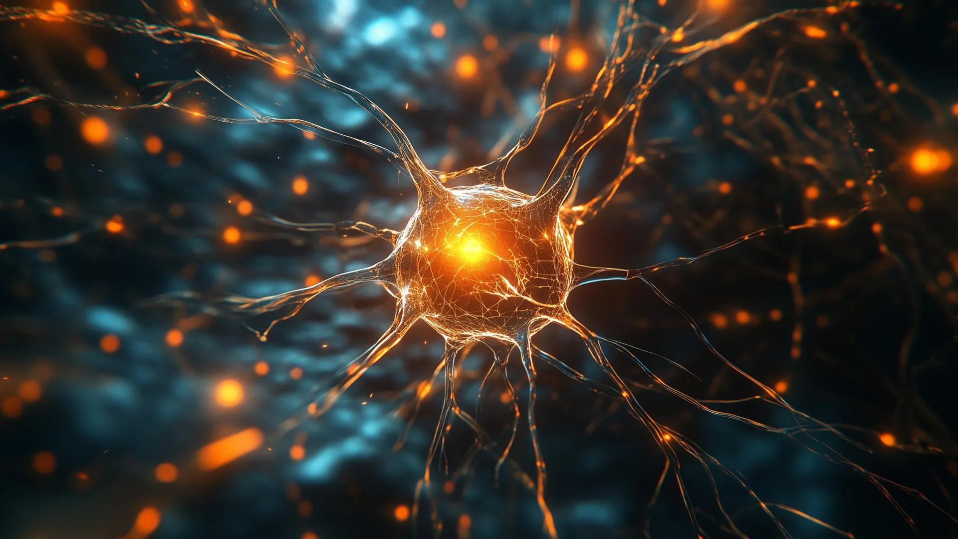
The multi-institutional project employed sophisticated mouse models to observe the brain’s inner workings during fear conditioning. To track the activity of astrocytes, which do not fire electrical impulses like neurons but instead communicate via internal calcium signaling, the team used fluorescent calcium sensors. These sensors glow when the cells become active, allowing researchers to use fiber photometry to "watch" the astrocytes in real-time as the subjects learned to associate a specific stimulus with a negative outcome.
The experiment was structured around three distinct phases: fear acquisition (learning to be afraid), fear recall (remembering the fear), and fear extinction (learning that the stimulus is no longer a threat). The data revealed a direct correlation between astrocyte activity and behavioral responses:
- Learning and Recall: As the mice learned to associate a tone with a mild foot shock, astrocyte activity in the basolateral amygdala (BLA) spiked. This heightened activity remained consistent when the mice were later exposed to the tone alone, indicating that the astrocytes were part of the "memory trace."
- Extinction: When the tone was repeatedly presented without the shock, the mice eventually learned that the danger had passed—a process known as extinction. During this phase, astrocyte activity significantly declined, suggesting these cells are essential for the "unlearning" process.
- Manipulation: To confirm a causal link, the researchers used chemogenetics to artificially increase or decrease astrocyte signaling. When astrocyte activity was boosted, the fear response became more intense and harder to extinguish. Conversely, inhibiting the astrocytes made the fear memories weaker and more easily forgotten.
Disrupting the Neural Architecture
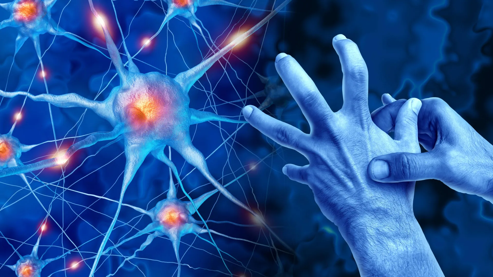
One of the most significant findings of the study was how the disruption of astrocytes fundamentally altered the behavior of neighboring neurons. The researchers observed that when astrocyte signaling was impaired, the surrounding neurons were unable to synchronize their firing patterns. This lack of coordination meant that the neurons could no longer effectively transmit "defense" signals to other parts of the brain, such as the periaqueductal gray, which initiates physical responses like freezing or fleeing.
This discovery suggests that neurons are not autonomous agents. Instead, they rely on a constant stream of regulatory input from astrocytes to maintain the integrity of a memory. Without the "star cells" providing the necessary chemical environment and feedback loops, the neural circuits responsible for fear memory essentially become de-calibrated.
A Broader Network: From the Amygdala to the Prefrontal Cortex
The research team also looked beyond the amygdala to see how astrocyte activity influenced the broader "fear circuit." They found that changes in astrocyte signaling in the amygdala had a ripple effect on the prefrontal cortex (PFC), the region of the brain responsible for high-level decision-making and executive function.
In a healthy brain, the PFC helps modulate the amygdala’s fear response, providing a "top-down" check on whether a fear is rational. The study indicated that astrocytes help facilitate this cross-talk. By guiding how fear-related signals reach the PFC, astrocytes may play a role in helping an organism choose the most appropriate reaction to a threat—whether that is to hide, fight, or recognize that the environment is actually safe.
This systemic involvement suggests that astrocytes are not just local regulators but are key nodes in a wide-reaching network that determines how we navigate a dangerous world.
Clinical Implications for PTSD and Anxiety Disorders
The discovery that astrocytes actively control fear extinction has profound implications for clinical psychology and psychiatry. Conditions such as Post-Traumatic Stress Disorder (PTSD) and various phobias are characterized by a failure of the fear extinction process. In patients with PTSD, the brain remains in a state of high alert long after a threat has vanished, and the "safety" memories formed during therapy often fail to stick.
Current pharmacological treatments for anxiety and PTSD largely focus on modulating neurotransmitters like serotonin or norepinephrine, which primarily target neuronal receptors. However, these treatments are often only partially effective.
"Understanding the role of astrocytes could reshape how scientists approach disorders linked to persistent fear," the study suggests. If astrocytes are the gatekeepers of fear extinction, then developing drugs that specifically target glial signaling—rather than just neuronal firing—could offer a new pathway for treatment. Such "gliotransmitter" therapies could potentially help "reset" the astrocyte activity in the amygdala, making it easier for patients to let go of traumatic memories.
The Path Forward: Mapping the Glial Frontier
The University of Arizona and NIH team are already planning the next phase of their research. While the current study focused on the amygdala, Halladay intends to investigate the role of astrocytes in other regions of the fear network, including the hippocampus (which provides context to memories) and the periaqueductal gray (which controls motor output).
"Understanding that larger circuit could help answer a simple question of why someone with an anxiety disorder might exhibit inappropriate fear responses to something that isn’t actually dangerous," Halladay said.
The timeline for this research suggests a burgeoning field of "astro-neuroscience." For decades, the focus was almost exclusively on the "grey matter" (neurons), while the "white matter" and glia were sidelined. This study marks a significant milestone in a decade-long trend of rediscovering the brain’s non-neuronal components.
Analysis of Scientific Impact
The findings published in Nature represent more than just a new data point; they represent a shift in the biological understanding of consciousness and emotion. By elevating the astrocyte from a passive observer to an active participant, the study challenges the "neuron-centric" view of the brain that has dominated medical textbooks for a century.
Independent experts in the field have noted that this research adds to a growing body of evidence suggesting that the complexity of the human brain may lie not just in the number of its neurons, but in the sophisticated interactions between different types of cells. As researchers continue to peel back the layers of how astrocytes influence everything from sleep to pain perception and now fear, the definition of a "brain circuit" is being rewritten to include these star-shaped sentinels.
In conclusion, the work of Halladay, Holmes, and Bukalo provides a new lens through which to view the traumatized brain. By demonstrating that astrocytes encode and maintain fear, the study opens the door to a future where mental health treatments are as multifaceted as the cells that populate our minds. The star-shaped cells, once thought to be the brain’s simple scaffolding, have finally taken center stage.


Leave a Reply