

The decline of the olfactory sense has long been observed as a precursor to the cognitive deterioration associated with Alzheimer’s disease, yet the underlying biological mechanisms have remained largely elusive until now. New research conducted by a collaborative team of scientists from the German Center for Neurodegenerative Diseases (DZNE) and Ludwig-Maximilians-Universität München (LMU) has identified a specific immunological process that causes the brain to mistakenly destroy its own odor-processing connections. The study, published in the journal Nature Communications, reveals that the brain’s immune system—specifically cells known as microglia—may prematurely prune nerve fibers essential for the sense of smell. By combining longitudinal animal studies with human brain tissue analysis and advanced neuroimaging, the researchers have provided a definitive link between neuroinflammation and the sensory deficits that often precede memory loss by years, if not decades.
The Biological Mechanism of Olfactory Pruning
At the heart of this discovery is the interaction between two distinct but connected regions of the brain: the olfactory bulb and the locus coeruleus. The olfactory bulb, located in the forebrain, serves as the primary processing station for sensory information gathered by receptors in the nasal cavity. The locus coeruleus, situated within the brainstem, acts as a regulatory hub, sending long, projecting nerve fibers to various parts of the brain, including the olfactory bulb. These fibers release norepinephrine, a neurotransmitter that modulates how the brain perceives and prioritizes sensory input.
The research team, led by Dr. Lars Paeger and Prof. Dr. Jochen Herms, discovered that in the early stages of Alzheimer’s disease, these projecting nerve fibers from the locus coeruleus begin to exhibit abnormal electrical activity. This hyperactivity appears to trigger a chemical change on the surface of the nerve cells. Specifically, a fatty molecule called phosphatidylserine, which is typically sequestered on the inner layer of the cell membrane, migrates to the outer surface.
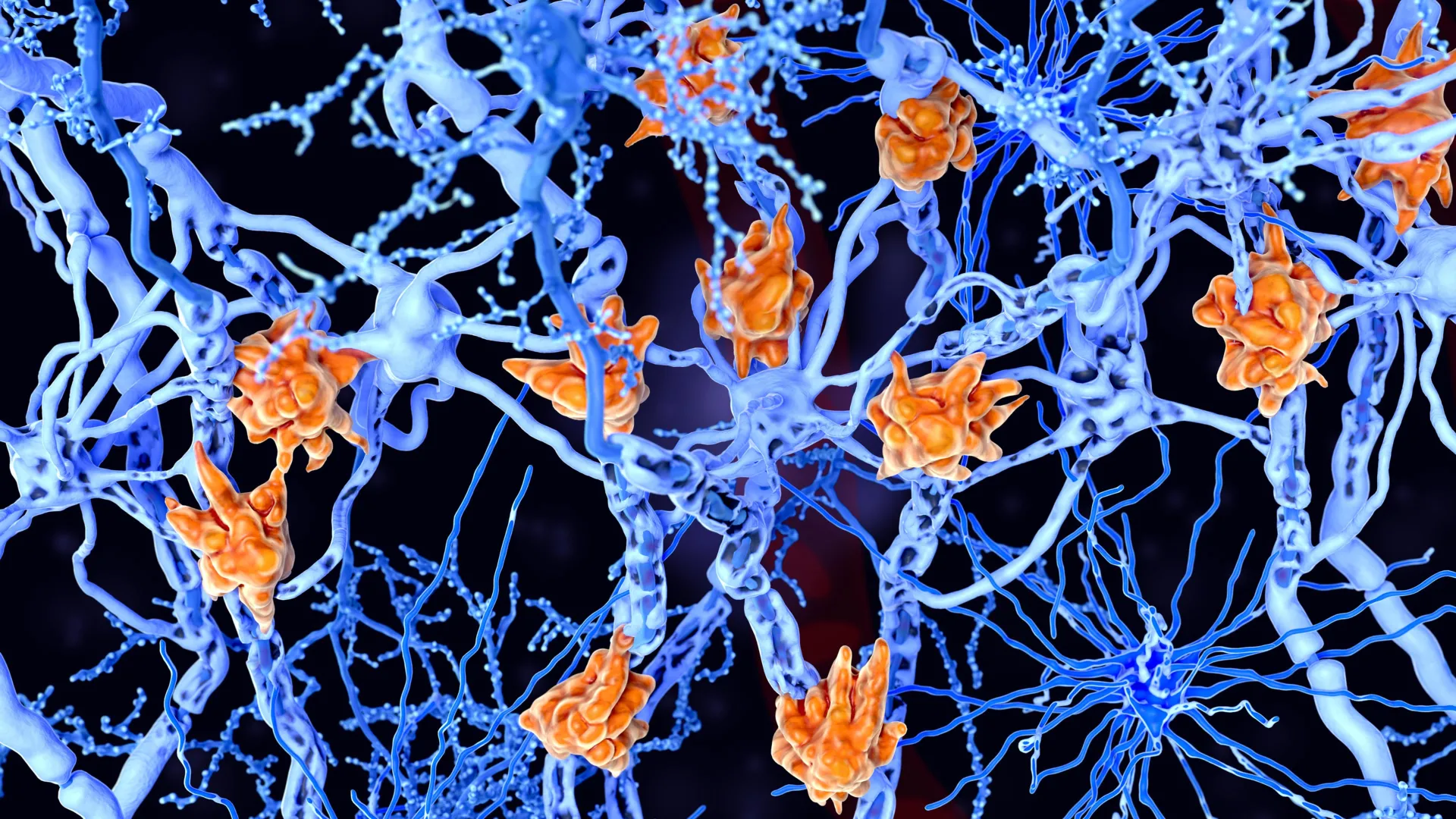
In the complex ecosystem of the brain, this translocation of phosphatidylserine acts as a potent "eat-me" signal. Microglia, the primary immune cells and "scavengers" of the central nervous system, are programmed to recognize this signal. Under normal circumstances, this process, known as synaptic pruning, is a vital part of brain development and maintenance, allowing the brain to remove weak or redundant connections to optimize neural efficiency. However, in the context of early-stage Alzheimer’s, the study suggests that this mechanism goes awry. The microglia misinterpret the "eat-me" signals on the hyperactive fibers of the locus coeruleus and begin to systematically break them down, effectively severing the regulatory link to the olfactory bulb and leading to a diminished sense of smell.
A Multi-Disciplinary Approach to Evidence
The strength of the study lies in its comprehensive methodology, which bridged the gap between laboratory models and clinical human data. To reach their conclusions, the researchers employed a three-pronged investigative strategy.
First, the team utilized transgenic mouse models designed to express the hallmarks of Alzheimer’s pathology. By monitoring these mice over time, the scientists were able to observe the chronological progression of nerve fiber degradation. They noted that the loss of connectivity between the locus coeruleus and the olfactory bulb occurred well before the accumulation of amyloid-beta plaques—the protein clumps traditionally associated with the later stages of the disease.
Second, the researchers performed post-mortem analyses on human brain tissue. By examining the olfactory bulbs and brainstems of deceased individuals who had been diagnosed with Alzheimer’s, they confirmed that the same patterns of nerve fiber loss and microglial activation observed in mice were present in human patients. This validation was crucial in proving that the "eat-me" signal mechanism was not merely a quirk of animal biology but a relevant factor in human neurodegeneration.
Finally, the study incorporated data from Positron Emission Tomography (PET) scans of living patients. These scans allowed the researchers to visualize the activity of microglia and the density of nerve connections in individuals with mild cognitive impairment (MCI) and confirmed Alzheimer’s. The PET data correlated strongly with the laboratory findings, showing that individuals with higher levels of microglial activity in the olfactory regions tended to have more significant deficits in their ability to identify odors.
The Chronology of Sensory Decline
For decades, Alzheimer’s research focused primarily on the "amyloid cascade hypothesis," which posits that the buildup of amyloid-beta plaques is the primary driver of the disease. However, many clinical trials targeting these plaques in late-stage patients have failed to yield significant cognitive improvements. This has led the scientific community to shift its focus toward the "prodromal" or preclinical phase of the disease—the period during which the brain is already undergoing changes but the patient has not yet developed dementia.
The DZNE and LMU study fits perfectly into this new paradigm by establishing a clear timeline for olfactory decline. The research indicates that the breakdown of the locus coeruleus-olfactory bulb connection is one of the earliest detectable events in the Alzheimer’s progression. Because the sense of smell is processed through a relatively direct pathway compared to complex memory or language functions, it may serve as a "canary in the coal mine" for neurodegeneration.
Historically, patients have often reported a loss of smell years before they began forgetting names or getting lost in familiar places. This study provides the biological "why" behind those anecdotal reports, suggesting that the immune system’s overreaction to neuronal hyperactivity is a fundamental early-stage driver of the disease.
Implications for Early Diagnosis and Therapy
The identification of an immunological cause for smell loss has profound implications for the future of Alzheimer’s care. Currently, one of the greatest challenges in treating neurodegenerative diseases is the timing of intervention. By the time a patient exhibits significant memory loss, a substantial portion of their neurons have already perished, making it difficult for treatments to reverse the damage.
"Our findings could pave the way for the early identification of patients at risk of developing Alzheimer’s," stated Prof. Dr. Joachim Herms. He noted that if a simple smell test could be validated as a reliable screening tool, it would allow clinicians to identify high-risk individuals much earlier. These patients could then undergo more intensive diagnostic testing, such as PET scans or cerebrospinal fluid analysis, to confirm the presence of Alzheimer’s markers before cognitive symptoms emerge.
This early detection is particularly relevant given the recent emergence of amyloid-beta antibodies, such as lecanemab and aducanumab. These therapies are designed to clear amyloid plaques from the brain, but clinical data suggests they are most effective when administered during the earliest stages of the disease. By using olfactory dysfunction as a biomarker, doctors could potentially initiate these treatments years earlier, significantly increasing the probability of preserving cognitive function and slowing the disease’s progression.
Expert Reactions and the Broader Scientific Context
While the DZNE and LMU study is a major step forward, it also opens up new questions for the field of neuroimmunology. Independent experts in the field have noted that the role of the locus coeruleus is particularly intriguing. As a major source of norepinephrine, the locus coeruleus is involved in the brain’s inflammatory response. The fact that its fibers are among the first to be pruned suggests that the loss of norepinephrine may actually accelerate neuroinflammation elsewhere in the brain, creating a vicious cycle of degeneration.
Dr. Lars Paeger emphasized that the study highlights the "double-edged sword" nature of the brain’s immune system. While microglia are essential for protecting the brain from infection and clearing debris, their "hyper-vigilance" in the presence of Alzheimer’s-related stress can lead to the destruction of healthy, functional tissue. This shift in perspective—viewing Alzheimer’s not just as a protein-misfolding disease but as a chronic inflammatory disorder—is gaining traction across the global research community.
Furthermore, the study suggests that future treatments might not only focus on clearing amyloid but also on modulating the "eat-me" signals on neurons. If a drug could be developed to prevent the translocation of phosphatidylserine or to "mask" the signal from microglia, it might be possible to protect the nerve fibers and preserve sensory and cognitive functions.
Future Outlook: From the Lab to the Clinic
The transition from these laboratory findings to a standardized clinical tool will require further validation. Large-scale longitudinal studies are needed to determine exactly how accurately smell loss can predict the onset of Alzheimer’s compared to other forms of dementia, such as Parkinson’s disease or Lewy body dementia, which also involve olfactory deficits.
However, the path forward is clearer than it was before. The integration of sensory testing into routine geriatric care could become a reality. Unlike expensive MRI or PET scans, olfactory testing is non-invasive, inexpensive, and quick to administer. If the immunological mechanism identified by the Munich-based researchers can be targeted, it could transform the sense of smell from a neglected symptom into a vital diagnostic and therapeutic window.
As the global population ages, the prevalence of Alzheimer’s disease is expected to rise sharply, placing an immense burden on healthcare systems. Insights such as those provided by the DZNE and LMU Munich team offer a glimmer of hope that by understanding the very first steps of the disease—down to the specific molecules on a nerve cell’s membrane—we may finally be able to intervene before the most devastating symptoms of the disease take hold. The study stands as a testament to the power of combining basic biological research with clinical observation, providing a roadmap for the next generation of Alzheimer’s diagnostics and treatments.


Leave a Reply