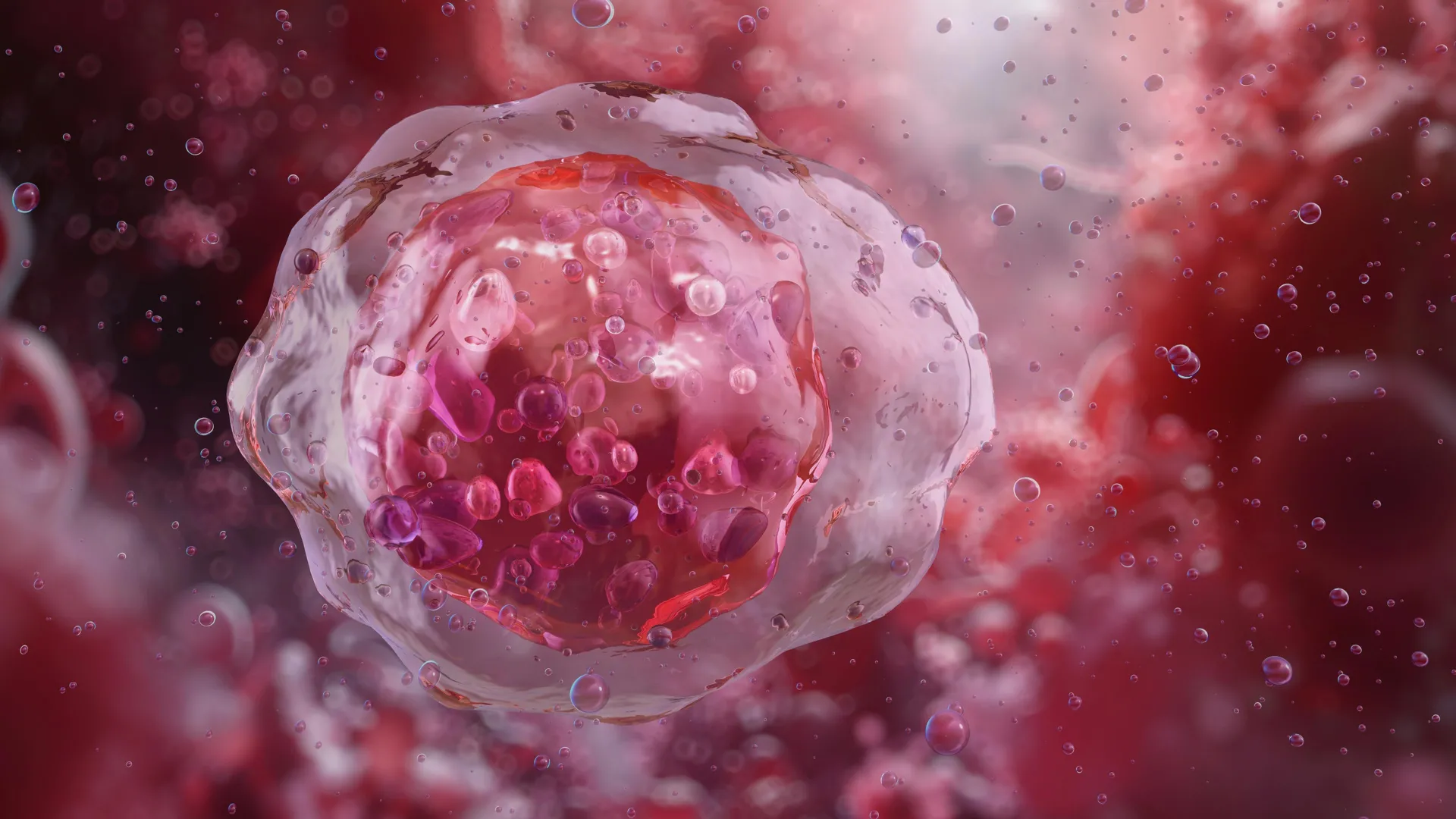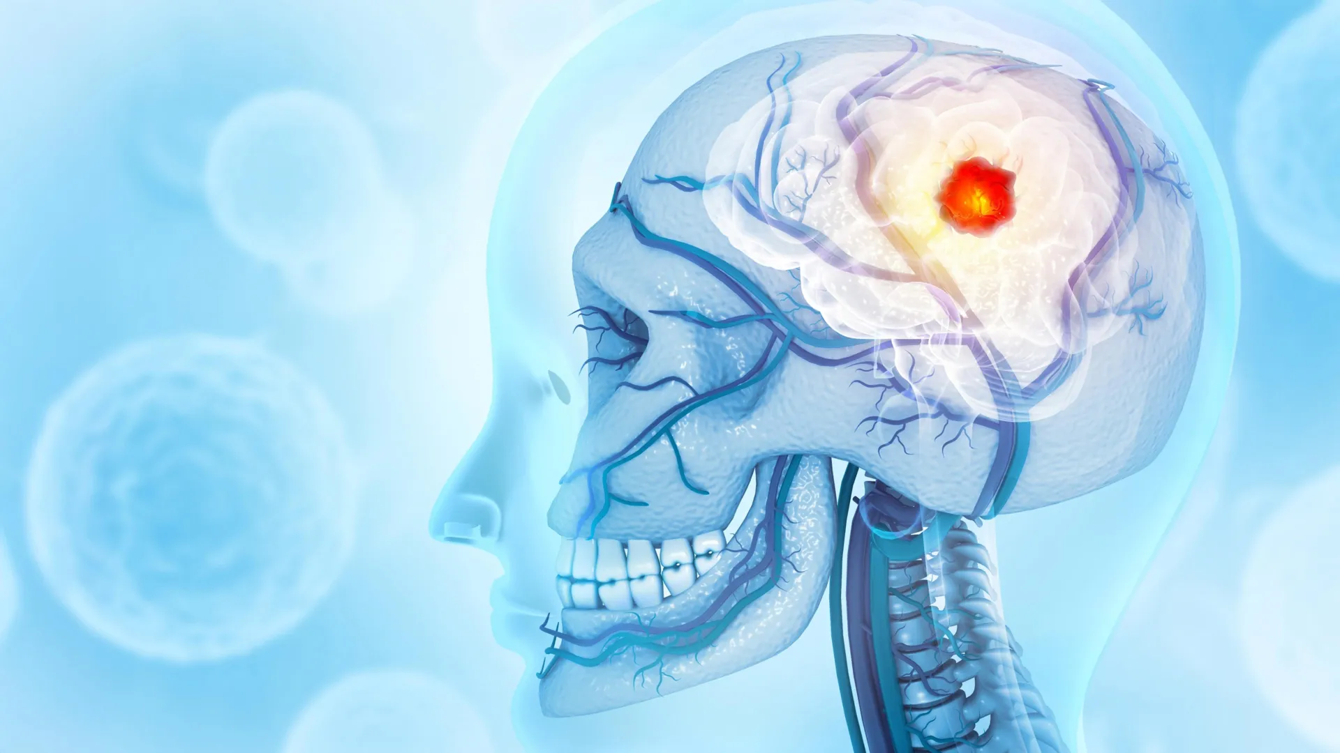
For generations, the foundational dogma of human physiology and hematology has remained absolute and unchallenged: the generation of blood cells, a relentless biological necessity required to sustain human life, is exclusively the domain of the bone marrow. Every medical student across the globe has been taught that the spongy interior of human bones serves as the singular factory where hematopoietic stem cells (HSCs) manufacture the roughly 200 billion red blood cells, trillions of platelets, and vast networks of immune cells needed daily to transport oxygen from the lungs to every peripheral organ. However, groundbreaking scientific research conducted at the University of California, San Francisco (UCSF) has fundamentally upended this long-standing paradigm. In a revelation that rewrites core medical textbooks, researchers have conclusively demonstrated that the human lung is not merely an passive gas-exchange organ, but an active, primary site of blood production.
Published in the esteemed scientific journal Blood, the study unveils the stunning presence of functional hematopoietic stem cells residing within human lung tissue. These resident stem cells are actively engaged in manufacturing red blood cells and megakaryocytes—the specialized progenitor cells responsible for producing the platelets essential for blood clotting and wound healing. This paradigm-shifting discovery not only transforms our fundamental understanding of human anatomy and physiology, but it also carries profound, potentially life-saving implications for the fields of regenerative medicine, oncology, and stem cell transplantation.
Bridging the Gap from Murine Models to Human Biology
The journey toward this landmark human discovery began several years earlier in the sophisticated laboratories of UCSF, spearheaded by Dr. Mark Looney, a professor of medicine and laboratory medicine, alongside his investigative team. In 2017, the research group published a seminal study involving murine models that sent shockwaves through the scientific community. By utilizing advanced microscopic imaging techniques and cellular tracking in mice, the UCSF team made the astonishing observation that approximately 50 percent of a mouse’s entire platelet production occurred not within the bone marrow, but directly inside the pulmonary vasculature of the lungs.
Encouraged and intrigued by these initial findings, the team pressed further into the murine biology, ultimately discovering that mouse lungs harbored versatile stem cells capable of generating the complete repertoire of blood constituents: red blood cells, megakaryocytes, and multiple distinct lineages of immune cells. Yet, a monumental scientific hurdle remained. Critics and skeptics within the broader hematological community rightly questioned whether these intriguing pulmonary phenomena were unique adaptations exclusive to rodents, or if they represented a deeply conserved, evolutionarily significant biological mechanism shared by humans.
Determined to bridge the critical gap between mouse models and human physiology, Dr. Looney’s laboratory embarked on a rigorous, multi-year investigative initiative. To answer the lingering question of human applicability, the researchers secured carefully managed, ethically donated samples of human lung tissue, human bone marrow, and peripheral blood. By establishing a direct comparative analysis across these diverse tissue types, the team set out to determine whether the human pulmonary landscape mirrored the surprising blood-producing capacity observed in their earlier murine studies.
Rigorous Methodology and Laboratory Verification
The investigative process required meticulous cellular screening and cutting-edge laboratory assays. When the UCSF researchers examined a volume of human lung tissue roughly equivalent to the size of a golf ball, they encountered a remarkable histological landscape. Interspersed securely between the delicate blood vessels of the lung parenchyma, they identified a robust population of stem cells that bore an unmistakable, striking resemblance to the classic hematopoietic stem cells traditionally isolated exclusively from bone marrow.
Quantitative analysis yielded an even greater surprise: these pulmonary hematopoietic stem cells were not isolated anomalies or transient cellular wanderers. Rather, they appeared at frequencies comparable to those found in traditional bone marrow reserves.
"The lung HSCs weren’t one-offs — they were a reliable presence in the lungs," noted Dr. Catharina Conrad, a postdoctoral scholar in Looney’s laboratory and the first author of the published study. "But we still needed to know that they were actually capable of making blood."
To definitively establish the functional competence of these newly identified pulmonary stem cells, the research team designed a series of rigorous in vitro experiments. They isolated HSCs from both human lung tissue and human bone marrow, placing them side-by-side into specialized petri dishes to observe their maturation trajectories under controlled laboratory conditions. The results were both definitive and illuminating. Both pulmonary and bone marrow HSCs thrived within this gold-standard environment, proving their viability. However, fascinating functional differences emerged between the two populations.
While bone marrow colonies tended to skew heavily toward the production of immune cells, the stem cell colonies derived from human lung tissue demonstrated a prolific capacity to manufacture red blood cells and megakaryocytes. To push the validation standards to the highest possible threshold, the team introduced human lung HSCs into immunodeficient, HSC-deficient mice. Remarkably, the human cells successfully engrafted and actively restored bone marrow function within the recipient subjects, confirming a deeply integrated functional synergy between the pulmonary system and the traditional hematopoietic network.
Historical Context and Institutional Support
This monumental scientific achievement was made possible through sustained, long-term institutional backing and the convergence of advanced cellular imaging technologies. The research was supported significantly by the National Heart, Lung, and Blood Institute (NHLBI), a prominent division of the National Institutes of Health (NIH). By funding exploratory research that crossed conventional disciplinary boundaries, the NHLBI enabled scientists to ask unconventional questions about organ interconnectedness and stem cell distribution.
Historically, the study of hematopoiesis has remained strictly compartmentalized within the study of hematology and bone marrow pathology. For nearly a century, medical consensus maintained that once embryonic development concluded, blood cell generation became localized primarily to the axial skeleton and long bones. While extramedullary hematopoiesis—the production of blood cells outside the bone marrow—was recognized as a pathological response to severe bone marrow failure, leukemia, or advanced myelofibrosis, the concept of a normal, healthy physiological reservoir of stem cells actively operating within a vital non-bone organ like the lung was unprecedented.
The UCSF discovery shifts this perspective from pathology to normal physiology. It suggests that the lungs and the bone marrow exist in a state of dynamic physiological partnership, actively communicating and potentially sharing stem cell traffic to maintain systemic homeostasis under varying physiological demands.
Clinical Implications for Stem Cell Transplants and Oncology
The ramifications of the UCSF discovery extend far beyond basic cellular biology, offering profound clinical implications for modern medicine—most notably in the realm of hematopoietic stem cell transplantation (HSCT). For decades, bone marrow transplants have served as a cornerstone, life-saving therapeutic intervention for patients battling aggressive hematological malignancies, including various forms of leukemia, lymphoma, and severe aplastic anemia. These procedures typically involve mobilizing stem cells from a donor’s bone marrow or peripheral blood, filtering them, and infusing them into a recipient whose diseased immune and blood-producing systems have been ablated through chemotherapy or radiation.
The revelation that functional hematopoietic stem cells reside abundantly within human lung tissue introduces the tantalizing possibility of utilizing the lungs as a novel, potent reservoir for life-saving stem cell harvesting.
"For decades, bone marrow transplants have been a lynchpin in the treatment of cancers like leukemia," stated Dr. Mark Looney. "The lung HSCs could prove to be a second and significant reservoir of these precious stem cells."
Perhaps the most astonishing piece of retrospective evidence uncovered during the study emerged when the researchers analyzed the cellular output of routine, contemporary bone marrow transplant preparations. Standard clinical protocols for these procedures typically begin with drawing peripheral blood from a donor and screening for circulating stem cells. When the UCSF team analyzed the molecular signatures of the stem cells routinely isolated during these standard procedures, they made a startling retrospective realization: nearly 20 percent of the stem cells collected and utilized in standard "bone marrow transplants" actually carried the distinct molecular signature of lung-derived HSCs.
This retrospective finding suggests that modern medicine has unknowingly been harvesting pulmonary stem cells alongside traditional bone marrow derivatives for years, entirely unaware of their true anatomical origin. Recognizing this reality opens up vast new avenues for optimizing transplant protocols, potentially improving donor yield, enhancing engraftment success rates, and offering alternative harvesting methodologies for patients with compromised bone marrow reserves.
Broader Medical Horizon and Future Research Directions
As the medical community digests the magnitude of this discovery, researchers emphasize that numerous critical questions remain unanswered. Why, from an evolutionary and physiological standpoint, do the lungs require their own localized blood-producing machinery? The anatomical reality of the pulmonary system provides a compelling functional clue. Because the lungs are intricately intertwined with the entire cardiac output and responsible for oxygenating the totality of human blood circulation, they are uniquely positioned as a frontline metabolic hub.
"The lungs are critical to blood circulation, so it’s tantalizing to see the lung HSCs as an emergency reservoir for red blood cell and platelet production," Dr. Looney observed.
Researchers hypothesize that pulmonary HSCs may function as a specialized "emergency reserve" or rapid-deployment cellular depot. In scenarios of acute systemic stress—such as severe hemorrhage, traumatic injury, systemic hypoxia, or overwhelming infection—this localized reservoir of stem cells can be rapidly activated to boost oxygen-carrying red blood cell output and platelet generation far more quickly than waiting for signals to propagate through the central skeletal system.
Furthermore, future investigations will likely explore whether distinct functional differences between bone marrow HSCs and lung HSCs can be harnessed for targeted therapeutic applications. If pulmonary stem cells possess a natural predisposition toward robust red blood cell and megakaryocyte generation, bioengineers and regenerative medicine specialists may learn to manipulate these cells to treat refractory anemias, severe clotting disorders, and acute lung injuries more effectively than ever before.
As clinical trials and follow-up studies are designed to explore these therapeutic frontiers, the textbook understanding of human anatomy has forever changed. The human lung is no longer viewed strictly as an isolated bellows for air exchange; it stands revealed as a dynamic, life-sustaining hematopoietic organ, quietly orchestrating the flow of human vitality from deep within the chest.