Researchers at the Johns Hopkins Kimmel Cancer Center and the Johns Hopkins Bloomberg School of Public Health have unraveled a critical mechanism by which rearranged genes drive the progression of a rare and aggressive form of kidney cancer. Published on April 22 in the scientific journal Cell Reports, the study sheds light on translocation renal cell carcinoma (tRCC), a malignancy that currently lacks standard, highly effective therapeutic interventions. Supported primarily by funding from the National Institutes of Health (NIH), the research reveals that aberrant fusion proteins assemble into microscopic liquid droplets within cell nuclei, acting as master switches that inappropriately activate genes responsible for tumor proliferation and metastasis.
The investigation opens a promising avenue for drug development. By demonstrating that the disruption of these liquid condensates halts the transcription of cancer-promoting genes, the Johns Hopkins team has identified a distinct vulnerability in a tumor type that has historically confounded oncologists. Beyond kidney cancer, the implications of this discovery extend to other fusion-gene-driven malignancies, offering a unified conceptual framework for targeting nuclear condensates across a spectrum of difficult-to-treat diseases.
Background and Context of Translocation Renal Cell Carcinoma
Translocation renal cell carcinoma is an uncommon subtype of kidney cancer that disproportionately affects children and young adults, though it can occur across all age groups. Unlike more common forms of adult renal cell carcinoma, which are frequently linked to Von Hippel-Lindau (VHL) gene mutations and clear cell histology, tRCC is characterized by chromosomal translocations involving the TFE3 gene located on the X chromosome.
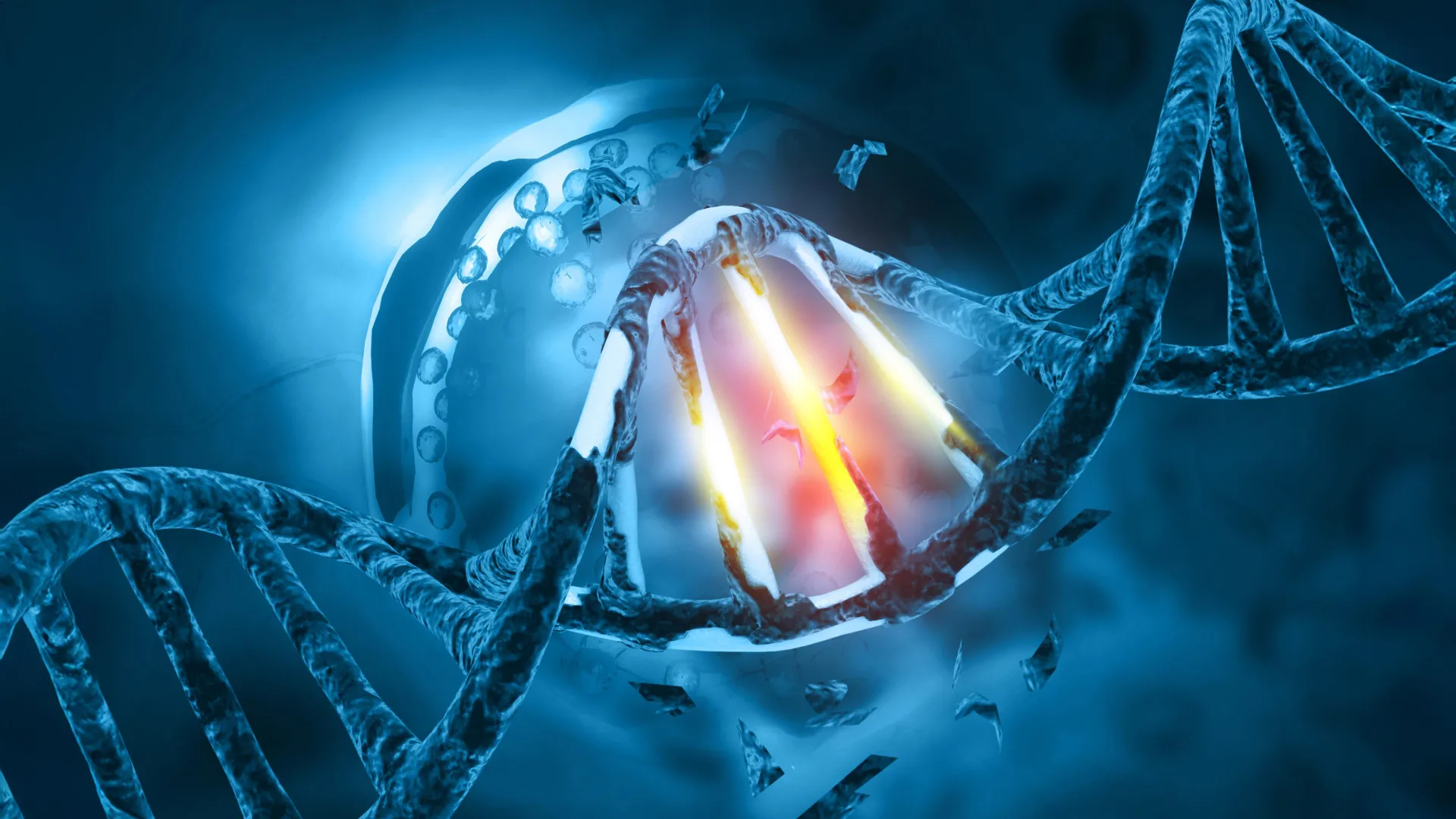
During chromosomal rearrangement, a segment of DNA breaks away and fuses with a portion of another gene. In the case of tRCC, the tail end of the TFE3 gene becomes inextricably joined with the beginning of any one of roughly 20 different partner genes. The most prevalent of these fusion partners are NONO and SFPQ, which collectively account for approximately 40 percent of all identified TFE3 fusions.
These genetic aberrations result in the production of chimeric TFE3 fusion proteins that do not exist in healthy cellular environments. While scientists have long recognized that these abnormal proteins are the primary catalysts for tRCC, the precise biochemical pathways by which they initiate and sustain tumorigenesis remained elusive. Traditional drug discovery paradigms aimed at inhibiting these proteins have faced immense hurdles because the proteins often lack traditional enzymatic binding pockets targeted by conventional small-molecule inhibitors. Consequently, clinicians have lacked standardized guidelines for treating advanced tRCC, frequently relying on regimens extrapolated from other renal cancer subtypes with limited success.
Chronology and Experimental Methodology
The path to the recent Cell Reports publication represents a multidisciplinary convergence of molecular biology, biophysics, and cancer genomics. The investigative effort was spearheaded by senior author Danfeng "Dani" Cai, Ph.D., an assistant professor of biochemistry and molecular biology at the Johns Hopkins Bloomberg School of Public Health whose laboratory specializes in the biophysical properties of cellular condensates. To decode the behavior of TFE3 fusion proteins, Cai collaborated with Eneda Toska, Ph.D., an assistant professor of oncology at the Johns Hopkins Kimmel Cancer Center.
The research workflow unfolded through a series of meticulous, step-by-step observations:
- Labeling and Visualization: Researchers introduced fluorescent tags to the TFE3 fusion proteins within cells derived from tRCC patients. Utilizing advanced microscopy, the team observed that the fusion proteins did not disperse evenly throughout the nucleus; instead, they coalesced into distinct, punctate dots.
- Condensate Identification: Further analysis confirmed that these dots were liquid condensates—concentrated assemblies of macromolecules interacting dynamically within a localized microenvironment without a surrounding membrane.
- Transcriptional Mapping: By evaluating the contents of these droplets, scientists discovered the presence of marker proteins typically associated with actively transcribed genes and transcriptional machinery, implying a direct role in gene activation.
- Chromatin Remodeling Analysis: Partnering with Toska, the team mapped the interaction between the fusion proteins and cellular DNA. They found that the condensates physically altered the chromatin architecture, opening tightly wound DNA segments to make them accessible for transcription.
- Structural Disruption Experiments: By systematically deleting segments of the TFE3 fusion proteins, researchers identified a specific coiled-coil structural domain essential for condensate formation. Removing this domain prevented the proteins from forming liquid droplets and effectively silenced their cancer-promoting transcriptional activity.
Supporting Data and Biophysical Insights
The study’s findings are anchored in the biophysics of liquid-liquid phase separation, a rapidly expanding field in cellular biology. Inside healthy cells, compartmentalization is traditionally achieved through membrane-bound organelles such as the nucleus, mitochondria, and endoplasmic reticulum. However, cells also utilize non-membrane-bound compartments—often referred to as biomolecular condensates—to concentrate specific proteins and nucleic acids to carry out rapid biochemical reactions.
In the context of tRCC, the fusion of TFE3 with partners like NONO and SFPQ endows the resulting proteins with altered valency and interaction domains. While normal full-length TFE3, NONO, and SFPQ proteins participate in standard transcriptional regulation, their fusion product exhibits an abnormally potent capacity to undergo phase separation.
The data demonstrated that these condensates act as hyper-efficient transcription hubs. By binding to regulatory regions of the genome, the droplets induce localized chromatin remodeling. DNA in eukaryotic cells is packaged into chromatin, winding tightly around histone proteins like beads on a string. Tightly packed regions (heterochromatin) are inaccessible and transcriptionally silent, whereas loosely wound regions (euchromatin) permit gene expression. Toska and her colleagues revealed that TFE3 fusion condensates chemically modify and reorganize the chromatin landscape, forcing open specific genomic sites that regulate cell proliferation and migration—the hallmark behaviors required for tumor growth and metastatic spread.
Official Statements and Investigator Perspectives
The implications of the Johns Hopkins study extend far beyond the specific confines of rare renal cell carcinomas. Investigators emphasize that the mechanistic insights gained from studying tRCC could illuminate pathogenic processes in a broader class of human cancers driven by similar genetic events.
"Other cancers, such as Ewing sarcoma and leukemia, are caused by fusion genes as well," notes senior author Danfeng Cai. "It’s possible that these fusion genes form similar droplets, or condensates, that regulate genes in these cancers and could react to similar treatment strategies."
This perspective highlights a unifying hypothesis in contemporary oncology: diverse chromosomal translocations across different tissue types may converge on a common biophysical mechanism—liquid-liquid phase separation—to drive aberrant gene expression.
Eneda Toska elaborates on the structural vulnerability uncovered by the team’s targeted protein deletions. "We found that these fusion proteins open and close different sites on the chromatin by making chemical modifications," Toska explains. "They bind, regulate and redesign the chromosome landscape, interacting with target genes that promote cell proliferation and movement—functions that cancer needs to grow and spread."
By pinpointing the coiled-coil domain as the structural linchpin required for condensate assembly, the researchers have provided structural biologists with a concrete target for rational drug design. Rather than attempting to block an elusive enzymatic site, future therapeutics could theoretically be engineered to disrupt the specific protein-protein interactions that allow these droplets to form and persist.
Broader Impact and Future Implications for Cancer Therapeutics
The publication of these findings in Cell Reports marks an important milestone in translational oncology, shifting the paradigm of how researchers conceptualize and potentially treat translocation-associated cancers.
Historically, transcription factors and fusion proteins resulting from chromosomal translocations have been classified as "undruggable" because they lack enzymatic activity that can be inhibited by conventional small molecules. They operate primarily through macromolecular protein interactions and spatial organization within the nucleus. The identification of liquid condensates as the functional engine of tRCC bypasses this traditional therapeutic roadblock.
If pharmacological agents can be developed to safely dissolve or prevent the formation of these pathological condensates without disrupting normal cellular housekeeping functions, clinicians could gain an entirely new class of anti-cancer drugs. Such strategies would hold immense promise not only for patients diagnosed with translocation renal cell carcinoma but also for individuals suffering from other fusion-gene-driven malignancies, including pediatric sarcomas and various leukemias.
The research team is already advancing to the next phase of investigation. Future studies will focus on cataloging the complete proteome within the TFE3 fusion condensates to identify additional co-factors, enzymes, or structural proteins that contribute to droplet stability and transcriptional activity. Once these auxiliary components are mapped, high-throughput drug screening campaigns can be deployed to identify small molecules capable of selectively dismantling the condensates.
Funding and Collaborative Scope
The breadth of this research reflects a highly collaborative enterprise involving multiple academic departments and specialized research centers. In addition to primary contributions from the Johns Hopkins Kimmel Cancer Center and the Johns Hopkins Bloomberg School of Public Health, the study benefited from the expertise of investigators from the Johns Hopkins University School of Medicine and the National Cancer Institute.
Financial support for the project was provided by a robust coalition of public and private funding bodies. Key contributors include the National Institutes of Health, specifically the National Institute of General Medical Sciences (grant R35GM142837); the National Cancer Institute (grants K22CA245487, R01CA276187, and K01CA245124); and the National Human Genome Research Institute (grants R01HG013409 and R01HG010889). Additional support was rendered by a Department of Defense Kidney Cancer Idea Development Award (grant W81XWH2210900), a Jayne Koskinas Ted Giovanis grant, and a Johns Hopkins Provost Catalyst Award.
As the scientific community evaluates these findings, the work serves as a testament to the power of basic biophysical research in illuminating the dark corners of rare diseases, ultimately paving the way for targeted therapies where none previously existed.