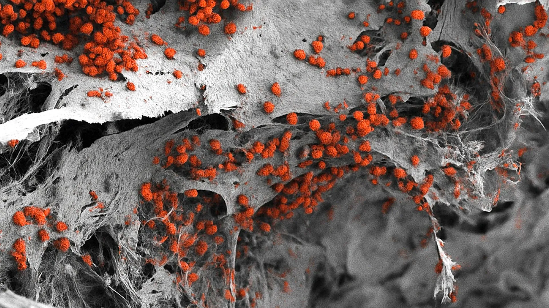
Researchers at the Indiana University School of Medicine have successfully engineered a groundbreaking imaging methodology designed to visualize the intricate cellular landscape of mouse bone marrow. Published recently in the peer-reviewed journal Leukemia, this innovation overcomes decades-long technical barriers inherent to bone marrow research. By leveraging the advanced multiplex imaging platform Phenocycler 2.0, the multidisciplinary scientific team has unlocked an unprecedented capability to observe up to 25 distinct cellular markers simultaneously within intact murine bone marrow tissue.
This technological leap is expected to dramatically accelerate the pace of therapeutic discovery for a wide array of pathologies, ranging from hematological malignancies and severe autoimmune disorders to complex musculoskeletal diseases. As pharmaceutical companies and academic institutions continually seek more accurate translational models, this novel visualization approach bridges a critical gap in preclinical research, offering a high-resolution window into one of the human body’s most inaccessible and vital tissues.
The Structural and Physiological Challenges of Bone Marrow Analysis
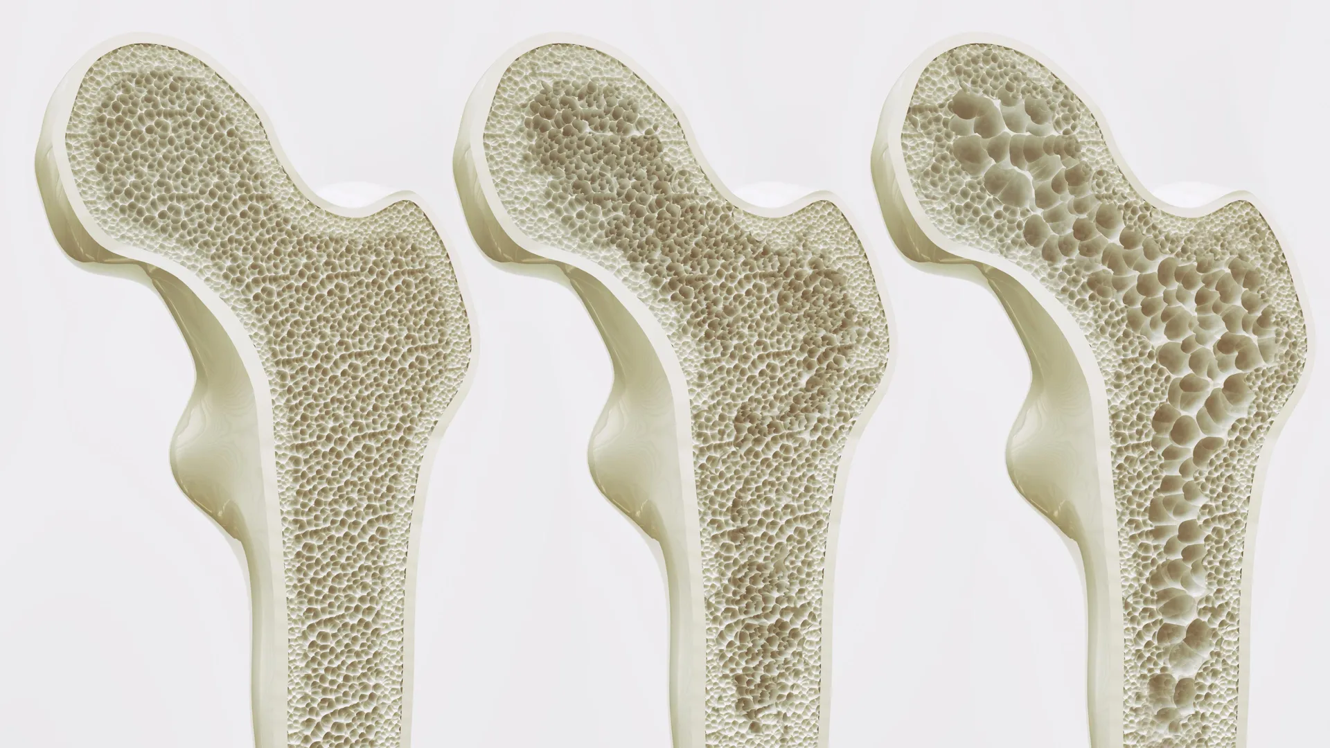
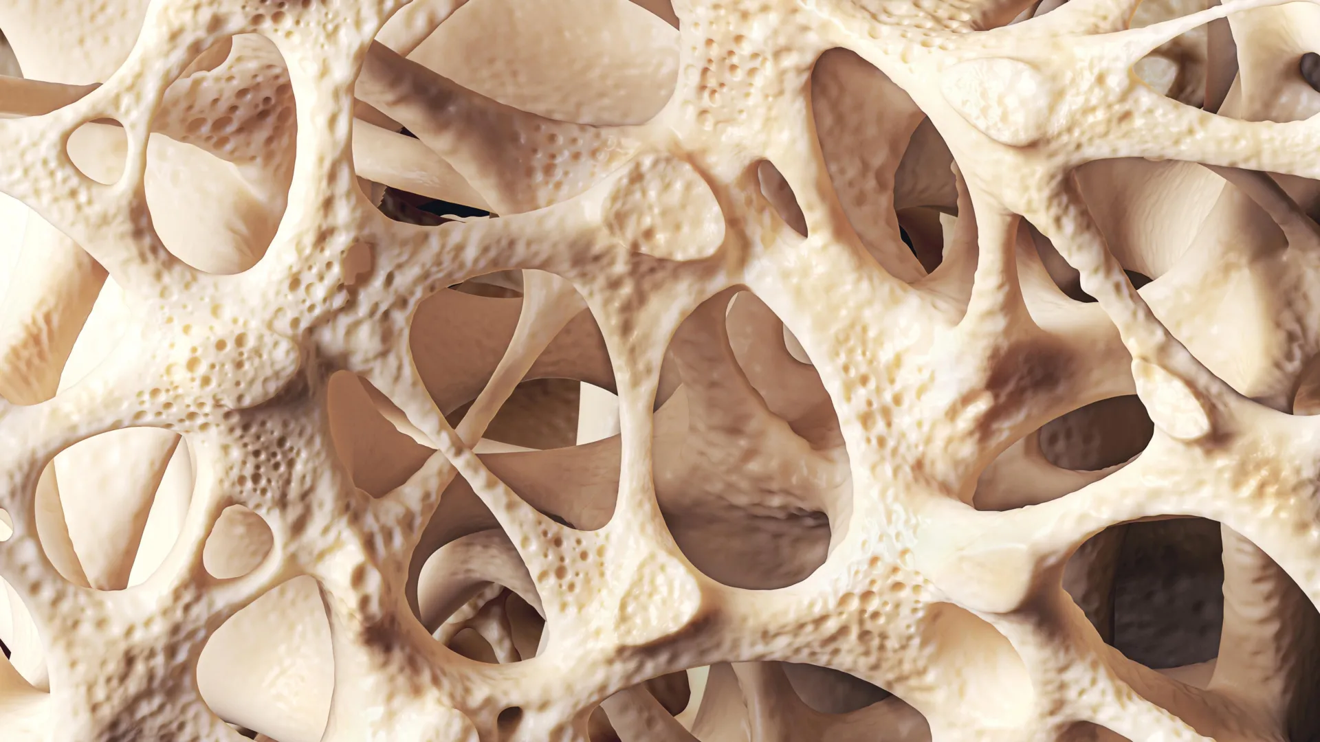
To fully appreciate the significance of the Indiana University team’s achievement, one must examine the unique physiological barriers that have historically stymied bone marrow research. Bone marrow is a soft, gelatinous tissue nestled securely within a dense, rigid matrix of cortical and trabecular bone. This dual composition creates a formidable physical obstacle for scientists attempting to study cellular architecture in situ.
Furthermore, bone marrow is the primary site of hematopoiesis—the continuous, highly regulated process responsible for the production of all circulating blood cells, including red blood cells, platelets, and various white blood cells that constitute the immune system. It also serves as the principal reservoir for hematopoietic stem cells (HSCs) and mesenchymal stem cells (MSCs), which are crucial for tissue regeneration, immune maintenance, and lifelong cellular turnover.
Because the marrow microenvironment is densely packed with a diverse and interacting array of cell types, extracellular matrix proteins, blood vessels, and signaling molecules, understanding how these components interact during disease progression has remained exceptionally difficult. Historically, researchers had to choose between methods that sacrificed spatial organization for molecular depth, or vice versa, leaving significant blind spots in the study of pathogenesis.
Limitations of Legacy Technologies: Flow Cytometry and Standard Fluorescence
Before the introduction of this advanced multiplexing technique, researchers relied primarily on traditional analytical tools such as flow cytometry and standard fluorescence microscopy. While these methodologies have driven countless biomedical discoveries over the past several decades, each possesses inherent limitations that restrict comprehensive tissue analysis.
Flow cytometry, long considered a gold standard for quantifying and characterizing cell populations, requires the mechanical or enzymatic dissociation of tissues into single-cell suspensions. While this allows for the rapid analysis of thousands of cells based on multiple surface and intracellular markers, it completely destroys the spatial architecture of the tissue. Researchers can determine which cell types are present and in what proportions, but they lose all contextual information regarding where those cells reside relative to one another, their proximity to blood vessels, or how they form localized niches within the bone marrow cavity.
Conversely, standard fluorescence imaging preserves spatial context, allowing scientists to visualize cells directly within histological sections. However, traditional fluorescence microscopy is severely constrained by spectral overlap—the phenomenon where the emission spectra of different fluorophores bleed into one another, typically limiting researchers to detecting no more than three or four cellular markers simultaneously. Consequently, capturing a holistic view of the diverse cell populations interacting within the complex bone marrow microenvironment was rendered virtually impossible using conventional microscopy alone.
Chronology of Innovation: Adapting the Phenocycler 2.0 for Murine Bone Marrow
The breakthrough achieved by the Indiana University School of Medicine team represents the culmination of targeted methodological adaptation. While the Phenocycler 2.0 platform—developed by Akoya Biosciences—had previously been validated and utilized for multiplex imaging in organs with less dense or more uniform structural properties, such as the human and murine spleen, kidney, and lymph nodes, its application to bone marrow had not been successfully optimized.
The research effort was spearheaded by the IU Cooperative Center of Excellence in Hematology (CCEH) in collaboration with the Herman B Wells Center for Pediatric Research. Over a multi-month development and optimization timeline, the team systematically addressed the technical challenges of staining and imaging decalcified, intact bone tissue without compromising epitope integrity or tissue morphology.
By systematically refining antibody panels, permeabilization protocols, and cycling conditions, the investigators successfully adapted the Phenocycler 2.0 workflow to the rigid demands of bone-encased tissue. This painstaking optimization process culminated in the ability to visualize a record-breaking 25 distinct cellular markers within a single intact murine bone marrow sample. The success of this methodology was formally documented and shared with the global scientific community through its publication in Leukemia, marking a major milestone in hematological research technology.
Expert Perspectives and Institutional Leadership
The implications of this technological advancement extend far beyond basic histological observation. Members of the research team have emphasized the profound utility of the tool for both academic investigation and pharmaceutical development.
"Bone marrow is difficult to study because it is gelatinous and encased in hard bone," noted Sonali Karnik, PhD, assistant research professor of orthopedic surgery at the IU School of Medicine and co-lead author of the study. "Since bone marrow plays an important role in blood and immune cell formation and houses valuable stem cells, our unique imaging approach offers a useful tool for a variety of research applications."
The value of utilizing murine models in this context is paramount. Mouse models are the cornerstone of preclinical biomedical research, serving as the primary vehicle through which scientists model human genetic mutations, therapeutic interventions, and disease progression. Because many human diseases manifest directly within the bone marrow niche, having a reliable, high-dimensional imaging modality for mice dramatically enhances translational potential.
"Because mouse models are widely used to study human diseases, this technique offers a promising new method for investigating a range of conditions like autoimmune diseases, leukemia and other disorders involving bone marrow," stated Reuben Kapur, PhD, co-senior author of the study, director of the IU School of Medicine’s Herman B Wells Center for Pediatric Research, and co-director of the IU Cooperative Center of Excellence in Hematology. Dr. Kapur’s extensive leadership in pediatric hematology underscores the clinical relevance of understanding bone marrow dynamics, particularly in the context of pediatric blood cancers and bone marrow failure syndromes.
Commercialization, Intellectual Property, and Future Development
Recognizing the immense commercial and translational value of this proprietary methodology, the Indiana University Innovation and Commercialization Office has moved swiftly to protect the intellectual property underlying the advancement. A provisional patent has been formally filed for the new imaging protocol, positioning Indiana University as a primary stakeholder in the future of high-dimensional tissue analysis.
Looking ahead, the research team is not resting on its initial success. The investigators are actively engaged in expanding the capabilities of the current assay. While the initial published study successfully demonstrated the simultaneous visualization of 25 cellular markers, ongoing optimization efforts are focused on broadening the marker panel. Future iterations of the panel aim to incorporate structural and functional features that were previously difficult to capture in tandem, including specific components of bone architecture, peripheral nerve networks, muscle tissue, and an even wider array of specialized immune and intercellular signaling cell types.
Expanding the marker panel will allow researchers to construct comprehensive, multi-layered spatial maps of the bone marrow microenvironment, tracking how structural remodeling of bone impacts hematopoiesis and how immune cells communicate with stem cell niches during pathological stress or pharmacological treatment.
Broader Impacts and Implications for Drug Development
The successful deployment of this high-plex imaging technique carries significant implications for the broader pharmaceutical and biotechnology sectors. In the realm of drug development, evaluating the efficacy and safety profile of novel therapeutics—particularly those targeting oncological conditions such as acute myeloid leukemia, multiple myeloma, and various lymphomas—often hinges on understanding how drugs penetrate and alter the bone marrow microenvironment.
Traditional endpoint assays, such as endpoint flow cytometry or terminal blood draws, provide limited insight into the spatial pharmacodynamics of a drug within deep tissues. By enabling high-resolution visualization of therapeutic agents, immune cell infiltration, and stem cell preservation within intact bone marrow, the Indiana University methodology provides drug developers with a powerful new pharmacodynamic biomarker tool. This capability can potentially streamline preclinical trial evaluations, reduce the number of animal subjects required through maximized data collection per sample, and accelerate the identification of promising drug candidates destined for clinical translation.
Furthermore, autoimmune diseases such as rheumatoid arthritis and systemic lupus erythematosus frequently involve complex systemic dysregulations originating in or mediated by bone marrow-derived cells. By shedding light on the localized cellular cross-talk within these microenvironments, the technique provides researchers with an invaluable platform to evaluate targeted immunotherapies designed to restore immune tolerance and halt tissue destruction.
Collaborative Foundation and Institutional Support
The breadth of this multidisciplinary study reflects the collaborative research culture fostered at the Indiana University School of Medicine. The publication in Leukemia acknowledges contributions from a diverse roster of co-authors spanning multiple departments and specialized research centers. In addition to Dr. Karnik and Dr. Kapur, the study’s authorial team includes Connor Gulbronson, Paige C. Jordan, Rahul Kanumuri, Baskar Ramdas, Ramesh Kumar, Melissa L. Hartman, Izza Khurram, Drew M. Brown, Karen E. Pollok, Pratibha Singh, and Melissa A. Kacena.
Financial backing for the project was provided by the National Institutes of Health (NIH), underscoring the federal commitment to advancing fundamental biomedical research tools that hold clear promise for public health improvement. Through continued institutional support, federal funding, and proactive commercialization strategies, the Indiana University School of Medicine team has established a robust foundation for the next generation of bone marrow research.