
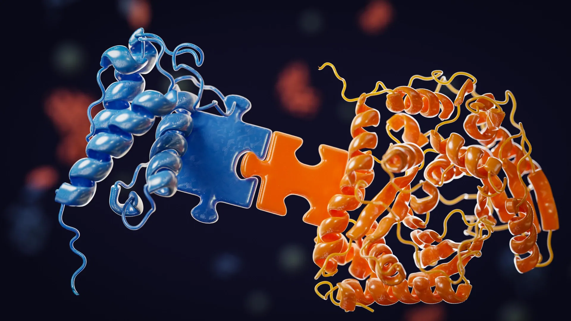
Researchers at Oregon Health & Science University (OHSU) have unveiled a groundbreaking discovery: a previously unknown internal system within cells that functions akin to atmospheric "trade winds," rapidly directing essential proteins to the cell’s leading edge. This revelation fundamentally reshapes our understanding of cellular locomotion, the intricate processes of wound healing, and the aggressive spread of cancer. The findings, published in the prestigious journal Nature Communications, challenge decades of established biological dogma regarding intracellular protein delivery.
For many years, the prevailing scientific model for how proteins navigate the complex cellular interior has been that of diffusion – a largely random process where molecules gradually drift until they reach their intended destination. This simplistic view, often depicted in biology textbooks, suggested that cells relied heavily on chance to orchestrate the movement of critical components. However, the OHSU team’s meticulous research now demonstrates that cells are far more sophisticated, actively generating directed fluid currents to propel proteins with remarkable speed and precision toward the dynamic front of the cell, the region responsible for extension, movement, and tissue repair.
Genesis of a Discovery: From Classroom Curiosity to Cellular Revelation
The genesis of this paradigm-shifting discovery can be traced back to an unexpected observation during a routine neurobiology course at the Marine Biological Laboratory in Woods Hole, Massachusetts. Catherine (Cathy) Galbraith, Ph.D., and James (Jim) Galbraith, Ph.D., co-corresponding authors of the study and established researchers in cell biology, were engaged in a standard laboratory experiment with students when they encountered an anomaly.
"It actually started out as an unexpected finding," Cathy Galbraith recounted. "We were just conducting an experiment with students in class." The experiment involved using a laser to temporarily render a strip of proteins at the rear of a living cell invisible, a common technique to track intracellular transport dynamics. As the experiment progressed, the researchers observed the appearance of a distinct, darkened band at the front edge of the cell – the very area that protrudes and drives cellular motility.
"We kind of did it for fun and then realized this gave us a way of measuring something that wasn’t able to be measured before," she elaborated. This unanticipated observation provided a novel avenue for investigation.
Unraveling the "Dark Band": The Wave of Actin and Directed Flow
Further detailed analysis of this "dark band" revealed it to be a concentrated wave of soluble actin, a critical protein indispensable for cell movement and structural integrity. This actin was being rapidly propelled forward, a phenomenon that contradicted the established diffusion model. Previously, scientists had assumed that actin primarily reached the leading edge through slow, random diffusion. The new findings unequivocally indicated a different, far more active mechanism at play.
"We realized the cartoon models in textbooks were missing a huge piece," Jim Galbraith stated, reflecting on the profound implications of their findings. "There had to be some kind of flow in the cell pushing things forward. Cells really do ‘go with the flow.’" This realization marked a pivotal moment, suggesting that cells possess an intrinsic, organized system for internal transport that far surpasses passive diffusion.
The Mechanism: Cellular Squeezing and "Atmospheric Rivers" Within
The OHSU team, having joined the university in 2013 after significant research at the National Institutes of Health (NIH), where they collaborated with Nobel Laureate Eric Betzig on cutting-edge imaging techniques at the Howard Hughes Medical Institute’s Janelia Research Campus, leveraged their expertise and advanced instrumentation to delve deeper.
Utilizing highly specialized imaging tools, they were able to visualize and characterize these newly identified directed fluid flows. The researchers drew an analogy between these internal cellular currents and "atmospheric rivers" – powerful, concentrated streams of moisture in the Earth’s atmosphere that transport vast quantities of water vapor over long distances. In the cellular context, these flows efficiently transport actin and a multitude of other proteins towards the front of the cell, at speeds significantly exceeding what diffusion alone could achieve.
"We found that the cell can actually squeeze at the back and target where it sends that material," Jim Galbraith explained, illustrating the active nature of the process. "If you squeeze half a sponge, the water only goes on that half. That’s basically what the cell is doing." This active "squeezing" or manipulation of the intracellular environment generates these directional currents.
Crucially, these flows appear to be nonspecific, meaning they can carry a diverse array of proteins simultaneously. This inherent efficiency creates a robust system that underpins fundamental cellular processes such as protrusion (extending the cell membrane), adhesion (attachment to surfaces), and rapid shape changes – all vital for cell migration, immune responses, and tissue regeneration.
A Novel Compartment: The Actin-Myosin Condensate Barrier
The study further identified that these directed flows are confined to a specialized region at the cell’s leading edge. This area is demarcated by an actin-myosin condensate barrier, a functional boundary that is not enclosed by a membrane like traditional organelles but acts as a physical partition. This barrier effectively channels the protein-rich fluid to the advancing edge, ensuring precise delivery and organization.
The researchers have termed this system a "pseudo-organelle" – a compartment that, while lacking a membrane, performs a critical organizing role in cellular behavior and function. This conceptualization highlights its importance as a distinct functional unit within the cell, albeit one with a unique structural basis.
Innovative Imaging Techniques: Visualizing the Unseen Currents
The ability to observe these internal cellular currents was contingent upon the development and application of novel imaging methodologies. The OHSU team modified a standard fluorescence-based technique. Instead of using a laser to quench fluorescence, they activated fluorescent molecules at a specific point and meticulously tracked their subsequent movement, effectively visualizing the flow.
One of their key experimental protocols was humorously named FLOP, an acronym for "Fluorescence Leaving the Original Point." Cathy Galbraith quipped, "It wasn’t a flop at all. It was the opposite. It is anything but a flop, because it worked." This innovative approach allowed them to quantify and visualize the directed movement of molecules within the cell in unprecedented detail.
Broader Implications: Cancer Metastasis and Therapeutic Targets
The implications of this discovery extend significantly into the realm of cancer biology. The rapid and efficient protein transport system identified by the OHSU team could provide a crucial explanation for the aggressive invasive capabilities of certain cancer cells.
"We know these highly invasive cells have this really cool mechanism to push proteins really fast, really rapidly where they need them at the front of the cell," Jim Galbraith explained. He further elaborated on the concept of cellular components being shared across different cell types, stating, "All cells have basically the same components inside, much like a Porsche and a Volkswagen have many of the same parts, but when those parts are assembled into the final machine, they behave and function very differently." This analogy underscores how subtle differences in the assembly and utilization of cellular machinery, such as this newly discovered transport system, can lead to dramatically different functional outcomes.
By understanding how cancer cells exploit this internal "trade wind" system more effectively or differently than normal cells, scientists may be able to develop novel therapeutic strategies aimed at impeding or halting cancer metastasis. "If you can understand the differences, you can target future therapies based on how cancer cells and normal cells work differently," he emphasized. This opens new avenues for precision oncology, focusing on the specific mechanisms that drive malignant cell invasion.
Collaborative Endeavor: Bridging Disciplines with Advanced Microscopy
This ambitious research was a testament to interdisciplinary collaboration, bringing together experts from engineering, physics, microscopy, and cell biology. Significant contributions were made by collaborators at the Janelia Research Campus in Virginia, including specialists in fluorescence correlation spectroscopy and advanced 3D super-resolution imaging.
"The instrumentation we needed doesn’t exist in most places," Cathy Galbraith noted. "Janelia had a one-of-a-kind setup that let us test and confirm what we were seeing." The study was heavily reliant on state-of-the-art imaging tools developed at Janelia, including iPALM (interferometric photoactivated localization microscopy), a technique renowned for its ability to resolve structures at the nanometer scale.
"iPALM allowed us to physically see the compartments," Jim Galbraith stated. "There’s no other light-based technique that could do that." This advanced imaging capability was instrumental in visualizing the membrane-less pseudo-organelle and the fluid dynamics within it.
Future Directions and Potential Impact
The researchers posit that this newly identified cellular transport system could have far-reaching implications across multiple scientific disciplines. Its influence is expected to be felt in cancer research, drug delivery mechanisms, regenerative medicine and tissue repair strategies, and the burgeoning field of synthetic biology.
"Just as small shifts in the jet stream can change the weather, small changes in these cellular winds could change how diseases begin or progress," Cathy Galbraith remarked, underscoring the profound impact of these subtle cellular mechanisms. "All you had to do was look," she concluded, reflecting on the presence of these flows. "The flows were there all along. Now we know how cells use them."
The study’s coauthors, in addition to the Galbraiths, include Brian English, Ph.D., from Janelia Research Campus, and Ulrike Boehm, Ph.D., formerly with Janelia and now affiliated with Carl Zeiss AG in Germany.
This pioneering research was supported by substantial funding from several leading scientific institutions, including the National Institute of General Medical Sciences of the National Institutes of Health (Award number R01GM117188), the U.S. National Science Foundation (Award numbers 2345411 and 171636), the W. M. Keck Foundation, the Howard Hughes Medical Institute Janelia Visiting Scientist Program, and the Howard Hughes Medical Institute. Specific aspects of the advanced imaging, such as the iPALM work, were partially supported by an award from the Advanced Imaging Center at Janelia, and the Super-resolution Imaging Microscopy (SIM) efforts received support from a Core Research Facilities grant from the OHSU School of Medicine.


Leave a Reply