
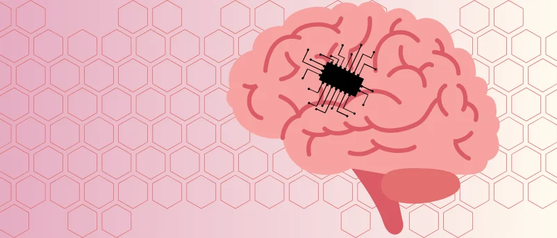
The realm of neural interfaces, critical for understanding and treating a myriad of neurological conditions, has long grappled with a fundamental challenge: the inherent variability of the human brain. Unlike many biological systems, the brain’s surface is uniquely convoluted, with distinct gyral patterns that differ from one individual to another. Recognizing this crucial anatomical reality, a groundbreaking study led by researchers at Penn State University (PA, USA) has introduced a revolutionary solution: 3D-printed soft electrodes, meticulously designed with a honeycomb-inspired architecture, engineered to precisely conform to an individual’s unique cerebral surface. This innovation, promising enhanced precision, biocompatibility, and efficacy, marks a significant leap towards truly personalized neuromodulation therapies and advanced neuroprosthetics.
The Inadequacy of Conventional Neural Interfaces
For decades, neural interfaces have been instrumental in advancing neuroscience and clinical applications, from deep brain stimulation for Parkinson’s disease to recording brain activity for brain-computer interfaces (BCIs). However, a persistent limitation has been their "one-size-fits-all" design. Traditional electrodes, typically rigid and standardized for mass production, fail to account for the intricate, individualized topography of the human cerebral cortex. The human brain’s surface is characterized by a complex pattern of folds (gyri) and grooves (sulci), a process known as gyrification. This unique "brain fingerprint" is influenced by a multitude of factors, including genetics, sex, age, weight, and height.
The mechanical mismatch between rigid electrodes and the dynamic, soft brain tissue often leads to several critical issues. Poor electrode-tissue contact can result in signal loss, reduced therapeutic efficacy, and compromised safety. Furthermore, the stiffness of conventional devices can trigger adverse foreign body responses, leading to inflammation, glial scarring, and encapsulation of the implant, ultimately diminishing long-term performance and potentially necessitating further interventions. These challenges underscore the urgent need for a more adaptable, patient-specific approach to neural interface design. As corresponding author Tao Zhou articulated, "This motivated us to create electrodes that are tailored for each individual, based on the structure of their brain."
HiPGE: A Bio-Inspired Revolution in Electrode Design
The innovation at the heart of this study is the development of a novel electrode dubbed HiPGE (Honeycomb-esque printable gel electrode). This device represents a paradigm shift, moving away from rigid, planar designs to a flexible, three-dimensional architecture inspired by the remarkable efficiency and resilience of honeycomb structures found in nature. The choice of honeycomb is not arbitrary; its hexagonal lattice provides an optimal balance of strength, flexibility, and material efficiency, allowing for robust yet pliant structures that can withstand mechanical stress while maintaining intimate contact with irregular surfaces.
Key to HiPGE’s success is its composition: ultra-soft hydrogels. These biocompatible, water-rich polymers closely mimic the mechanical properties of brain tissue, thereby minimizing the mechanical mismatch that plagues conventional electrodes. The combination of bio-inspired architecture and advanced material science enables HiPGE to mold seamlessly to the complex, individualized 3D models of patients’ brains, a capability that standard electrode designs cannot achieve. This foundational innovation lays the groundwork for devices that are not only more effective but also safer and more durable within the delicate neurological environment.
A Meticulous Process: From MRI to 3D Printing
The development of personalized HiPGEs is a testament to an interdisciplinary approach, integrating advanced medical imaging, computational modeling, and sophisticated additive manufacturing. The researchers established a novel neural interface platform that meticulously transforms patient-specific anatomical data into functional, custom-fit electrodes.
The process commenced with the acquisition of high-resolution Magnetic Resonance Imaging (MRI) scans from 21 patients. These scans served as the foundational dataset, allowing researchers to reconstruct precise 3D models of each individual’s cerebral cortex. Each model faithfully captured the unique gyral complexity – the intricate folds and grooves – that define an individual’s brain surface. This initial step highlighted the profound variability in brain anatomy, reinforcing the imperative for personalized solutions.
Following 3D reconstruction, the team employed MRI-based surface curvature analysis and region-of-interest mapping. This computational phase was critical for designing electrodes that were not merely generic shapes but were specifically contoured to fit the unique geometry and functional areas of each individual cerebral cortex. This level of anatomical precision is unprecedented in neural interface design, moving beyond generalized templates to truly bespoke solutions.
Computational Validation: Ensuring Performance Before Production
Before physical fabrication, the designed electrode geometries underwent rigorous computational validation using finite element analysis (FEA). FEA is a powerful simulation technique that predicts how a material or design reacts to real-world forces, heat, fluid flow, and other physical effects. In this context, FEA was used to optimize the device geometry, ensuring both structural integrity and optimal functional performance.
The results of the FEA were highly encouraging. Simulations revealed that the stiffness of HiPGE closely matched that of brain tissue, a crucial factor in minimizing adverse tissue reactions and maximizing long-term stability. This contrasted sharply with rigid control electrodes, which exhibited significant mechanical mismatch. Furthermore, distance contour maps and strain distribution analysis demonstrated that HiPGE maintained significantly closer contact with brain tissue and induced minimal tissue deformation when compared to conventional, stiffer controls. These findings provided strong in silico evidence that the personalized hydrogel electrodes would offer superior physical integration with the brain.
Complementing FEA, in silico modeling was also utilized to assess the electrical performance of the personalized devices. These simulations predicted that the HiPGE system would achieve near-perfect connectivity to electrical signals within the brain, suggesting high-fidelity electrophysiological recording capabilities. This comprehensive computational validation phase was instrumental in refining the designs and confirming the theoretical advantages of the personalized, honeycomb-inspired approach before proceeding to costly and time-consuming physical fabrication.
Bringing Designs to Life: 3D Printing and Anatomical Conformity

With the designs computationally validated, the next step involved physical fabrication. Five patients were randomly selected from the initial cohort, and their complementary electrodes were meticulously 3D printed using a technique called direct ink writing. This additive manufacturing method allows for the precise deposition of hydrogel "ink" in complex, pre-designed patterns, layer by layer, to create the intricate honeycomb structure of the HiPGE.
To visually and physically assess the conformity of these personalized neural interfaces, the researchers also 3D-printed high-fidelity models of the selected patients’ brains. Placing the custom-made HiPGEs onto their respective brain models provided compelling evidence of their precise conformity. The personalized electrodes exhibited an exceptional match to the unique shapes and spatial arrangements of the individual cortical surfaces, visually confirming the success of the integrated design and fabrication workflow. This step underscored the transformative potential of combining patient-specific imaging with advanced 3D printing for medical device development.
Pre-Clinical Success: Superior Performance in Animal Models
The ultimate test of any novel medical device lies in its biological performance. The research team moved to in vivo studies, testing their personalized electrode system in rat models. Similar to the human application, personalized electrodes were designed and created to precisely fit the unique brain anatomy of individual rodents.
In a crucial comparative experiment, each rat was fitted with a personalized HiPGE on the left side of its brain and a conventional electrode on the right side. This direct, intra-animal comparison provided robust data on the relative performance of the two systems. The results were unequivocal: the personalized HiPGE system consistently exhibited superior electrical activity recording performance. This indicates that the improved anatomical conformity and mechanical matching translated directly into better signal acquisition, a critical factor for both diagnostic monitoring and therapeutic neuromodulation.
Beyond functional performance, biocompatibility is paramount for long-term neural implants. After four weeks of implantation, fluorescent imaging of excised brain tissue from the rats revealed no obvious immune response to the implanted HiPGE. This finding is profoundly significant, as a robust foreign body response is a major cause of implant failure and complications with conventional, rigid electrodes. The absence of a significant immune reaction suggests that the soft, tissue-matching properties of the hydrogel-based HiPGE significantly reduce the biological burden on the brain, paving the way for safer and more stable long-term implants.
Transformative Implications for Neurological Care
The findings from Penn State University establish a truly transformative framework for neural interface engineering, with profound implications for the future of neurological diagnosis, treatment, and enhancement. The seamless conformity of these 3D-printed electrodes to cortical contours, combined with their ability to eliminate mechanical mismatch and provide high-fidelity electrophysiological recording with minimal adverse effects, addresses long-standing challenges in the field.
Personalized Neuromodulation Therapies: Conditions such as epilepsy, Parkinson’s disease, chronic pain, and severe depression often benefit from neuromodulation, where electrical stimulation is used to alter neural activity. The precision offered by personalized HiPGEs could revolutionize these treatments. By achieving optimal electrode-tissue contact, therapies could be delivered with greater accuracy and efficiency, potentially reducing side effects and improving therapeutic outcomes for individual patients. Tailoring the interface to a patient’s unique brain could allow for more targeted stimulation, optimizing efficacy and safety.
Advanced Neuroprosthetics: For individuals with paralysis or limb loss, neuroprosthetics offer the promise of restoring function through direct brain control. The ability to record brain signals with unprecedented fidelity and stability could significantly enhance the performance and intuitive control of robotic limbs, exoskeletons, and communication devices. The improved biocompatibility also suggests that these implants could function reliably for extended periods, reducing the need for revision surgeries.
Enhanced Disease Monitoring: Early detection and continuous monitoring are vital for managing progressive neurological diseases like Alzheimer’s or tracking recovery after stroke or traumatic brain injury. HiPGEs could provide a more stable and accurate platform for long-term brain activity monitoring, allowing clinicians to detect subtle changes, track disease progression, and personalize interventions more effectively. Tao Zhou emphasized this future direction: "We are looking to further improve this technology to optimize the electrodes to monitor for specific diseases. In the future, we would really like to work with patients to see how this approach could support brain monitoring and disease treatment in clinical settings."
Addressing Current Limitations and Future Directions
While the potential is immense, the journey from laboratory breakthrough to widespread clinical application involves navigating several critical steps and addressing ongoing challenges. Scalability and cost-effectiveness of personalized 3D printing will be crucial. While direct ink writing offers precision, optimizing the manufacturing process for larger-scale production while maintaining cost efficiency will be essential for making these personalized devices accessible.
Regulatory hurdles are also a significant consideration. Any novel implantable medical device must undergo rigorous testing and approval processes by regulatory bodies such as the FDA. Demonstrating long-term safety, reliability, and efficacy in human clinical trials will be the next major phase for this technology. The transition from animal models to human patients requires extensive validation.
Furthermore, ongoing research will likely focus on integrating even more advanced functionalities into these personalized electrodes. This could include incorporating drug delivery capabilities, more sophisticated sensing mechanisms (e.g., chemical sensing), or wireless power and data transmission to minimize invasive components. The foundational framework established by the Penn State team provides a robust platform for such future enhancements.
A New Era of Neurological Intervention
This pioneering research from Penn State represents a pivotal moment in the evolution of neural interface technology. By embracing the unique complexity of individual brain anatomy and drawing inspiration from nature’s elegant designs, the development of honeycomb-inspired 3D-printed soft electrodes ushers in a new era of personalized neurological care. The promise of higher precision, enhanced biocompatibility, and superior performance holds the potential to profoundly improve the lives of millions suffering from neurological disorders and to unlock unprecedented capabilities in neuroprosthetics and brain-computer interfaces. As the scientific community continues to push the boundaries of bioengineering, the vision of truly personalized medicine for the brain moves ever closer to reality.


Leave a Reply