
In the rapidly evolving landscape of life sciences, Polymerase Chain Reaction (PCR) stands as a foundational molecular biology technique, indispensable for genetic analysis. What began as a revolutionary method for amplifying DNA has diversified into a powerful suite of technologies: endpoint PCR, quantitative real-time PCR (qPCR), and digital PCR (dPCR). Far from being competing platforms, these methods are highly complementary, each offering distinct advantages that enable researchers to tackle a broad spectrum of scientific challenges, from basic detection to ultra-sensitive quantification. Their strategic integration is paramount for optimizing experimental design, enhancing data quality, and accelerating scientific discovery across various disciplines, including basic research, clinical diagnostics, and applied genomics.
A Brief History of a Molecular Revolution
The Polymerase Chain Reaction was first conceived by Kary Mullis in 1983, a discovery that earned him the Nobel Prize in Chemistry in 1993. Mullis’s elegant idea—to use repeated cycles of DNA synthesis to exponentially amplify specific DNA sequences—revolutionized molecular biology. Prior to PCR, isolating and studying specific DNA fragments was a laborious and often impossible task. PCR made it possible to generate millions to billions of copies of a particular DNA segment from a minute starting sample, transforming fields from forensics to infectious disease diagnostics. Early PCR, now referred to as endpoint PCR, relied on gel electrophoresis for visualization and qualitative assessment of amplification. The subsequent evolution of PCR technologies saw the development of qPCR in the 1990s, introducing real-time quantification capabilities, and more recently, dPCR in the 2000s, pushing the boundaries of sensitivity and absolute quantification. This chronological progression reflects a continuous drive within the scientific community for greater precision, sensitivity, and throughput in genetic analysis.
Endpoint PCR: The Enduring Foundation
Endpoint PCR remains a cornerstone technique, valued for its simplicity, robustness, and cost-effectiveness. Its primary function is qualitative analysis, confirming the presence or absence of a target DNA sequence. The process involves three main steps repeated cyclically: denaturation (separating DNA strands), annealing (primers binding to target sequences), and extension (DNA polymerase synthesizing new strands). After 25-40 cycles, the amplified products are typically visualized using gel electrophoresis, where DNA fragments are separated by size and stained, allowing researchers to observe specific bands corresponding to the amplified target.
Endpoint PCR’s applications are vast and varied. In clinical microbiology, it’s routinely used for initial screening of pathogens, such as bacteria or viruses, where a simple "yes/no" answer regarding infection is sufficient. In molecular cloning, it confirms the successful insertion of a gene into a plasmid. Forensic science relies on endpoint PCR for DNA fingerprinting, amplifying minute amounts of DNA from crime scenes to generate profiles for identification. Furthermore, in agricultural biotechnology, it’s employed for detecting genetically modified organisms (GMOs) or identifying plant diseases. Its key advantages lie in its accessibility, minimal equipment requirements, and straightforward interpretation. However, its qualitative nature means it cannot accurately quantify the initial amount of target DNA, and its sensitivity can be limited compared to its more advanced counterparts.
Quantitative Real-Time PCR (qPCR): Precision in Real-Time
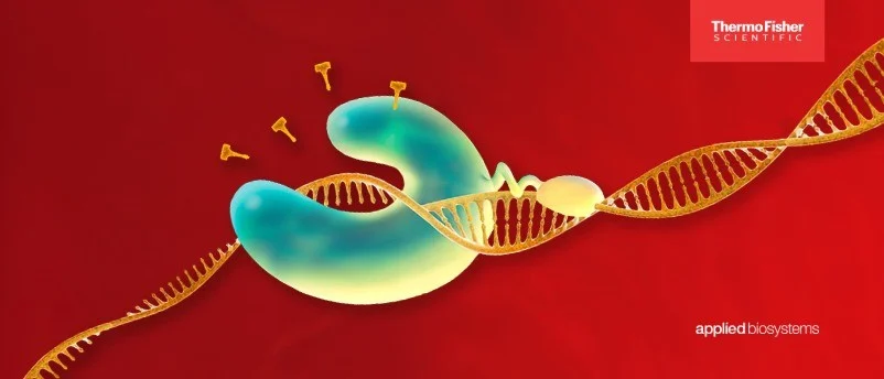
Building upon the foundation of endpoint PCR, quantitative real-time PCR (qPCR) marked a significant leap forward by enabling the quantification of nucleic acids in real-time. Introduced in the late 1990s, qPCR revolutionized gene expression analysis by allowing researchers to monitor the accumulation of PCR products during the amplification process, rather than just at the end. This is achieved through the use of fluorescent dyes or probes that emit a signal proportional to the amount of amplified DNA. Common methods include SYBR Green, which binds non-specifically to double-stranded DNA, and TaqMan probes, which are sequence-specific and offer higher specificity.
The critical metric in qPCR is the threshold cycle (Ct value), which is the cycle number at which the fluorescent signal crosses a defined threshold. A lower Ct value indicates a higher initial amount of target nucleic acid. By comparing Ct values to a standard curve generated from samples of known concentrations, researchers can perform relative quantification, determining the fold change in gene expression between different samples (e.g., treated vs. untreated cells, healthy vs. diseased tissue).
qPCR has become indispensable in numerous fields. In oncology, it’s used to quantify oncogene expression or tumor suppressor gene activity, providing insights into cancer progression and response to therapy. In infectious disease diagnostics, qPCR precisely measures viral load (e.g., HIV, HCV) or bacterial burden, crucial for monitoring disease severity and treatment efficacy. It’s also vital in drug discovery for target validation and assessing drug efficacy at the transcriptional level. The technique offers high sensitivity, allowing detection of low-copy targets, and significantly improves throughput compared to endpoint PCR, making it a workhorse for laboratories worldwide. However, qPCR typically provides relative quantification, requiring normalization with reference genes and careful experimental design to ensure accurate comparisons.
Digital PCR (dPCR): The Gold Standard for Absolute Quantification
The latest evolution in the PCR landscape is digital PCR (dPCR), which offers unparalleled sensitivity and the ability to perform absolute quantification of nucleic acids without the need for a standard curve. Introduced in the 2000s, dPCR fundamentally differs from traditional PCR and qPCR by partitioning the sample into thousands or even millions of individual, minute reactions. Each partition contains either zero or one (or a few) target molecules. After amplification, the partitions are analyzed for the presence or absence of fluorescence. By counting the number of positive partitions and applying Poisson statistics, the absolute concentration of the target nucleic acid in the original sample can be calculated with high precision.

dPCR is particularly adept at detecting extremely low-abundance targets, rare mutations, and subtle copy number variations (CNVs). Its applications are expanding rapidly, especially in clinical diagnostics and advanced research. In oncology, dPCR is emerging as the gold standard for liquid biopsy, detecting circulating tumor DNA (ctDNA) in blood samples for early cancer detection, monitoring treatment response, and identifying minimal residual disease (MRD) after therapy, often at levels undetectable by qPCR. For example, dPCR can detect a single mutated DNA molecule among thousands of wild-type molecules. In prenatal diagnostics, it can quantify fetal DNA in maternal blood for non-invasive prenatal testing (NIPT). Furthermore, dPCR is crucial for validating CRISPR-Cas9 gene editing efficiency and for accurately quantifying viral loads in highly sensitive contexts. Its inherent robustness to inhibitors, due to the dilution effect across many partitions, also makes it advantageous for challenging sample types. While dPCR offers superior sensitivity and absolute quantification, it typically involves higher instrumentation costs and lower throughput compared to qPCR for large-scale screening applications.
The Synergistic Power: Integrating PCR Workflows
A profound understanding among researchers and industry leaders, such as Thermo Fisher Scientific, is that endpoint PCR, qPCR, and dPCR are not competing technologies but rather a complementary toolkit. The optimal choice of PCR method depends critically on the specific experimental goals, the required level of sensitivity, and the need for qualitative, relative, or absolute quantification.
Consider a typical workflow in biomarker discovery or clinical diagnostics:
- Initial Screening: Researchers might begin with endpoint PCR to quickly screen a large number of samples for the presence or absence of a potential biomarker or pathogen. Its cost-effectiveness and speed make it ideal for broad, preliminary sweeps. For example, identifying individuals positive for a particular genetic variant.
- Relative Quantification and Validation: For samples identified as positive, qPCR would then be employed to accurately quantify the expression levels of the biomarker or the viral/bacterial load. This provides precise relative data, allowing researchers to compare expression changes between different patient groups or treatment conditions. For instance, determining if the genetic variant leads to increased or decreased gene expression.
- Ultra-Sensitive Absolute Quantification: In cases requiring the highest sensitivity or absolute quantification—such as detecting minimal residual disease after cancer treatment, quantifying rare mutations in liquid biopsies, or precisely determining gene copy numbers—dPCR becomes the indispensable tool. It can confirm and quantify targets that might be at or below the detection limits of qPCR, providing definitive absolute concentrations. This might involve tracking the absolute copy number of the variant in circulating cell-free DNA to monitor disease progression or recurrence.
This layered approach ensures that resources are utilized efficiently, moving from broader, less expensive qualitative assessments to highly precise, sensitive quantitative analyses as needed. Experts in the field consistently emphasize that a well-designed experiment often leverages the strengths of multiple PCR platforms to achieve comprehensive and robust results.
The Unsung Hero: Critical Role of Sample Preparation
The success of any PCR experiment, regardless of the platform, hinges significantly on the quality and integrity of the starting nucleic acid sample. Poor sample preparation is a leading cause of experimental failure, leading to false negatives, inaccurate quantification, and irreproducible data. The process of preparing samples for PCR involves several critical steps:
- RNA Extraction: For gene expression analysis, high-quality RNA extraction is paramount. RNA is highly susceptible to degradation by ubiquitous RNases. Effective extraction methods (e.g., silica-based columns, magnetic beads, phenol-chloroform) are designed to yield intact, pure RNA, free from proteins, lipids, and other cellular contaminants that can inhibit downstream enzymatic reactions. The integrity of RNA is typically assessed using gel electrophoresis or spectrophotometric methods (e.g., A260/280 ratio).
- DNase Treatment: When working with RNA for gene expression studies, genomic DNA (gDNA) contamination is a significant concern. gDNA can be amplified by PCR primers, leading to false-positive signals or inaccurate quantification. DNase treatment, using enzymes like RNase-free DNase I, specifically degrades DNA without affecting RNA, ensuring that only cDNA (synthesized from RNA) is amplified in subsequent PCR steps.
- cDNA Synthesis via Reverse Transcription: Since PCR enzymes typically use DNA templates, RNA must first be converted into complementary DNA (cDNA) through a process called reverse transcription. This step uses a reverse transcriptase enzyme, along with primers (oligo-dT for mRNA, random hexamers for total RNA, or gene-specific primers), to synthesize a DNA strand from an RNA template. The efficiency of this conversion directly impacts the accuracy of subsequent qPCR or dPCR quantification.
- Removal of PCR Inhibitors: Biological samples often contain substances that can inhibit the activity of DNA polymerase, leading to reduced amplification efficiency or complete reaction failure. Common inhibitors include heme (from blood), humic acids (from soil), heparin (anticoagulant), polysaccharides, and various chaotropic salts used in extraction buffers. Effective sample purification protocols are designed to remove these inhibitors. In some cases, diluting the sample or using specific PCR master mixes formulated to tolerate inhibitors can mitigate their effects. Ignoring inhibitor presence can lead to underestimated target concentrations or completely missed detections.
Leading scientific equipment providers, such as Thermo Fisher Scientific, develop comprehensive kits and reagents for each of these sample preparation steps, recognizing their critical role in ensuring data accuracy and experimental success across all PCR platforms.
Broader Impact and Future Directions
The combined power of endpoint PCR, qPCR, and dPCR extends across nearly every facet of modern life sciences, driving innovation and improving human health. In clinical diagnostics, they enable rapid and accurate diagnosis of infectious diseases, personalized cancer treatment strategies based on genetic profiles, and non-invasive prenatal screening. In drug discovery and development, they are essential for identifying therapeutic targets, screening compounds, and monitoring drug efficacy and safety. Forensic science continues to rely heavily on PCR for human identification, while environmental monitoring uses these techniques to detect pollutants and pathogens in water and soil. Agricultural biotechnology benefits from PCR for crop improvement, disease resistance, and food safety.
Looking ahead, the field of PCR technology continues to evolve. Miniaturization and automation are making PCR workflows more efficient and accessible. Integration with other high-throughput technologies, such as next-generation sequencing (NGS), is creating even more powerful analytical pipelines. The development of portable PCR devices for point-of-care diagnostics and field applications is expanding its reach. Furthermore, advancements in data analysis, including artificial intelligence and machine learning, are enhancing the interpretation of complex PCR data.
The strategic leveraging of endpoint PCR, qPCR, and digital PCR together represents a sophisticated approach to molecular biology. By understanding the unique strengths and optimal applications of each technique, researchers are empowered to design robust experiments, generate high-quality data, and ultimately accelerate the pace of scientific discovery, pushing the boundaries of what is possible in genetic analysis. The collaborative and complementary nature of these PCR platforms underscores their indispensable role in navigating the complexities of the biological world and addressing the pressing challenges of today and tomorrow.


Leave a Reply