Researchers at Princeton University (NJ, USA) have introduced a groundbreaking methodology that combines advanced microscopy techniques with artificial intelligence to map the intricate relationship between the shape dynamics of cellular substructures and a cell’s response to various pharmaceutical compounds. This innovative approach promises to revolutionize drug development by providing unprecedented insights into the mechanisms of drug action and identifying novel markers of cellular health and disease. The study, spearheaded by Cliff Brangwynne, focused on biomolecular condensates, particularly the nucleolus, and revealed previously unobserved cellular reactions to established drugs, including the discovery of entirely new morphological states within the cell.
Unveiling the Secrets of Biomolecular Condensates
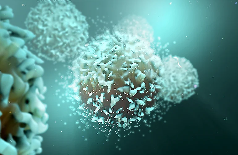
At the heart of this research lies the study of biomolecular condensates – tiny, membrane-less organelles within living cells that organize cellular processes through a phenomenon known as liquid-liquid phase separation. These dynamic droplets play critical roles in a multitude of cellular functions, including gene transcription, RNA processing, and protein synthesis. Their functional integrity is paramount for cellular health, and dysregulation in their formation, composition, or dynamics has been increasingly linked to a range of devastating diseases, including neurodegenerative disorders such as Alzheimer’s and Amyotrophic Lateral Sclerosis (ALS), as well as various forms of cancer.
Cliff Brangwynne, the June K. Wu ’92 Professor of Chemical and Biological Engineering and the principal investigator of this seminal study, articulated the fundamental challenge addressed by their work: “The central problem in biology is how do you get emergent structure from individual molecular interactions.” He emphasized that understanding these emergent properties is crucial for deciphering cellular behavior and its response to external stimuli, such as therapeutic agents. The key innovation, according to Brangwynne, was “to develop a way to learn from the images and classify the patterns that are emergent,” allowing researchers to move beyond qualitative observations to a more systematic and quantifiable understanding of cellular architecture.
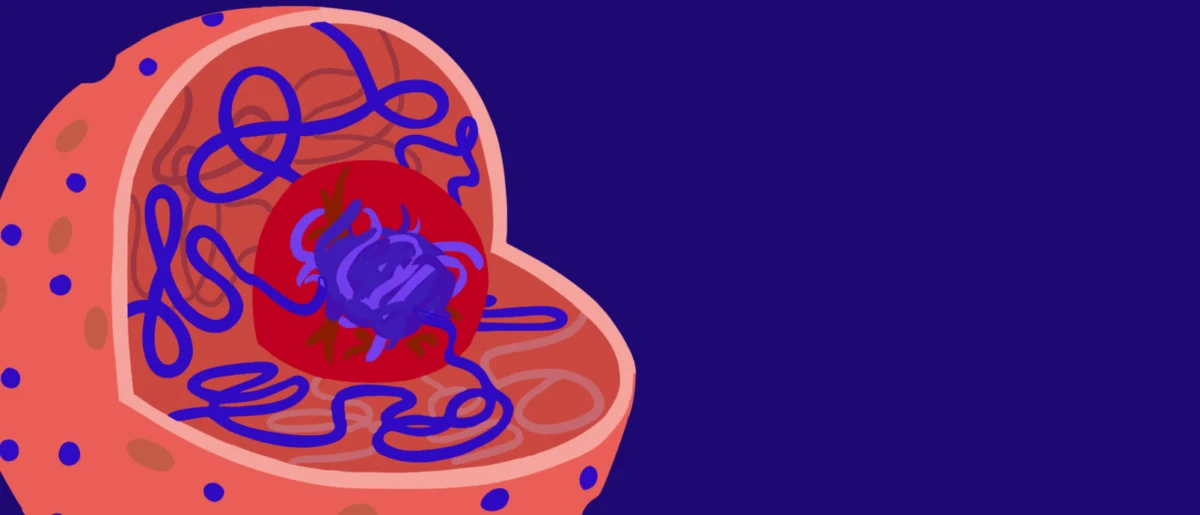
The research specifically homed in on the nucleolus, a prominent biomolecular condensate primarily responsible for the assembly of ribosomes, the cell’s protein-building machinery. The nucleolus is known for its highly dynamic nature, rapidly adapting its size, shape, and internal organization in response to cellular stress, nutrient availability, and various signaling pathways. This inherent plasticity makes it an ideal candidate for studying drug-induced cellular changes.
A New Paradigm in Cellular Imaging and Analysis
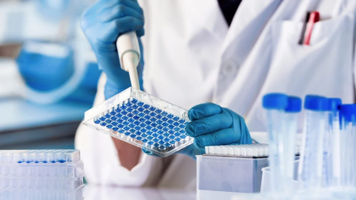
To achieve their objectives, Brangwynne’s team employed state-of-the-art advanced microscopy techniques to capture high-resolution images of nucleolar shape changes in hundreds of human cells. These experiments were conducted under a carefully controlled range of drug conditions, allowing for a systematic assessment of dose-dependent and drug-specific effects on nucleolar morphology. The sheer volume and complexity of the image data generated from such experiments present a significant challenge for human interpretation, even for highly trained cell biologists, as subtle but significant changes can easily be overlooked.
This is where artificial intelligence, specifically machine learning, became an indispensable tool. The researchers developed a bespoke machine-learning algorithm, a neural network, designed to process and interpret these intricate microscopic images. The neural network was initially trained on tens of thousands of images depicting three well-established nucleolar forms: healthy, spherical nucleoli, and two known atypical forms—the ‘cap’ shape and the ‘beaded necklace’ shape. These atypical morphologies have previously been linked to specific cellular stress responses and disruptions in RNA processing pathways. For instance, the cap shape is often associated with treatments that interfere with the synthesis of ribosomal RNA (rRNA), essential for ribosome assembly, while the beaded necklace shape can arise from drugs that disrupt other critical RNA-related processes.
Anita Donlic, a postdoctoral researcher, and Troy Comi, a research software engineer, both from Brangwynne’s lab, were instrumental in the development and training of this sophisticated neural network. Their meticulous work enabled the AI to not only recognize known patterns but also to identify and classify new ones with remarkable accuracy and efficiency.

Unveiling Unexpected Morphologies and Mechanisms of Action
Upon completion of the initial training phase, the team embarked on a comprehensive drug screening panel. Human cells were exposed to a diverse array of pharmaceutical compounds at varying concentrations, and the neural network was deployed to visually measure and quantify the resulting changes in nucleolar morphology. The AI successfully correlated different drug concentrations with varying degrees of change in the prevalence and characteristics of both cap and necklace shapes, providing a quantitative framework for assessing drug-induced cellular stress.
Crucially, the neural network yielded several unexpected and significant discoveries. For example, the AI identified that two known anti-cancer drugs, whose primary mechanisms of action were believed to be well-understood, induced the formation of cap-shaped nucleoli. This phenomenon had not been previously reported for these specific drugs, as noted by Donlic, the paper’s first author. This finding strongly suggests that these drugs may exert previously unappreciated effects on nucleolar function, potentially opening new avenues for understanding their therapeutic efficacy or identifying novel targets for intervention. This discovery highlights the AI’s ability to uncover subtle, yet biologically significant, cellular responses that might escape traditional observation methods.
Perhaps the most striking discovery involved a third drug, topotecan, a chemotherapy agent commonly used in cancer treatment. The neural network detected a completely novel nucleolus shape in response to topotecan, a morphology that researchers subsequently labeled the ‘flower’ shape. As Brangwynne recounted, “No one’s seen this flower morphology before. The network flagged it as not fitting neatly into the other three categories.” This classification by the AI as an entirely new category underscored its capacity for unbiased discovery, moving beyond preconceived notions of cellular states.
Further investigation by Donlic revealed that the flower shape was induced by the loss of Topoisomerase 1 (TOP1), an enzyme known to be inhibited by topotecan and crucial for DNA replication. More importantly, her work uncovered a previously unknown role for TOP1 in maintaining nucleolar organization through its regulation of RNA processing. This discovery not only provides a deeper understanding of topotecan’s mechanism of action but also sheds new light on the intricate regulatory networks governing nucleolar structure and function. The ability to link a specific drug-induced morphology to a precise molecular mechanism represents a significant leap forward in cell biology and pharmacology.
Broader Impact and Future Implications for Drug Development
These findings collectively point towards the development of a robust and highly scalable system for monitoring and evaluating cellular responses to drugs at an unprecedented single-cell level. The methodology’s power extends beyond the nucleolus; the team successfully tested their neural network on other biomolecular condensates involved in RNA processing, observing similar dose-and-response results. This included nuclear speckles, which serve as critical hubs for messenger RNA (mRNA) activity, and condensates associated with the respiratory syncytial virus (RSV). This demonstration of broader applicability underscores the versatility and transformative potential of their AI-driven approach.
The implications for the pharmaceutical industry and clinical medicine are profound.
- Accelerated Drug Screening: The ability to rapidly and accurately classify complex cellular responses at a high throughput offers a powerful tool for early-stage drug screening, potentially identifying promising drug candidates more efficiently and cost-effectively.
- Deeper Understanding of Drug Mechanisms: By revealing subtle morphological changes and linking them to specific molecular pathways, the platform can provide unparalleled insights into how drugs exert their effects, helping to refine drug design and predict potential off-target effects.
- Discovery of Novel Drug Targets: The identification of new cellular states and the underlying molecular players (like TOP1’s role in nucleolar organization) can uncover entirely new biological pathways amenable to therapeutic intervention.
- Development of New Biomarkers: Drug-induced morphological signatures could serve as novel biomarkers for assessing drug efficacy, predicting patient response, or monitoring disease progression, paving the way for more personalized and precise treatment strategies.
- Repurposing Existing Drugs: The discovery that known anti-cancer drugs induce cap shapes, previously unassociated with them, suggests the potential for repurposing existing medications for new indications based on their newly uncovered cellular effects.
The research also underscores a critical limitation of traditional biological analysis. As Donlic aptly concluded, “You could be missing other important features. Things that could tell you there’s new biology.” The human eye, no matter how trained, is inherently limited in its capacity to process vast amounts of complex, multidimensional image data and discern subtle, emergent patterns. AI, conversely, excels at identifying these latent features, offering a powerful complement to human intuition and expertise.
The convergence of advanced bioimaging technologies with sophisticated artificial intelligence represents a new frontier in biomedical research. This work from Princeton University exemplifies how interdisciplinary approaches can unlock molecular-level mysteries that have long eluded scientists, ultimately accelerating the pace of discovery and forging new pathways for the development of life-changing therapies. The Omenn-Darling Bioengineering Institute, directed by Brangwynne, stands at the forefront of such innovations, fostering the collaborative environment necessary to tackle complex biological challenges. The insights gained from deciphering the dynamic language of cellular architecture, translated by AI, are poised to reshape our understanding of health and disease, offering a glimpse into a future where drug development is more precise, efficient, and ultimately, more effective.