In a significant breakthrough for neurobiology, researchers at the Yale School of Medicine have identified two specific membrane proteins that act as a "gateway" for the spread of Parkinson’s disease within the brain. This discovery, published in the journal Nature Communications, provides a long-sought explanation for how the disease progresses from a localized cluster of damaged cells to a systemic neurological condition. By identifying the molecular mechanism used by toxic proteins to invade healthy neurons, the research team has opened a potential new front in the battle against neurodegenerative disorders, shifting the focus from merely managing symptoms to potentially halting the disease in its tracks.
The Pathological Hallmark: Alpha-Synuclein and Neuronal Decay
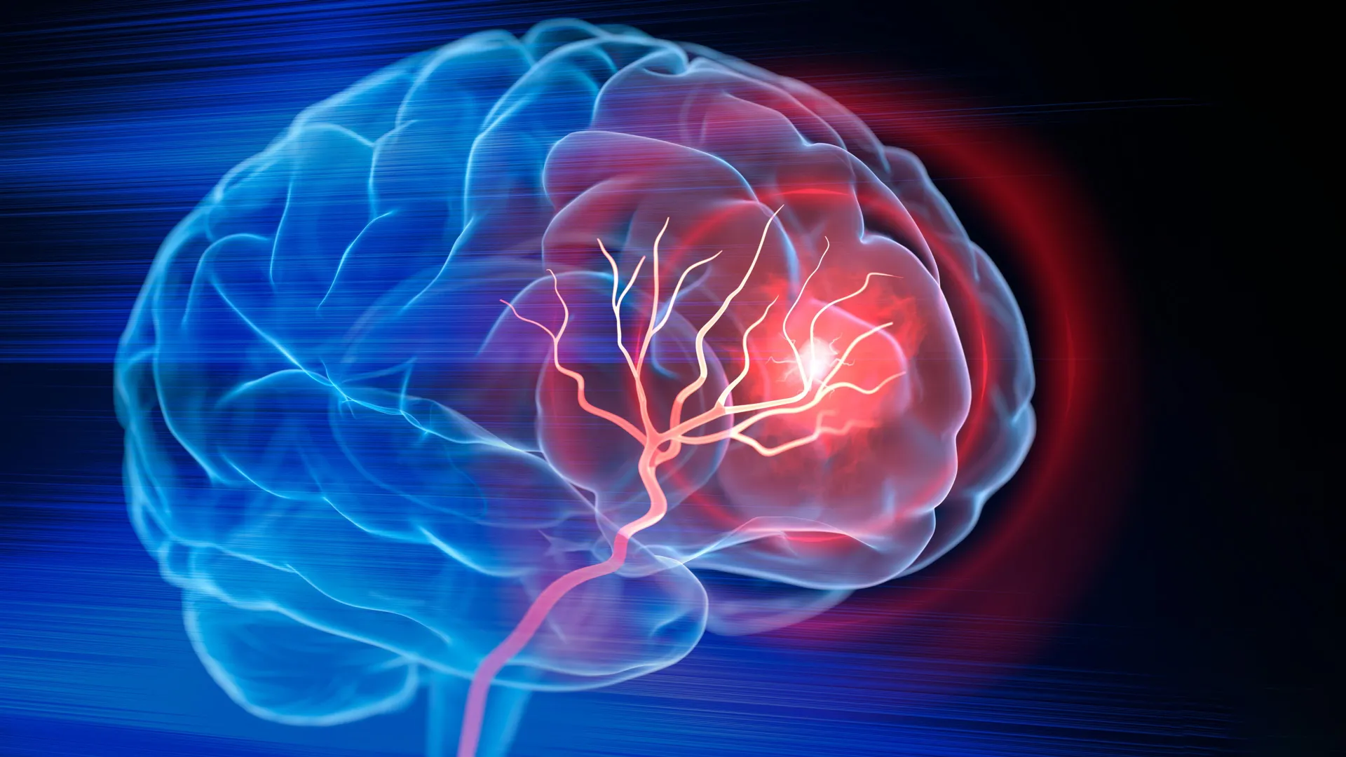
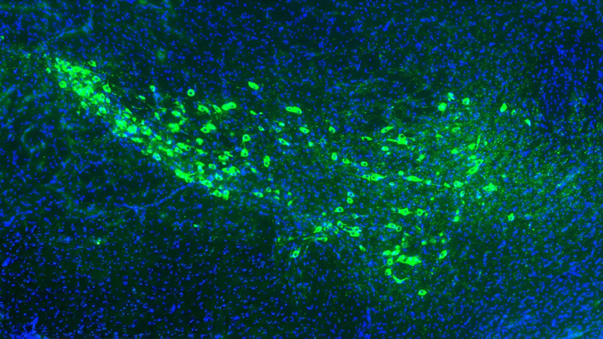
Parkinson’s disease is characterized by the progressive loss of dopaminergic neurons, particularly in the substantia nigra, a region of the midbrain responsible for motor control and reward. For decades, the scientific community has recognized that the primary driver of this destruction is a protein known as alpha-synuclein (α-synuclein). In a healthy brain, alpha-synuclein plays a role in neurotransmission, but in patients with Parkinson’s, the protein misfolds into a toxic shape.
These misfolded proteins aggregate into clumps called Lewy bodies. As the disease advances, these toxic aggregates appear to "infect" neighboring cells. When a neuron becomes overwhelmed by alpha-synuclein and dies, it releases the misfolded proteins into the extracellular space. These proteins are then taken up by healthy adjacent neurons, where they act as templates, forcing the healthy alpha-synuclein in those cells to also misfold. This chain reaction creates a "prion-like" spread that moves through the brain’s circuitry, leading to the worsening of motor and cognitive symptoms.
Despite knowing that alpha-synuclein moves between cells, scientists have struggled to identify the specific "locks" on the surface of healthy neurons that allow this "poison" to enter. Without identifying these entry points, developing a therapy to stop the spread remained an elusive goal.
The Yale Study: A High-Throughput Search for Entry Points
The research team at Yale, led by Stephen Strittmatter, MD, PhD, the Vincent Coates Professor of Neurology and chair of the Department of Neuroscience, embarked on a massive screening project to solve this mystery. Recognizing that the brain contains thousands of different surface proteins, the team utilized a high-throughput approach to narrow down the candidates.
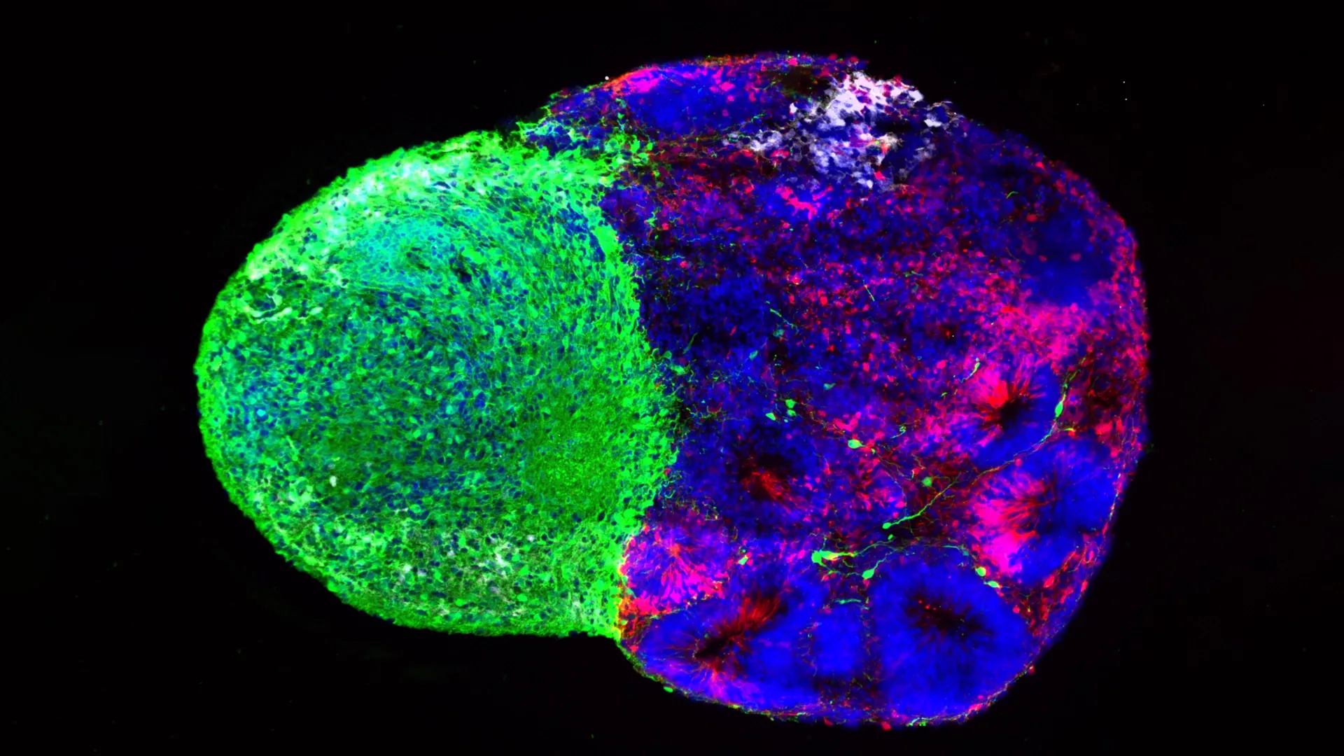
The researchers engineered 4,400 distinct groups of cells, each designed to express one specific human surface protein. They then exposed these cells to misfolded alpha-synuclein to see which, if any, would bind to the toxic protein. The vast majority of the proteins—more than 4,380—showed no interaction whatsoever. However, a small subset of 16 proteins demonstrated a clear affinity for alpha-synuclein.
Upon further analysis, two proteins stood out due to their high concentration in the substantia nigra and their role in motor neuron function: metabotropic glutamate receptor 4 (mGluR4) and neural proliferation differentiation and control protein 1 (NPDC1). These two proteins were found to be particularly effective at capturing extracellular alpha-synuclein and transporting it across the cell membrane into the internal environment of the neuron.
Validating the Discovery: Evidence from Animal Models
To confirm that mGluR4 and NPDC1 were indeed responsible for the disease’s progression, the Yale team conducted a series of experiments using mouse models of Parkinson’s disease. These experiments were designed to test whether removing these "gatekeeper" proteins could protect the brain from the spread of alpha-synuclein.
In the control group, mice with normal levels of mGluR4 and NPDC1 were injected with misfolded alpha-synuclein. As expected, the toxic protein spread rapidly through their brains, leading to significant neuronal death and the development of classic Parkinsonian symptoms, such as tremors and impaired coordination.
In contrast, the researchers used genetic engineering to create mice that lacked either the mGluR4 or NPDC1 protein. When these mice were exposed to the same toxic alpha-synuclein, the results were dramatic. The spread of the protein was significantly curtailed, and the mice remained largely asymptomatic. In a second model designed to mimic advanced Parkinson’s, the removal of these genes not only slowed the progression of symptoms but also significantly increased the survival rate of the animals.
These findings suggest that mGluR4 and NPDC1 are not just incidental observers but are critical partners in the transmission of the disease. While they normally serve essential functions—mGluR4, for instance, helps regulate neurotransmitter release—their structure inadvertently provides a docking site for the misfolded alpha-synuclein.
The Growing Public Health Challenge of Neurodegeneration
The urgency of the Yale study is underscored by the rising prevalence of Parkinson’s disease in the United States and globally. According to the Parkinson’s Foundation, approximately 1.1 million Americans currently live with the disease, a number expected to rise to 1.2 million by 2030. Each year, nearly 90,000 new cases are diagnosed, making it the second most common neurodegenerative disorder after Alzheimer’s disease.
The economic impact is equally staggering. A study published in the journal Movement Disorders estimated that the total economic burden of Parkinson’s disease in the U.S. is approximately $52 billion per year, including direct medical costs and indirect costs such as lost wages for patients and family caregivers.
As the "Baby Boomer" generation ages, the medical community is bracing for what some have called a "silver tsunami" of neurological conditions. Age is the primary risk factor for Parkinson’s; while early-onset cases exist, the majority of diagnoses occur in individuals over the age of 60.
"We have an aging population. How we can stop or slow neurons from dying is an enormous problem," Dr. Strittmatter noted. "This is really the time to make some inroads into figuring out how to slow it down."
Chronology of Parkinson’s Research and the Shift in Focus
The history of Parkinson’s research has evolved through several distinct phases:
- 1817: James Parkinson publishes "An Essay on the Shaking Palsy," the first formal clinical description of the disease.
- 1960s: The discovery of dopamine deficiency leads to the development of Levodopa (L-Dopa), which remains the "gold standard" for managing symptoms.
- 1997: Researchers identify alpha-synuclein as the primary component of Lewy bodies, shifting the focus to protein misfolding.
- 2003: The Braak Hypothesis is proposed, suggesting that Parkinson’s begins in the gut or nose and spreads through the brain via a specific anatomical path.
- 2020s: Research shifts toward "disease-modifying therapies" (DMTs) that aim to stop the spread of pathology rather than just replacing lost dopamine.
The Yale study represents a pinnacle of this latest phase. While L-Dopa and other treatments like Deep Brain Stimulation (DBS) provide relief for tremors and rigidity, they do nothing to prevent more neurons from dying. Eventually, as more cells perish, the effectiveness of these symptomatic treatments wanes. A therapy based on the Yale findings would represent a fundamental shift toward curative or preventative care.
Implications for Future Therapeutics
The identification of mGluR4 and NPDC1 provides pharmaceutical researchers with concrete targets for drug development. There are several potential avenues for translating this research into human medicine:
- Small Molecule Inhibitors: Scientists could develop drugs designed to bind to mGluR4 or NPDC1, blocking the site where alpha-synuclein attaches without interfering with the protein’s healthy functions.
- Monoclonal Antibodies: Targeted antibodies could be engineered to "shield" these receptors, preventing the toxic protein from gaining entry.
- Gene Therapy: Advanced techniques such as CRISPR could potentially be used to modify the expression of these proteins in high-risk areas of the brain.
However, the researchers caution that mGluR4 is a vital receptor in the brain, and simply "turning it off" could have side effects. The challenge for future clinical trials will be to find a way to block the toxic interaction with alpha-synuclein while preserving the receptor’s role in normal signaling.
A Broader Impact on Neurology
The implications of this study may extend beyond Parkinson’s disease. Other neurodegenerative conditions, including Alzheimer’s and Amyotrophic Lateral Sclerosis (ALS), also involve the spread of misfolded proteins (tau and beta-amyloid in Alzheimer’s; TDP-43 in ALS).
The methodology used by the Yale team—screening thousands of surface proteins to find entry points—could serve as a blueprint for identifying how these other toxic proteins invade healthy cells. If a common mechanism or a shared set of receptors is found, it could lead to a universal class of drugs designed to protect the brain from protein-based degradation.
As the scientific community digests these findings, the focus will now move toward the development of compounds that can replicate the protective effects seen in the Yale mouse models. While human clinical trials are likely years away, the discovery provides a clear roadmap for a journey that, until now, was being navigated in the dark.
"If we understood how it gets into neurons, we could perhaps block or slow down the progression of the disease," Strittmatter said. With the identification of mGluR4 and NPDC1, that understanding has arrived, marking a new chapter in the effort to conquer Parkinson’s disease.