Before birth, the human brain is assembled through an enormous series of cellular choices, and at the center of this intricate orchestration are radial glia—a specialized class of stem cells responsible for the structural expansion and functional complexity of the human cerebral cortex. New research published in the journals Cell and Science has provided an unprecedented look at how these progenitor cells navigate their developmental journey. By identifying the critical roles of metabolic processing and direct physical cellular signaling, scientists at the University of California, Los Angeles (UCLA), have unveiled the sophisticated decision-making mechanisms that dictate how the human brain attains its unique architecture, offering potential breakthroughs for understanding neurodevelopmental disorders and the aberrant growth patterns seen in brain cancers.
The Role of Radial Glia in Human Neurogenesis
Radial glia act as the primary architects of the developing brain. During the prenatal period, these cells undergo extensive division to produce the neurons and support cells that constitute the cerebral cortex, the region responsible for higher-order functions such as cognition, memory, and language. Evolutionary biology suggests that the massive expansion of the human cortex—a hallmark of our species—is directly tied to the unique proliferative behavior of these radial glia. While the majority of these cells reach the end of their lifecycle before birth, they retain a haunting resonance in clinical medicine; similar cell populations have been observed to re-emerge in malignant brain tumors, where they contribute to aggressive growth and treatment resistance.
Aparna Bhaduri, an assistant professor of biological chemistry at the David Geffen School of Medicine at UCLA, characterizes these cells as the linchpin of human development. "Radial glia are the coolest cells that have ever existed," Bhaduri notes. "They are key to making us human. However, they are also at the center of many neurodevelopmental and neuropsychiatric disorders, as well as cancer. Understanding how they make their developmental decisions is the first step toward understanding how these conditions arise."
The Metabolic Atlas: A New Lens on Development
In a landmark study published in Cell, a collaborative team led by Bhaduri and Heather Christofk—with co-first authors Jessenya Mil and Jose Soto—constructed a comprehensive metabolic map of the developing human cortex. By analyzing donated human tissue alongside advanced brain organoids, the researchers challenged the long-held assumption that metabolism is a passive, background process.
The investigation revealed that radial glia are highly sensitive to their metabolic environment, specifically relying on the pentose phosphate pathway to fuel rapid division. When the researchers experimentally modulated glucose availability or disrupted this pathway, the stem cells altered their output, prematurely shifting production toward inhibitory neurons—a cell type that typically appears in later stages of cortical development. This finding suggests that nutrient availability and metabolic health during gestation may be critical environmental determinants of structural brain formation, potentially linking maternal nutrition and metabolic health directly to the architectural integrity of the fetal brain.
Thalamic Projections and Physical Cellular Signaling
Parallel research published in Science, led by first author Claudia Nguyen, explored the extrinsic signals that guide radial glia. Scientists have long observed that the thalamus—the brain’s deep-seated relay station—sends long, wire-like projections toward the cortex early in development, well before functional synaptic connections are established. The mystery of why these fibers arrive so early has persisted for decades.
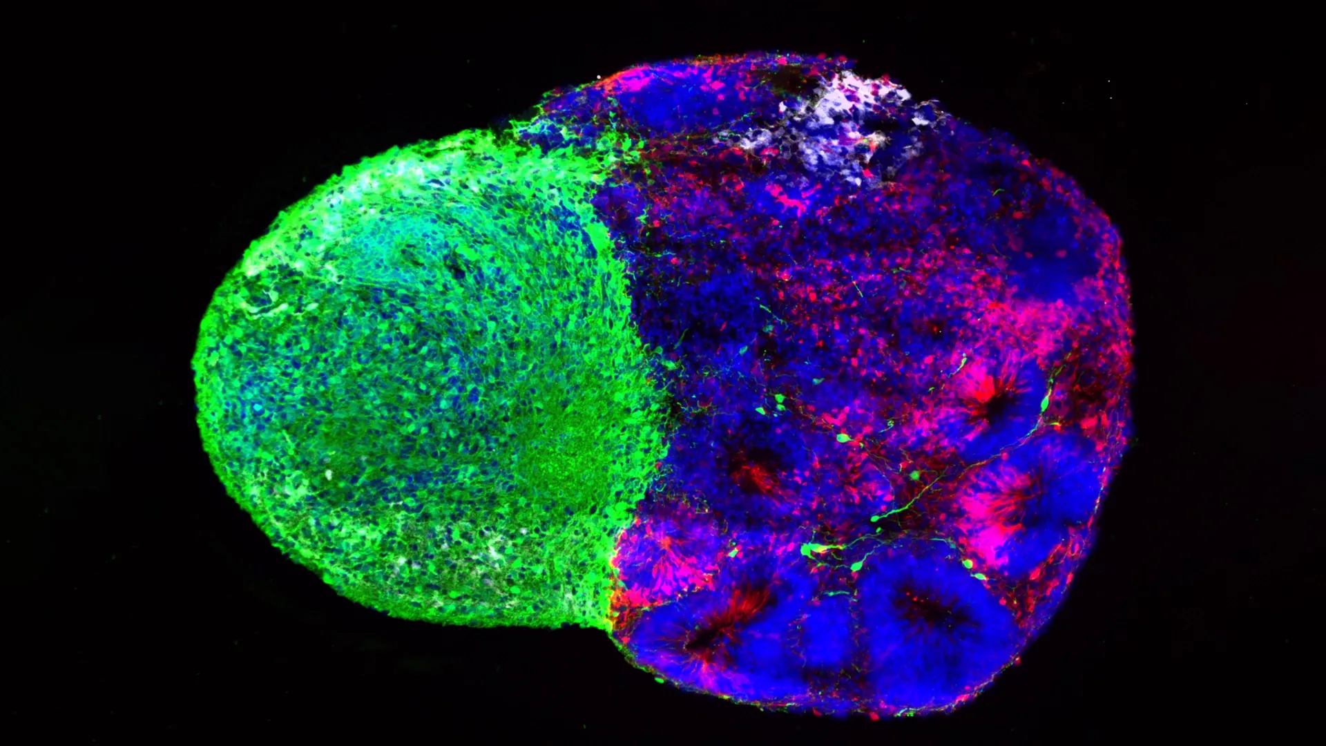
Utilizing innovative "assembloid" technology—a method that allows researchers to grow different regions of the brain in proximity to study their interactions—the UCLA team discovered that these thalamic projections make physical contact with radial glia. This physical interaction serves as a signaling mechanism, triggering the stem cells to increase the production of excitatory neurons, particularly those destined for the upper layers of the cortex. These upper-layer neurons are significantly more numerous in humans than in other primates or rodents, suggesting that this physical interaction is a distinctively human developmental trait. The discovery of this "contact-based signaling" provides a new framework for understanding how disparate parts of the brain communicate to ensure a synchronized growth pattern.
Linking Development to Neurodevelopmental Disorders
The implications of these findings extend into the pathology of neurodevelopmental conditions. The researchers focused specifically on the gene NRXN1, which is heavily involved in the formation of neural connections and has been strongly linked to autism spectrum disorder. By creating assembloids derived from cells carrying an NRXN1 mutation, the team demonstrated that the thalamic signals were fundamentally altered. The disrupted signals led to an imbalance in the ratio of stem cells to their progeny, effectively changing the trajectory of cortical formation. This provides a tangible biological mechanism through which genetic predispositions can manifest as structural irregularities in the brain during early development.
The Evolution of Brain Organoid Technology
The ability to perform this research is a testament to the rapid maturation of organoid technology over the last decade. Ten years ago, the study of human-specific neural development was largely restricted to animal models, which fail to capture the nuances of human cortical expansion. Today, human stem cell-derived organoids and assembloids allow for the modeling of complex, human-specific developmental timelines. These systems provide a high-resolution window into the "black box" of prenatal brain growth, enabling researchers to manipulate specific variables—such as glucose concentration or genetic expression—without the ethical and logistical constraints associated with human subjects.
Broader Impact and Future Directions
The synthesis of these two studies suggests that the radial glia do not function in isolation; they are instead the recipients of a constant stream of information from their internal metabolic state and their physical environment. The findings serve as a call to action for the scientific community to re-evaluate metabolism and cell-to-cell physical interaction as "active drivers" of neural development.
For the medical community, the research offers a potential paradigm shift in how we approach the study of neurological disease. By "looking under the hood" of these cellular decisions, clinicians and researchers hope to eventually map how vulnerability to certain conditions is established in the womb. Furthermore, the link between these early developmental stem cells and cancer suggests that the same mechanisms governing growth in the healthy brain may, when hijacked, drive the formation of brain tumors.
As researchers move forward, the integration of these findings into clinical practice remains a distant but promising goal. The data provided by the metabolic atlas and the identification of thalamic contact points offer a foundation for future studies into how environmental factors—ranging from maternal stress to metabolic disorders—shape the trajectory of human cognition.
"Ultimately, these studies give us a glimpse under the hood of how these cells make decisions," Bhaduri concluded. "Understanding those decisions is a first step toward understanding normal brain development, disease vulnerability and, potentially, how similar stem-cell programs operate in brain cancer."
This extensive research effort was made possible through the support of numerous organizations, including the National Institutes of Health, the National Science Foundation, the Brain & Behavior Research Foundation, the Alfred P. Sloan Foundation, the Rose Hills Foundation, the Esther A. & Joseph Klingenstein Fund, the Simons Foundation, the Chan Zuckerberg Initiative, the NIH BRAIN Initiative Cell Atlas Network, the International Foundation for Ethical Research, the UCLA Broad Stem Cell Research Center, and the UCLA Health Jonsson Comprehensive Cancer Center. As the scientific community digests these findings, the focus will likely turn toward translating these cellular insights into preventative strategies and targeted interventions for the most complex disorders of the human brain.