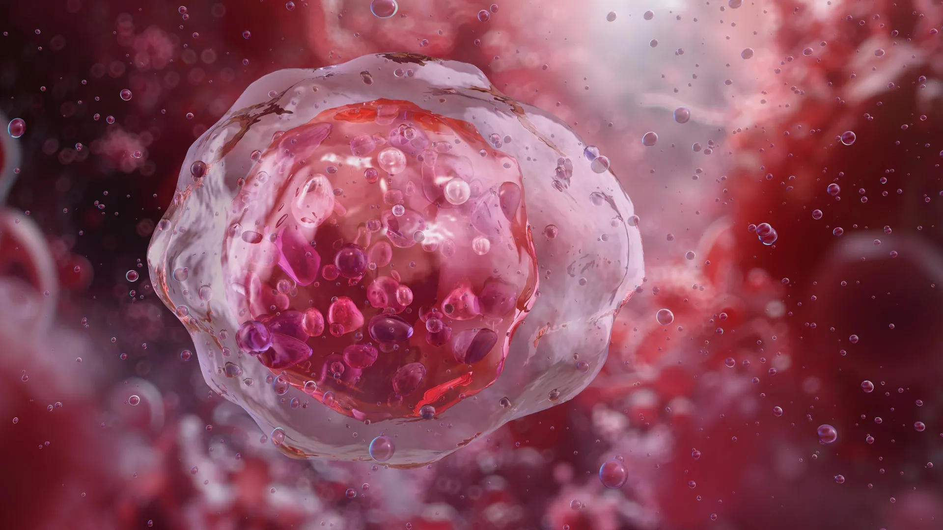
In a discovery that upends decades of textbook biology, researchers at Stanford Medicine have revealed that glucose—long understood universally as the primary metabolic fuel for living cells—harbors a critical, previously unrecognized identity. According to a landmark study published online March 21 in Cell Stem Cell, glucose serves as a master regulator of tissue differentiation, the fundamental biological process through which unspecialized stem cells transform into the diverse, mature cell types that compose human organs and tissues.
Rather than functioning solely as a chemical battery broken down to release energy, intact glucose molecules physically bind to hundreds of intracellular proteins. In doing so, glucose directly modulates protein function and dictates which genes within the human genome are expressed, and when. This revelation challenges the foundational paradigm of cellular metabolism and opens entirely new avenues for investigating, diagnosing, and treating conditions ranging from diabetes to cancer.
The findings were so counterintuitive and radical that the research team spent several years executing exhaustive follow-up experiments to verify their data before bringing the work to public attention. Led by senior author Dr. Paul Khavari, the Carl J. Herzog Professor in Dermatology and chair of dermatology at the Stanford School of Medicine, alongside lead author and research scientist Dr. Vanessa Lopez-Pajares, the study establishes a universal role for glucose across multiple human tissue types and mammalian models.
A Serendipitous Scientific Detour
The path to this discovery was entirely unintentional. When Khavari and Lopez-Pajares initiated their research project, their objective was not to investigate glucose, but rather to identify novel biomolecules responsible for driving cellular differentiation in human skin. To achieve this, the team deployed a high-throughput screening approach coupled with advanced mass spectrometry. They tracked the fluctuating abundance of thousands of distinct biomolecules within human skin stem cells as those cells transitioned into mature keratinocytes, the predominant cell type found in the outermost protective layer of human skin.
The underlying hypothesis was straightforward: biomolecules that experience a significant surge in concentration during the differentiation process are statistically likely to play a causal role in facilitating that cellular transition.
Upon analyzing the screening data, the researchers isolated 193 suspect molecules, many of which possessed established biological links to skin differentiation. However, occupying the second-highest spot on the list of elevated molecules was an unexpected compound: glucose.
This finding completely defied conventional biochemical expectations. Stem cells typically exhibit high rates of division and substantial energy demands, whereas cells undergoing terminal differentiation divide less rapidly, require less energy, and eventually progress toward senescence and programmed death. Consequently, the researchers initially anticipated that intracellular glucose levels would plummet during differentiation. Instead, measurements demonstrated that glucose concentrations increased markedly as epidermal stem cells matured into keratinocytes.
To ensure this observation was not an artifact of a single testing method, the team validated the results using multiple orthogonal techniques. They measured the direct cellular uptake of fluorescent and radioactive glucose analogs, and deployed biological sensors engineered to emit red or green fluorescence in the presence of physiological concentrations of glucose. As cellular differentiation advanced, the fluorescence intensity increased correspondingly.
Subsequent testing in other human cell lineages—including developing fat, bone, and white blood cells—as well as in genetically engineered murine models expressing fluorescent glucose sensors, revealed the identical biological pattern. Across every tissue examined, intracellular glucose accumulated as cells matured, pointing to a systemic, body-wide regulatory function.
Uncoupling Metabolism from Gene Regulation
To determine the mechanism driving this intracellular accumulation, the team analyzed the transport kinetics of the cells. The experiments revealed that the localized surge in glucose was the dual result of accelerated cellular import—driven by an upregulation of specialized glucose-transport proteins—combined with decreased export.
Crucially, metabolic assays confirmed that this intracellular influx of glucose was not accompanied by a corresponding rise in glycolysis; the sugar molecules were not being catabolized into metabolic byproducts to generate adenosine triphosphate (ATP).
To test whether glucose’s newly discovered regulatory role was independent of its metabolic function, the researchers cultured human skin organoids—three-dimensional, engineered skin tissues grown in liquid media designed to mimic the structural and cellular architecture of native skin—in environments deficient in normal glucose. Under low-glucose conditions, the organoids failed to differentiate properly. Genomic analysis revealed that the expression of more than 3,000 genes was disrupted, many of which encode proteins essential for normal cutaneous development.
When these glucose-starved organoids were subsequently replenished with a non-metabolizable glucose analog—a synthetic molecule that the cells cannot break down for energy—normal tissue differentiation resumed completely. These non-metabolizable analogs supported cellular maturation just as effectively as regular glucose. This empirical proof demonstrated definitively that glucose acts as a structural signaling ligand, exercising regulatory control entirely separate from its role as an energy substrate.
Historical Clues and Mechanistic Insights
While the Stanford study is the first to comprehensively map this phenomenon, retrospective analysis reveals that subtle hints of glucose’s regulatory capacity have existed in scientific literature for years. For instance, embryonic stem cells—pluripotent cells capable of giving rise to every tissue type in the human body—frequently lose their developmental plasticity and differentiation potential when cultivated in environments with chronically elevated glucose concentrations. This impairment is now understood to be the result of aberrant signaling that forces premature differentiation and strips the cells of their stemness.
Furthermore, clinical observations involving diabetic patients have long demonstrated that chronic hyperglycemia impairs physiological wound healing and tissue regeneration, processes that fundamentally rely on precise cellular differentiation.
At the molecular level, Lopez-Pajares, Khavari, and their colleagues mapped the precise sequence of events following glucose influx. Once transported inside the cell, glucose binds directly to hundreds of intracellular proteins. Among these is IRF6, a critical transcription factor involved in skin development. The binding of glucose alters the physical conformation of the IRF6 protein, modifying its capacity to bind to DNA and activate genes required for differentiation.
Rather than operating through a highly targeted, localized signaling cascade—the traditional mechanism by which hormones or growth factors trigger specific cellular responses—glucose functions as a global broadcast signal. When intracellular glucose concentrations rise, they increase uniformly and simultaneously throughout the cellular environment. Khavari likened the process to a fire alarm activating in a firehouse, prompting a coordinated, immediate response across the entire cellular machinery.
Broader Implications for Diabetes and Oncology
The implications of this discovery extend deeply into pathology, offering fresh theoretical frameworks for understanding complex human diseases characterized by metabolic and developmental dysregulation.
In the context of oncology, cancer is fundamentally a disease defined by failed or arrested cellular differentiation, wherein immature cells continue to proliferate uncontrollably without maturing into specialized, functional tissue. Interestingly, certain synthetic glucose analogs have previously shown therapeutic promise in preclinical and clinical trials as experimental anticancer agents. While these compounds were originally designed under the assumption that they would starve tumor cells of metabolic energy, the new Stanford findings suggest an alternative mechanism of action: these analogs may act by binding to proteins within immature cancer cells, driving them to differentiate and halting their unchecked proliferation.
Similarly, the findings provide a vital springboard for future investigations into metabolic disorders. Dysregulated blood sugar levels affect hundreds of millions of individuals worldwide, particularly patients suffering from type 1 and type 2 diabetes. Understanding how fluctuations in systemic glucose impact cellular differentiation and tissue repair may lead to novel therapeutic strategies designed to mitigate the long-term systemic complications associated with chronic hyperglycemia.
As the scientific community begins to digest these findings, researchers emphasize that this study serves as a compelling reminder that small biomolecules once assumed to be passive metabolic intermediates can exert profound control over gene expression and cell fate.
Funding for the research was provided by the National Institutes of Health, under grants R01AR043799, AR045192, K01AR070895, and P30CA124435, alongside support from the U.S. Department of Veterans Affairs Office of Research and Development. As this nascent field of study expands, the Stanford Medicine team plans to continue probing the complex, dual-natured biology of glucose in both healthy physiology and disease pathogenesis.