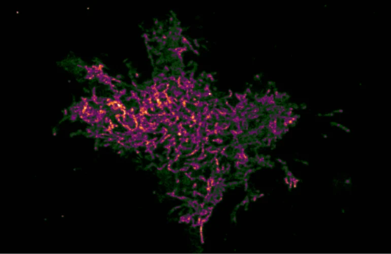
For decades, the field of molecular biology has grappled with a significant limitation in cellular visualization: the inability to accurately map the spatial distribution and temporal dynamics of enzyme activity at the nanometer scale. Traditional negative biosensors—tools designed to detect specific biochemical processes—have long been hampered by a "signal-loss" paradox. When these sensors detect enzymatic activity, they lose their fluorescence, making it impossible to distinguish between a region of high activity and a region where the sensor is absent or non-functional. Researchers at the University of Illinois Chicago (UIC) have now effectively dismantled this barrier with the development of a pioneering technique known as Fluctuation Increase Negated by Intra-Chain Interaction (FINICI).
The findings, published in the Proceedings of the National Academy of Sciences, represent a paradigm shift in how scientists observe the inner workings of a living cell. By fundamentally altering the optical readout of biosensors from a negative signal to a positive feedback loop, the FINICI platform allows researchers to map biochemical events with unprecedented resolution, revealing complex landscapes of enzyme activity previously obscured by the limitations of conventional light microscopy.
The Mechanics of FINICI: A Technical Breakthrough
The core innovation of the FINICI platform lies in its ability to manipulate the electronic state of a chromophore—the part of a molecule responsible for its color and fluorescence. Through the use of precisely engineered electrostatic interactions between fluorescent proteins, the technique reversibly modifies the potential of the chromophore.
According to Gary Mo and Kriti Srivastava, the primary researchers behind the project, the interaction works by physically restricting the chromophore, which in turn prevents electron delocalization and limits the number of possible electronic transitions. This suppression of fluorescence and "blinking" is the key to the system’s sensitivity. By flipping the readout, the researchers have created a tool that essentially shines a light on activity rather than letting it fade into the background.
This capability is particularly vital for super-resolution microscopy. Because the method allows for visualization at a resolution below the classical diffraction limit of light—approximately 200 nanometers—scientists can now observe cellular structures that were once considered invisible or indistinct. This allows for the mapping of enzymes within microdomains as small as 150 to 200 nanometers, providing a granular view of cellular decision-making processes.
Validation and Comparative Efficacy
To ensure the reliability of their new platform, the UIC team subjected FINICI to a rigorous series of validation tests, measuring it against the industry’s "gold standard": Förster resonance energy transfer (FRET). FRET has historically been the preferred method for monitoring protein-protein interactions and enzyme activity due to its ability to measure distance at the molecular level.
The comparative analysis yielded striking results. In cells where receptor expression was notably low, FRET often failed to detect any response, likely due to a lack of sufficient signal-to-noise ratio. In contrast, the FINICI platform remained highly sensitive, successfully mapping enzymatic activity in those same low-expression environments. Furthermore, to mitigate the risk of false positives, the researchers utilized extensive biological controls, including cells lacking the specific target enzyme or receptor, to verify that the observed signals were exclusively the result of genuine biochemical activity rather than artifacts or noise.
Mapping Cellular Architecture: Src Kinase, Syk Kinase, and cGMP
The team utilized FINICI to investigate three critical signaling components: Src kinase, Syk kinase, and cyclic guanosine monophosphate (cGMP). Each of these molecules plays a fundamental role in cell physiology, from cancer progression and motility to immune response.
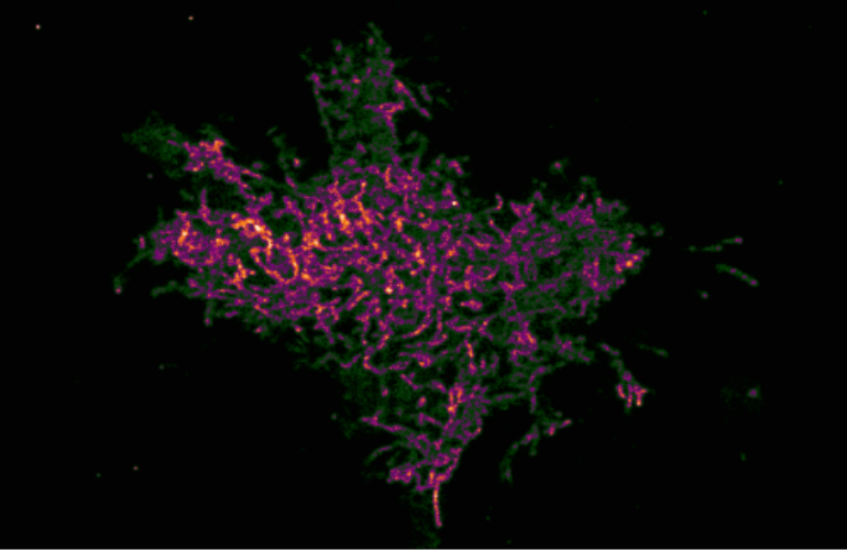
The observations regarding Src kinase were particularly illuminating. Linked to cancer cell migration and metastasis, Src kinase was found to exhibit localized, transient bursts of activity across the cell membrane, specifically within cholesterol-rich lipid rafts. Unlike traditional, bulk-cell measurements that average out these signals, FINICI revealed a dynamic environment where some of these activity zones appeared briefly and dissolved, while others persisted. This suggests that the cell membrane is not a static container but a highly active, compartmentalized landscape where enzymatic "hotspots" are tightly regulated.
The study of Syk kinase yielded similar insights. While the researchers confirmed that the enzyme is anchored by adaptor proteins, they discovered that this anchoring occurs predominantly away from the cell membrane, at the internal scaffolding of immune cells. This spatial specificity suggests that the cell employs different mechanical strategies—membrane-based lipid control for Src and scaffold-based protein control for Syk—to govern its internal operations. Finally, the imaging of cGMP demonstrated that the formation of nanodomains is not a terminal event; rather, the cell maintains a dynamic background that allows it to tune the efficacy of these signaling clusters in real-time.
Implications for Pharmaceutical Development
The potential impact of FINICI on drug discovery cannot be overstated. Current high-throughput screening processes often rely on whole-cell measurements, which can overlook the subtle, localized failures of a drug candidate. The research team noted that in trials using a "cousin" to the FINICI platform, known as FLINC, they tested a kinase inhibitor at concentrations far exceeding its IC50—the concentration required to inhibit 50% of the enzyme’s activity.
Even at these high dosages, the drug failed to completely shut down the enzyme. The researchers observed that phosphorylation continued to occur within microdomains approximately 250 nanometers in size. It was only when they introduced a peptide that disrupted the anchoring complex that the microdomains were fully abolished. This finding suggests that many drug candidates may be failing in clinical trials not because they lack potency, but because they cannot penetrate or disrupt these highly specialized, compartmentalized enzyme microdomains.
"We believe that drug discovery with FINICI will have more information, like a transcriptomic screen," Mo and Srivastava stated. By providing a "spatial map" of drug efficacy, pharmaceutical researchers could move beyond simply asking if a drug works, and instead ask: where does it work, how does it impact localized cellular compartments, and what are the spatial patterns of the remaining enzymatic activity?
Future Directions and Technological Evolution
As the technology matures, the UIC team is shifting its focus toward scaling the assay for broader use. One of the primary challenges in cellular imaging is the sheer volume of data produced; to combat this, the researchers are currently integrating automation and machine-learning-based pattern recognition into their microscopy workflows. These tools will allow for faster, more objective analysis of complex cellular patterns.
Furthermore, the team is working on "multiplexing," or the ability to observe multiple cellular observables simultaneously. By expanding the number of targets that can be visualized at once, FINICI could provide a holistic view of the signaling pathways that drive complex diseases. The researchers are also exploring the limits of spatial resolution. While they have currently resolved compartments between 150 and 200 nanometers, it remains an open question whether cells utilize even smaller, sub-diffraction structures to regulate their behavior.
Beyond Pharmacology: A Broader Utility
While the primary application currently lies in drug development and molecular biology, the implications of FINICI extend into materials science and bioengineering. The researchers suggest that the technique could be adapted to detect heterogeneity in advanced biomaterials. By highlighting locations of internal stress or mechanical compression within a material, FINICI could provide a new way to monitor the integrity and performance of synthetic tissues or bio-hybrid devices.
In the context of modern biomedical research, the development of FINICI highlights a growing trend toward "spatial biology"—the recognition that the position of a molecule is just as important as its identity. By transforming how we visualize the activity of enzymes, the researchers at the University of Illinois Chicago have opened a new window into the cellular world, turning once-invisible processes into clear, measurable data points. As this technology enters the mainstream of lab-based research, it promises to reshape our understanding of how life is organized at the smallest possible scales, offering a new, precise tool in the ongoing battle against complex, localized diseases.