The construction of the human brain before birth is arguably the most complex architectural feat in biology. At the heart of this process lies a specialized class of stem cells known as radial glia. These cells serve as both the foundation and the scaffold for the cerebral cortex—the wrinkled, outer layer of the brain responsible for higher-order functions such as consciousness, memory, language, and executive decision-making. Recent breakthroughs from the University of California, Los Angeles (UCLA) have shed new light on how these enigmatic cells orchestrate the development of the human mind, revealing that their decision-making process is far more dynamic and responsive to external stimuli than previously understood.
For decades, the prevailing view of radial glia was that of a relatively static "mother cell," dutifully producing neurons in a predetermined sequence. However, two landmark studies published in the journals Cell and Science suggest that these cells are sophisticated biological processors, constantly integrating metabolic data and physical environmental signals to determine the final composition of the cortex. By utilizing advanced brain organoid technology—three-dimensional tissue cultures that mimic the structure of the developing human brain—researchers have finally begun to peek under the hood of human neural development, offering potential pathways for treating neurodevelopmental disorders and even understanding the resurgence of stem-cell-like properties in certain brain cancers.
The Architectural Role of Radial Glia
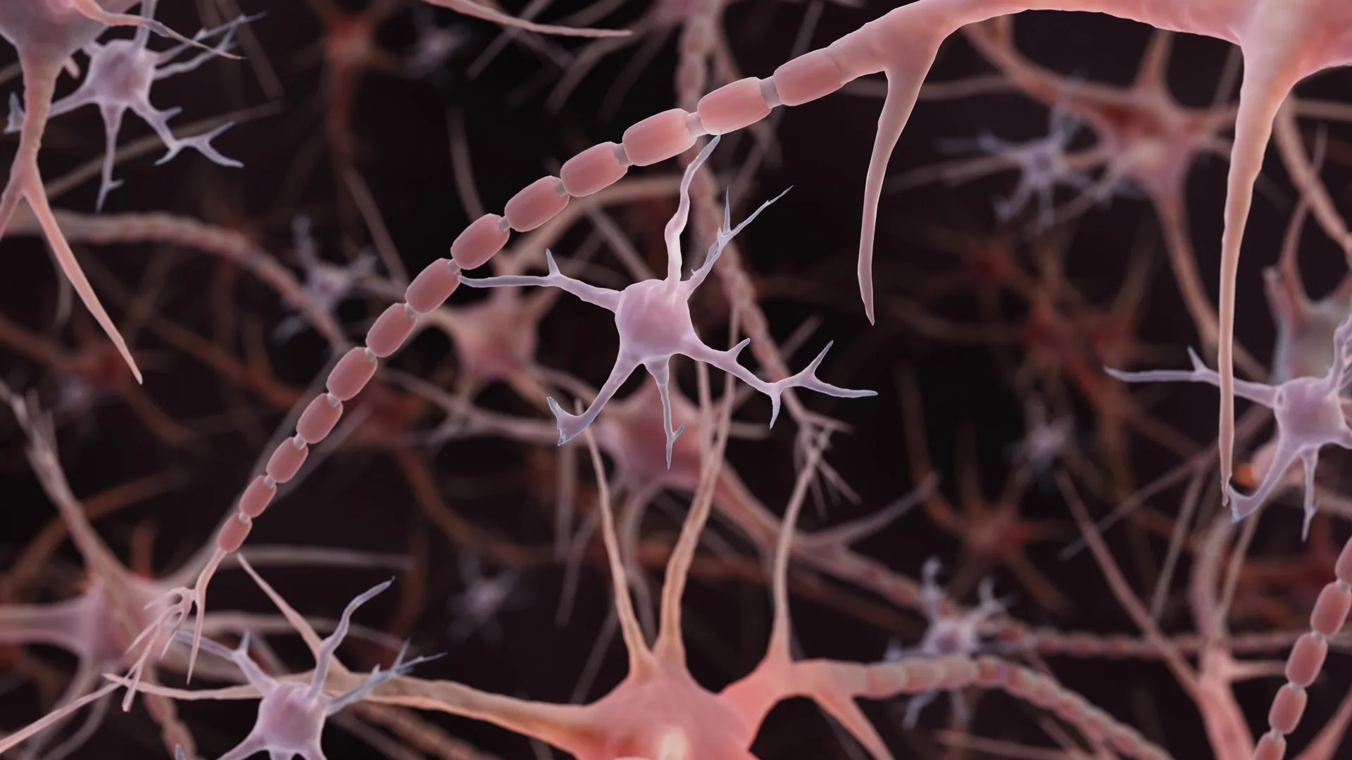
Radial glia are the primary progenitors of the cerebral cortex. During the embryonic period, these cells undergo asymmetric division, producing either another radial glial cell to maintain the stem cell pool or a specialized neuron. This delicate balance is what dictates the size and complexity of the brain. The expansion of the human cortex—which is significantly larger and more folded than that of our closest primate relatives—is widely attributed to the unique behaviors and longevity of these progenitor cells.
Despite their critical importance, radial glia largely disappear or differentiate into other cell types before birth. Their re-emergence in adult pathologies, particularly in aggressive glioblastomas, remains one of the most compelling mysteries in oncology. Scientists suspect that when these cells "wake up" in a tumor environment, they may be hijacking the same developmental programs used during gestation, allowing cancer cells to proliferate with the same rapid, unchecked efficiency as fetal brain cells.
Metabolism as a Developmental Governor
The study published in Cell, led by researchers at the UCLA Broad Stem Cell Research Center and the Jonsson Comprehensive Cancer Center, challenges the long-held assumption that cellular metabolism is a mere passive byproduct of cell division. By creating a high-resolution metabolic atlas of the developing human cortex, the team demonstrated that metabolic pathways act as a regulatory switch for stem cell fate.
The research highlights the significance of the pentose phosphate pathway, a metabolic route that diverts glucose to create the building blocks required for rapid DNA synthesis and cell division. The study found that radial glia rely heavily on this specific pathway to fuel their proliferative phase. When researchers manipulated glucose availability or inhibited the enzymes driving this pathway, the stem cells prematurely pivoted, shifting their output from the production of progenitor cells to the generation of inhibitory neurons.
This shift is significant because inhibitory neurons typically appear later in the developmental timeline. By altering the metabolic environment, the researchers were essentially able to "age" the stem cells prematurely. This suggests that maternal health, dietary intake, and metabolic disorders during pregnancy could have a more direct, granular impact on the structural integrity of the fetal brain than previously recognized. If the metabolic fuel provided to these stem cells is suboptimal, the "programming" of the cortex could be altered, potentially predisposing the offspring to various neuropsychiatric conditions.
Thalamic Projections and the Physical Language of Development
While the metabolic study explored internal chemical signals, the research published in Science investigated how the developing brain communicates across its own geography. Focusing on the thalamus—a deep-brain structure often described as the brain’s "relay station"—the research team discovered that thalamic neurons extend long, wire-like axons into the cortex much earlier than traditional models suggested.
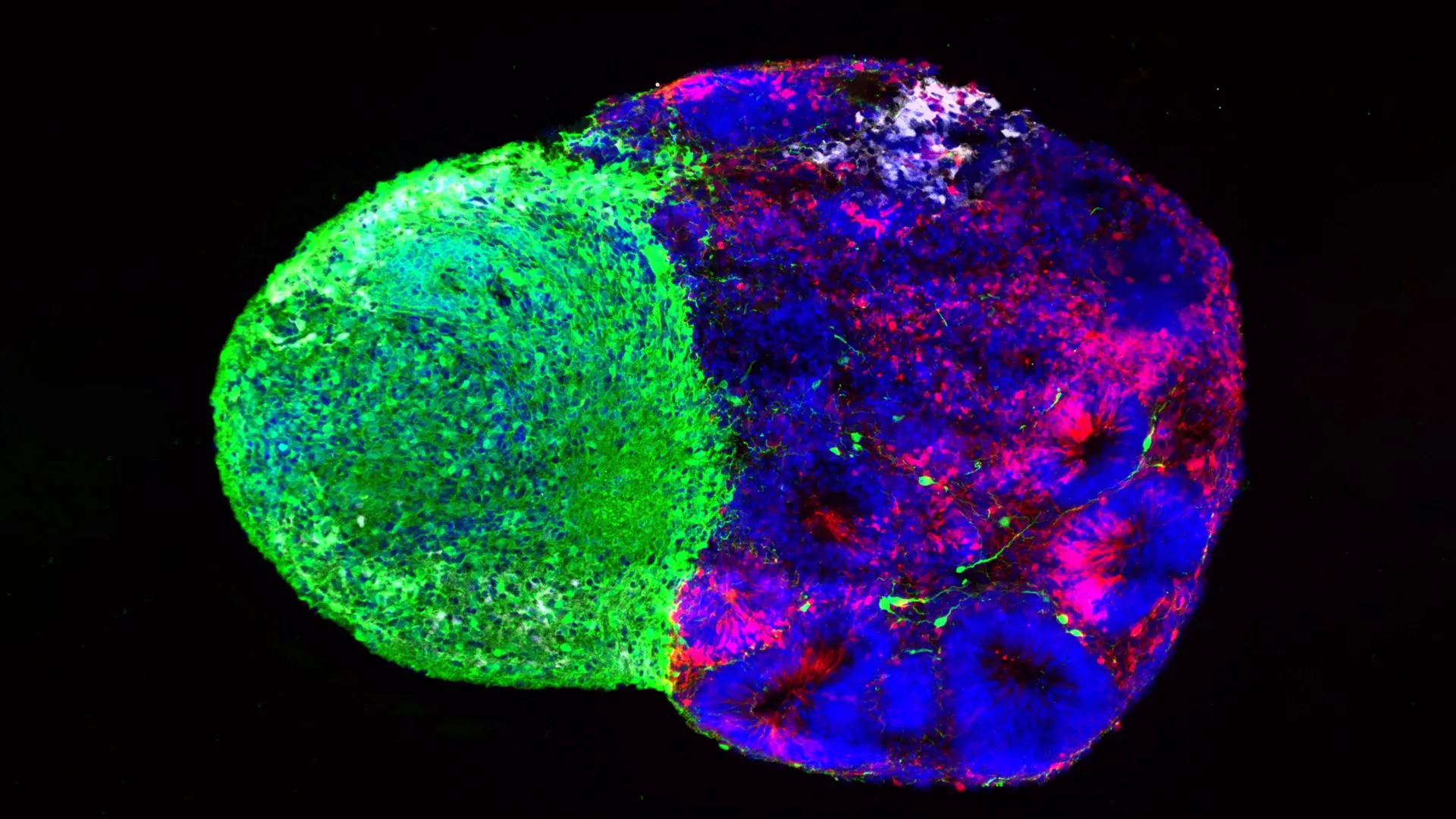
Historically, neuroscientists observed these projections but believed they remained inactive until the cortex was sufficiently developed to receive them. The UCLA study, utilizing "assembloids"—a sophisticated technique where two different types of organoids are fused together to observe their interaction—demonstrated that these projections physically contact radial glia long before functional synapses are formed.
This physical touch is not accidental; it is instructive. The contact between thalamic axons and radial glia triggers the stem cells to increase the production of excitatory neurons, particularly those destined for the upper layers of the cortex. These upper-layer neurons are the most evolved components of the human brain, facilitating the complex neural circuits required for higher cognitive function. The study posits that this direct, physical "handshake" between the thalamus and the cortex is a distinctively human developmental feature, likely absent or significantly different in rodents, which explains why animal models have historically struggled to fully replicate human cortical expansion.
Genetic Vulnerability and the NRXN1 Link
The researchers further deepened their analysis by examining the role of the gene NRXN1 (Neurexin 1). Known to be critical for synaptic function, mutations in NRXN1 are frequently identified in patients diagnosed with autism spectrum disorder (ASD). By creating assembloids derived from the cells of patients carrying these mutations, the team observed that the thalamic signals were fundamentally disrupted.
The altered signals failed to effectively "instruct" the radial glia, leading to an imbalance in the ratio of stem cells to neurons. This finding provides a mechanistic bridge between genetic predisposition and structural brain abnormalities. It suggests that autism and other neurodevelopmental disorders may not just be problems of mature synaptic connectivity, but rather the result of a "domino effect" that begins with early-stage cellular miscommunication during the embryonic phase.
Implications for Modern Neuroscience and Medicine
The ability to study these phenomena in a laboratory setting represents a paradigm shift in developmental biology. For decades, the "black box" of human gestation was nearly impossible to study directly due to ethical constraints and the lack of human-specific experimental models. Today, brain organoid and assembloid technologies provide a window into the human brain that was previously limited to retrospective post-mortem studies or, at best, limited rodent models.
The implications of this research are twofold. First, it provides a foundation for the field of "metabolic psychiatry," suggesting that future therapeutic or preventative interventions might one day address neurodevelopmental risks through the management of metabolic pathways in the uterine environment. Second, it offers a new framework for cancer research. If radial glia are indeed the drivers of aggressive brain tumors, identifying the specific metabolic or signaling triggers that cause these cells to activate could lead to therapies that "force" cancer cells back into a dormant or differentiated state, rendering them less harmful.
Dr. Aparna Bhaduri, who led these efforts, emphasizes that the goal is not merely to observe but to understand the logic of the cell. "These cells are the architects of our identity," she noted in recent discussions regarding the findings. "By deciphering the instructions they follow, we are moving closer to understanding the vulnerabilities that lead to disease."
Conclusion: A New Era of Developmental Mapping
The dual findings regarding metabolic regulation and physical axonal contact underscore a vital truth: the developing brain is a highly communicative, integrated system. Radial glia do not work in a vacuum; they exist in a constant, bidirectional dialogue with their environment.
As technology continues to advance, the integration of these metabolic and physical maps will be essential for creating a comprehensive "atlas" of human brain development. Such a resource will be invaluable for researchers seeking to untangle the complex genetic and environmental threads that contribute to the spectrum of human neurological health. By shifting the focus from viewing these processes as passive background noise to recognizing them as active, regulatory drivers, the scientific community is taking a crucial step toward unlocking the mysteries of the human cortex, potentially transforming our approach to both the origins of intelligence and the treatment of the brain’s most persistent disorders.