For over a century, the standard textbook illustration of a neuron has remained largely static: a sleek, uniform cylinder extending from the cell body, acting as a passive conduit for electrical impulses. This long-held model of the axon, the primary signaling cable of the nervous system, is currently undergoing a radical revision. Recent findings from Johns Hopkins Medicine, supported by subsequent research in human tissue, suggest that the microscopic wiring of the brain is not a smooth, featureless tube, but rather a dynamic structure that frequently resembles a string of tiny, repeating pearls. This discovery, centered on the physical geometry of axons, challenges fundamental assumptions about how information travels through the human brain.
The Paradigm Shift in Neuroanatomy
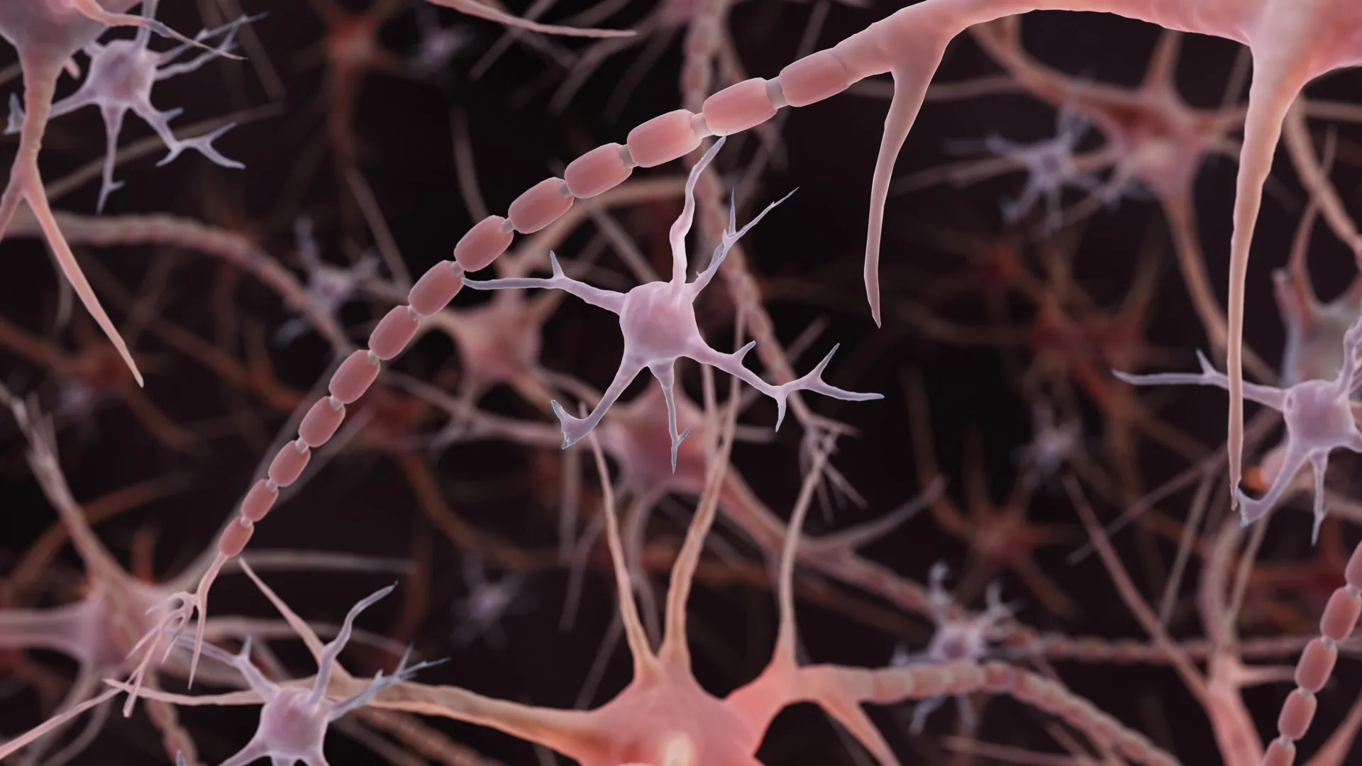
The traditional view of the axon depicts it as a narrow, consistent wire, punctuated only by occasional, large swellings known as synaptic varicosities, where neurotransmitters are stored and released. While these large bulbs have long been understood as essential components of synaptic communication, the "pearls" identified by the Johns Hopkins team are distinct. These smaller, repeating swellings—referred to as non-synaptic varicosities—appear along the length of the axon in healthy, functioning neurons.
This discovery did not emerge from a single observation, but rather from a rigorous re-evaluation of how biological samples are prepared for microscopy. Historically, researchers have utilized chemical fixation and dehydration—a process akin to turning a grape into a raisin—to prepare brain tissue for electron microscopy. While effective for observing large structures, this process often obscures the subtle, nanoscale details of the axonal membrane.
By employing high-pressure freezing—a technique that preserves cellular structures in their native, hydrated state—researchers were able to capture the authentic, beaded architecture of the axons. The comparison is significant: if standard microscopy creates a "raisin," high-pressure freezing preserves the "grape," revealing that the undulating, pearled shape is a natural, inherent feature of these neural pathways.
Chronology of Discovery: From Worms to Humans
The investigation into these structural undulations began with serendipitous observations in the nervous systems of the nematode C. elegans. Dr. Shigeki Watanabe, an associate professor of cell biology and neuroscience at the Johns Hopkins University School of Medicine, noticed that the axons of these worms exhibited a consistent, beaded pattern. Following a technical discussion with Dr. Graham Knott, an expert in high-resolution electron microscopy, the team initiated a broader study to determine whether this architecture was unique to invertebrates or a conserved feature of mammalian biology.
The project reached a milestone in late 2024 when the team published their findings in Nature Neuroscience. The study, led by graduate student Jacqueline Griswold, confirmed the existence of these "nanoscale pearls" in both laboratory-grown mouse neurons and tissue samples from adult mice.
The trajectory of this research accelerated significantly in 2025. In November of that year, a study led by Chelsy R. Eddings, with contributions from the Watanabe lab, was published online in the journal Neuron. This work moved the findings into the clinical realm, identifying similar pearled structures within human cortical tissue harvested during epilepsy surgeries. This cross-species verification shifted the conversation from a niche anatomical observation to a fundamental question of human neurobiology: if the brain’s wiring is inherently pearled, how does this geometry dictate the velocity and integrity of our thoughts?
Mechanical Physics and the "Traffic Jam" Theory
To understand why an axon would adopt such a complex, undulating shape, the research team turned to theoretical biophysics. By collaborating with Dr. Padmini Rangamani, a professor of pharmacology at the University of California San Diego, the team developed mathematical models to simulate the physical properties of the axonal membrane.
The models revealed that the pearled structure is not maintained by a rigid internal scaffold, as once hypothesized, but is instead a result of the membrane’s own physical tension and composition. Experiments confirmed that the degree of "beading" is highly sensitive to the concentration of cholesterol within the membrane. When cholesterol was removed, the membrane became more fluid, the pearls altered their shape, and the electrical signals traversing the axon slowed down.
Dr. Watanabe suggests that the dimensions of these beads serve as a regulatory mechanism for neural signaling. "A wider space in the axons allows ions to pass through more quickly and avoid traffic jams," Watanabe noted in the 2024 announcement. This suggests that the axon acts more like a complex hydraulic system than a simple copper wire. The geometry of the pearls may modulate the flow of ions—the fundamental currency of brain activity—effectively "tuning" the signal as it moves from one part of the brain to another.
Physiological Responsiveness and Neural Plasticity
Perhaps the most provocative finding is the discovery that these structures are not static. The research team subjected mouse neurons to high-frequency electrical stimulation, mimicking the intense activity of a firing brain. Following this stimulation, the pearl-like regions underwent measurable physical changes: they expanded in length by an average of 8% and in width by 17%.
Crucially, these structural changes persisted for at least 30 minutes, and the corresponding electrical signals remained slowed for over an hour. This indicates that the axon is an active, responsive participant in synaptic plasticity. If the very structure of the wire changes in response to the signal it carries, then the brain’s "hardware" is constantly reconfiguring itself on a millisecond-by-millisecond basis to optimize communication.
Distinguishing Health from Pathology
A significant challenge for the field remains distinguishing these normal, functional beads from the pathological beading long associated with neurodegenerative diseases, such as Parkinson’s disease or Alzheimer’s. In clinical settings, axonal beading has historically been treated as a hallmark of neuronal degeneration, where it is often accompanied by membrane degradation and a breakdown of the internal protein skeleton.
The Johns Hopkins study highlights a critical distinction: unlike the chaotic, damage-induced swelling observed in disease states, the non-synaptic varicosities observed by Watanabe’s team are orderly, repeating, and physiologically functional. The challenge for future research is to determine the "breaking point"—the threshold at which a healthy, pearled axon transitions into a state of structural failure. Understanding this transition could provide new biomarkers for the early detection of neurodegenerative conditions before irreversible damage occurs.
Broader Implications and Ongoing Research
The implications of these findings extend beyond basic anatomy. The National Institute of Mental Health (NIMH) has recognized the potential of this research, awarding a Multiple Principal Investigator grant to Watanabe and Rangamani to further explore the interplay between physical axonal geometry and neural signaling. The ongoing project aims to refine computational models that can predict how different environmental factors or disease states influence the axon’s shape and, consequently, its signaling efficiency.
Furthermore, the scientific community is beginning to adopt new tools to investigate this phenomenon. In July 2026, researchers at the University of Edinburgh utilized high-throughput automated imaging in zebrafish to screen nearly a thousand chemical compounds for their effects on axon diameter. By identifying 33 compounds that could modulate axonal width, researchers have established a pharmacological roadmap for studying how neural wiring can be physically altered.
This multi-pronged approach—combining mathematical modeling, high-resolution electron microscopy, and chemical screening—marks a transition toward a more nuanced understanding of the brain. The field is moving away from a "static-wire" model and toward a "fluid-dynamic" model of the nervous system.
Conclusion: A New Frontier in Neuroscience
The discovery that the brain’s wiring is inherently pearled and physically responsive represents a significant shift in neurobiology. By challenging the 100-year-old dogma of the smooth, uniform axon, researchers have opened a new door into the study of brain function. While many questions remain—such as how these structures vary across different regions of the human brain or how they are affected by aging—the evidence gathered since 2024 confirms that the axon is a sophisticated, tunable component of our cognitive machinery.
As researchers continue to map the relationship between these microscopic pearls and the speed of neural communication, they may eventually uncover how these physical structures contribute to complex processes like learning, memory, and the emergence of neurological disease. For now, the "string of pearls" stands as a vital, if previously overlooked, reminder that in the architecture of the human brain, form and function are inextricably linked at the nanoscale.