The aftermath of an ischemic stroke has long represented a clinical dead-end in modern neurology; once brain tissue dies due to oxygen deprivation, the damage is traditionally considered irreversible. However, a breakthrough study conducted by biomedical engineers at Duke University, recently published in the journal Cell Biomaterials, introduces a sophisticated injectable biomaterial that may fundamentally shift the paradigm of stroke recovery. By transforming the "dead space" of a stroke cavity into a regenerative hub, this new technology has demonstrated the ability to recruit immune cells, foster vascularization, and restore motor function in preclinical models.
The Persistent Clinical Challenge of Ischemic Stroke
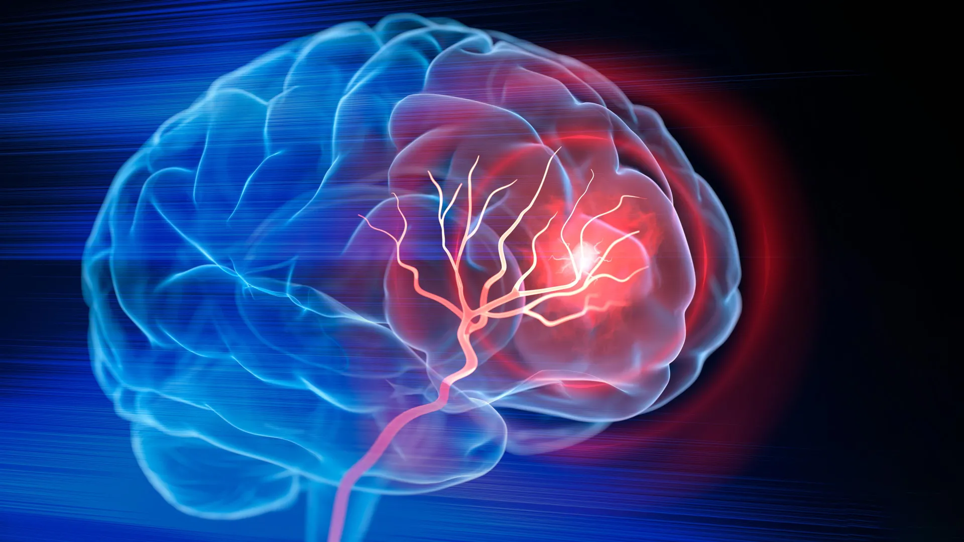
Ischemic strokes—which account for approximately 87% of all stroke cases—occur when a thrombus or embolus obstructs an artery, cutting off vital blood flow to the brain. Current standard-of-care treatments, such as the administration of tissue plasminogen activator (tPA) or mechanical thrombectomy, are highly effective at restoring blood flow if administered within the critical "golden hour" window. However, these interventions only salvage tissue that is currently oxygen-deprived but still viable.
For the millions of patients who experience significant tissue death (infarction), current medicine offers little more than physical and occupational therapy. While these rehabilitative efforts help patients compensate for lost functions by training surviving brain circuits, they do not address the physical void left behind by the stroke. This cavity serves as a physical barrier to healing, as the brain’s extracellular environment is often hostile to neurogenesis and axonal regrowth.
Engineering the Regenerative Scaffold: The MAPS Technology
The research team, led by Tatiana Segura, the Robert Plonsey Distinguished Professor of Biomedical Engineering at Duke, aimed to address this void not by replacing the tissue, but by providing a structural roadmap for the body’s own regenerative machinery. The solution lies in a material known as MAPS, or microporous annealed particle scaffolds.
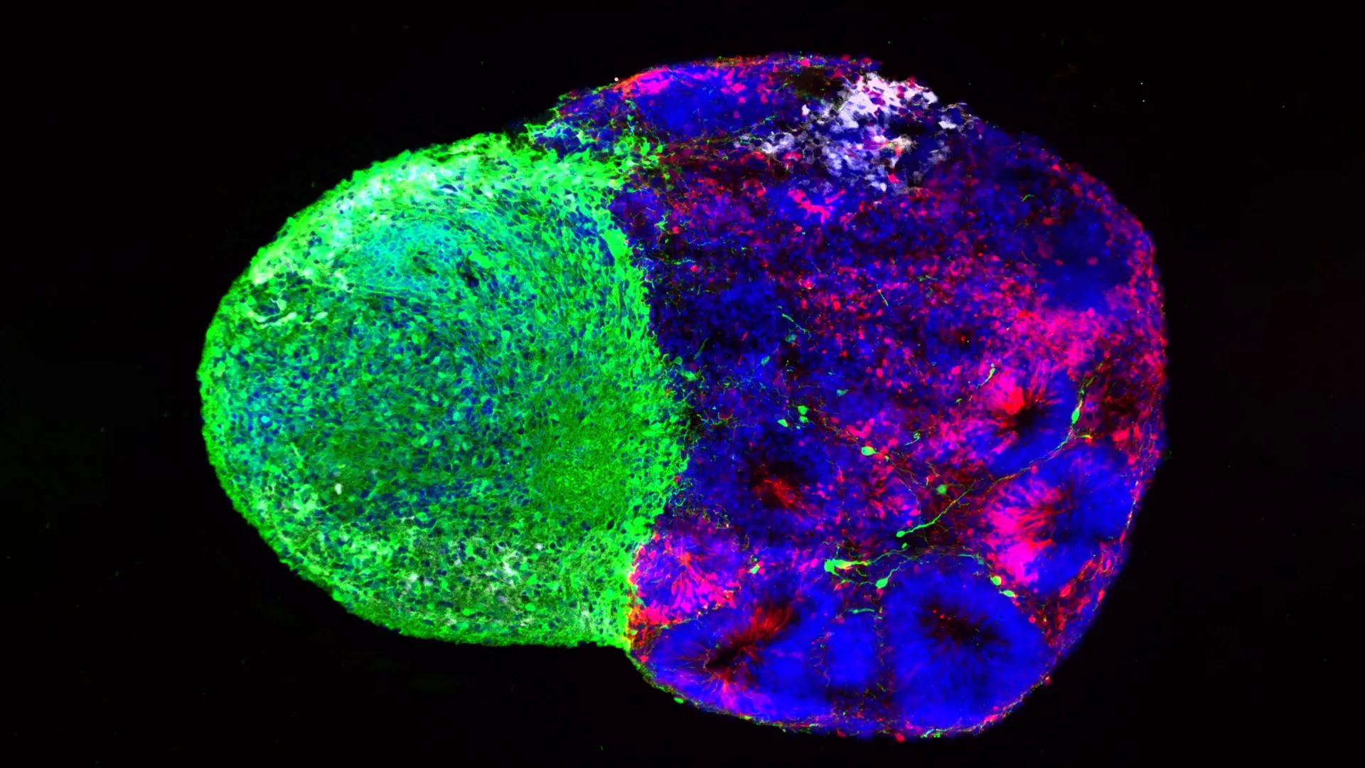
Unlike traditional solid implants, MAPS are composed of thousands of individual hydrogel microparticles that spontaneously assemble into a porous, injectable structure. This architecture is crucial; the open pores create an interconnected network that serves as a temporary scaffolding, allowing endogenous cells to migrate into the injured zone. By creating a synthetic "niche," the engineers are essentially building a bridge across the necrotic cavity, facilitating cellular migration and communication.
Harnessing the Immune System for Tissue Repair
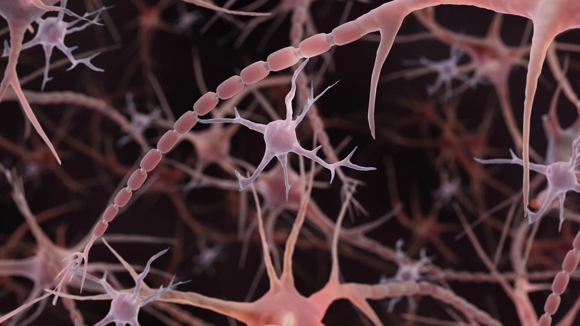
Perhaps the most significant innovation in the Duke study is the strategic use of extracellular vesicles (EVs). Astrocytes, the star-shaped glial cells that provide structural and metabolic support to neurons, are known to communicate via these small, lipid-bound packages containing proteins, lipids, and genetic material.
In the study, the researchers harvested EVs from lab-grown astrocytes and tethered them to the surface of the MAPS microparticles. This chemical immobilization ensures that the signaling molecules—specifically those designed to recruit immune cells—remain concentrated within the scaffold. This prevents the "dilution" effect that typically occurs when drugs or signals are injected freely into the brain.
The signaling combination of IL-4 (interleukin-4) and C1q was found to be particularly potent. When introduced via the scaffold, these molecules successfully recruited immune cells, including macrophages and neutrophils, into the lesion site.
Rethinking the Role of Neutrophils in Neuro-Inflammation
The study’s findings regarding neutrophils have challenged long-held neurobiological assumptions. Historically, neutrophils have been characterized as agents of chaos in the post-stroke brain, often associated with acute inflammation, oxidative stress, and secondary tissue damage.
However, the Duke team’s data suggests that the reputation of the neutrophil may be overly simplistic. By analyzing the temporal dynamics of the injury, the researchers discovered that when these cells are recruited into a supportive, engineered environment at specific post-injury stages, they appear to shift from a destructive role to a constructive one.
When the researchers depleted the neutrophil population in the experimental mice, they observed a significant decline in vascular formation and a reduction in the remodeling of the scaffold. This suggests that in the presence of the correct biochemical cues, neutrophils become essential architects of the healing process, helping to clear debris and signal for the formation of new blood vessels.
Empirical Outcomes: Motor Function and Recovery
The biological success of the scaffold was confirmed through behavioral testing. Mice treated with the optimized MAPS-EV combination showed a marked recovery in motor function compared to control groups. Using a grid-walking test—a standard metric for neurological impairment—researchers measured the frequency of forelimb placement errors.
By the eighth week post-treatment, the motor performance of the treated mice was statistically indistinguishable from that of healthy, uninjured control mice. Furthermore, these improvements were sustained for the duration of the observation period, indicating that the structural and vascular changes induced by the scaffold led to long-term functional stability.
A critical control group in the study involved injecting the EVs without the porous scaffold. The results were clear: without the MAPS framework to act as a physical anchor and concentration point for the signaling molecules, the repair process failed. This confirms that the scaffold’s architecture is not merely a delivery vehicle, but a functional component of the healing process.
Implications and the Path to Clinical Translation
While the results are highly encouraging, the transition from murine models to human clinical trials is a complex, multi-year process. Currently, the EVs used in the study are derived from primary rat astrocytes. To move toward human applications, the Segura laboratory is now focusing on EVs produced by human induced pluripotent stem cell (iPSC)-derived astrocytes. This shift not only addresses ethical and biological compatibility concerns but also provides a more scalable, reproducible source for the treatment.
Beyond the source of the EVs, researchers must address the challenges of delivery and safety. The brain is an incredibly delicate organ, and any surgical intervention carries risks of further injury or infection. Future studies will need to determine the optimal timing for the injection and assess the long-term integration of the scaffold within the complex environment of the human brain.
The implications of this research extend far beyond stroke recovery. The ability to "engineer the injured space" could theoretically be applied to other forms of brain injury, including traumatic brain injury (TBI) and neurodegenerative conditions where tissue loss occurs.
"You do not restore an ecosystem simply by containing the initial damage," Professor Segura noted in her summary of the work. "You have to create the conditions that allow life to return." By treating the stroke cavity as an environment to be managed rather than a void to be filled, the Duke University team has established a new frontier in regenerative medicine. If the results can be replicated in larger, human-scale models, this injectable scaffold could eventually provide a path for restoring the functional integrity of the brain after the most devastating of strokes.