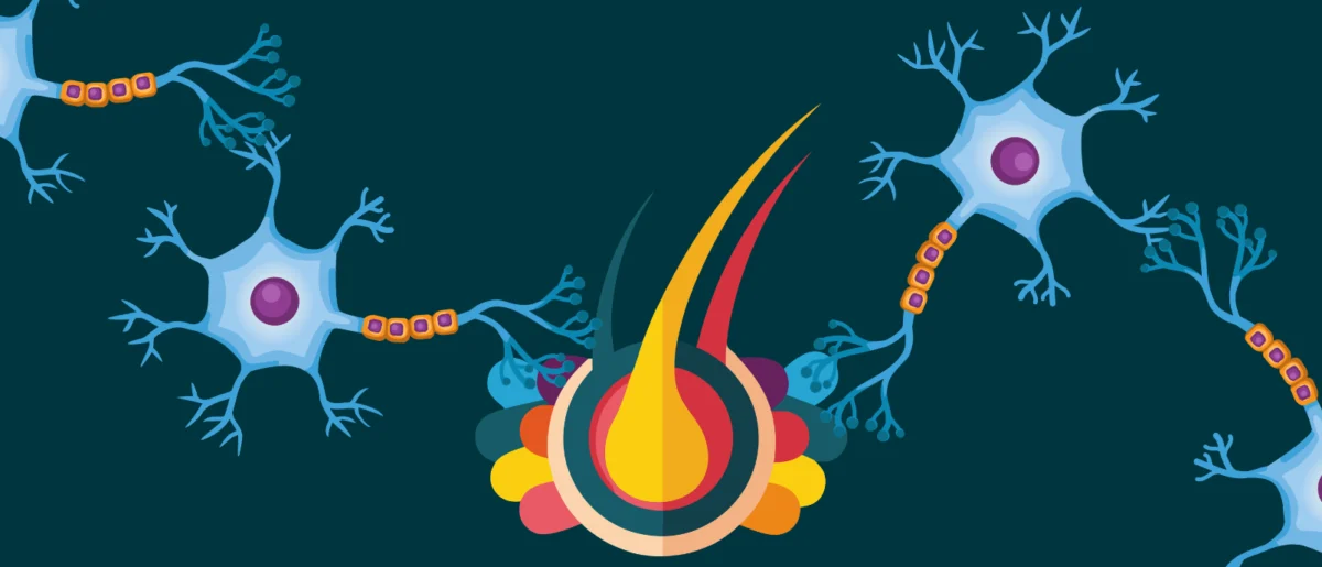
A groundbreaking discovery by researchers at Johns Hopkins University (MD, USA) has identified a previously unknown set of neurons located in an evolutionarily ancient region of the vertebrate brain that plays a critical role in directing attention by filtering out distractions. This revelation, published on June 22, 2026, challenges long-held assumptions about the neural mechanisms underlying selective spatial attention and opens promising new avenues for understanding and treating attention-related disorders such as Attention-Deficit/Hyperactivity Disorder (ADHD) and Autism Spectrum Disorder (ASD). The findings suggest that the ability to focus amidst a chaotic environment is not solely governed by the highly developed prefrontal cortex, as traditionally believed, but is also orchestrated by a fundamental, conserved brainstem circuit present across a vast array of species, from fish to humans.
Challenging the Prefrontal Cortex Paradigm
For decades, the scientific community has largely attributed the complex cognitive function of selective attention to the prefrontal cortex, a region of the brain that is particularly prominent and highly developed in humans and other primates. This area is known for its executive functions, including planning, decision-making, working memory, and inhibition. The prevailing model suggested that the prefrontal cortex acted as the brain’s "command center" for directing focus, suppressing irrelevant stimuli, and prioritizing sensory information. While its role in higher-order attentional processes remains undeniable, this new research introduces a fundamental component operating at a more ancestral level of brain organization.
The established view, however, presented a significant evolutionary conundrum. Many animals, including birds, reptiles, and fish, exhibit sophisticated attentional capabilities – the ability to hunt prey, avoid predators, or navigate complex environments – despite lacking a prefrontal cortex comparable in development to that of mammals, particularly primates. This discrepancy prompted neuroscientists to question whether a more ancient, conserved mechanism might be at play. Lead author Ninad Kothari, a postdoctoral fellow in the university’s Department of Psychological and Brain Sciences, articulated this core question: "If we really go back in evolution, for hundreds of millions of years, birds have had this ability, fish have had this ability. And they do not typically have a highly developed prefrontal cortex, so how does the brain solve this problem? We were able to identify an evolutionarily old region in the brainstem which affords this ability."
The Discovery in the Brainstem: An Evolutionary Perspective
The breakthrough emerged from research conducted by Shreesh Mysore, a neuroscientist who studies neural circuits tied to behavior, and his team at Johns Hopkins. Their investigation into the brainstem, a phylogenetically ancient part of the brain responsible for vital functions like breathing, heart rate, and sleep, revealed a circuit of inhibitory neurons critical for attention. This region, situated at the base of the brain, acts as a crucial relay station for information flowing between the brain and the body, and its involvement in such a complex cognitive process as selective attention marks a significant paradigm shift.
The impetus for investigating the brainstem in mammals stemmed from earlier comparative studies. Mysore and other scientists had previously observed remarkable attentional abilities in non-mammalian vertebrates like birds, frogs, and turtles, species whose brain structures diverge significantly from those of primates. These observations strongly hinted at the existence of a common, evolutionarily conserved neural substrate for attention that predates the sophisticated cortical expansions seen in mammals. By focusing their attention on the brainstem in mice, a mammalian model, the researchers aimed to identify whether this ancient attentional "engine" was also preserved and functional in higher vertebrates. The success of this approach not only confirmed the existence of such a circuit but also demonstrated its profound impact on attentional behavior.
Experimental Design and Compelling Findings
To precisely pinpoint the function of these newly identified neurons, the research team designed a sophisticated behavioral task for mice that mirrored human-like attention challenges. Mice were trained to focus on visual cues presented directly in front of them on a screen, while simultaneously ignoring distracting visual information appearing to the side. Successful completion of the task, indicated by the mouse touching the screen with its nose at the location signaled by the primary cue, earned them a reward. This setup allowed the researchers to quantitatively assess the mice’s ability to selectively attend to relevant information and filter out irrelevant distractions.
The critical phase of the experiment involved temporarily silencing the activity of the identified brainstem neurons. Using advanced neuroscientific techniques, likely involving optogenetics or chemogenetics which allow for precise control over neuronal activity, the researchers were able to reversibly inhibit these specific neurons. The results were stark and immediate: when these brainstem neurons were inactivated, the mice’s performance on the attention task drastically deteriorated. As Kothari reported, "When we inactivate these neurons, the mice become hyper distractible." The animals struggled to ignore the side distractions, frequently choosing the incorrect location and failing to earn rewards.

Crucially, the team conducted rigorous control experiments to rule out alternative explanations for the observed behavioral changes. They verified that silencing these neurons did not impair the mice’s basic motor movements or their ability to perceive visual objects. The only observable deficit was specifically related to their capacity for selective attention. "The only thing impaired was their ability to take the competing pieces of information, compare them, and pay attention to the location with the most important information," Mysore commented, underscoring the specificity of the neurons’ role. The profound and reversible nature of this effect—where mice became distractible when the neurons were silenced and regained their focus when the neurons were reactivated—provided compelling evidence for the direct involvement of this brainstem circuit in attentional control. Senior author Shreesh Mysore emphasized the fundamental role of this region, stating, "This part of the brain is like an attentional selection engine. It helps solve the question: ‘What is most important information I should pay attention to right now?’"
Implications for Attention Disorders: ADHD and Autism
The discovery holds immense promise for advancing our understanding and treatment of neurological conditions characterized by attentional dysregulation, such as ADHD and certain forms of ASD. ADHD, a neurodevelopmental disorder affecting millions worldwide, is defined by persistent patterns of inattention, hyperactivity, and impulsivity. A core symptom of ADHD is an increased susceptibility to distractions, even faint ones, which significantly impairs an individual’s ability to sustain focus on tasks. The experimental results in mice directly mirrored this clinical hallmark. Mysore noted, "A hallmark of ADHD is that even faint distractors draw attention away—and that’s exactly what we see here when these neurons are silenced. But the very next day, when the neurons are turned back on, the same animal can ignore distractors again, even very strong ones." This direct correlation suggests a potential common underlying mechanism.
Similarly, individuals with ASD often exhibit atypical attentional patterns, including difficulties with shifting attention, hyperfocus on specific stimuli, or an inability to filter out sensory overload, leading to significant challenges in social interaction and communication. If the identified brainstem circuit proves to be dysfunctional or improperly regulated in these human conditions, it could provide a much-needed biological target for therapeutic intervention.
Currently, treatments for ADHD primarily involve stimulant medications that increase neurotransmitter levels (like dopamine and norepinephrine) in the prefrontal cortex, alongside behavioral therapies. While effective for many, these treatments are not universally beneficial and can have side effects. The identification of a distinct, evolutionarily conserved attentional circuit could pave the way for entirely new classes of targeted pharmacological agents or even non-pharmacological interventions, such as specific neurostimulation techniques, that directly modulate the activity of these brainstem neurons. This specificity could lead to treatments with fewer side effects and greater efficacy for a broader range of patients.
Future Directions and Broader Impact
The Johns Hopkins team is now embarking on the crucial next steps of their research. A primary goal is to fully elucidate the precise mechanisms by which these brainstem neurons control spatial attention across different vertebrates. Understanding the intricate neural pathways and molecular signaling involved will be essential for translating these findings into clinical applications.
Perhaps the most exciting immediate next step is to investigate the presence and function of these neurons in humans. While "all the evidence to date suggests that these neurons exist in humans too," as Mysore concluded, their specific role in human selective spatial attention needs to be definitively established. "An exciting hypothesis is that they play a crucial role," he added. Advanced neuroimaging techniques, such as functional magnetic resonance imaging (fMRI) or magnetoencephalography (MEG), could be employed to study the activity of these deep brainstem structures during attentional tasks in human volunteers.
Furthermore, the team plans to measure the activity and structural integrity of these neurons in individuals diagnosed with ADHD and autism. If their function is indeed impaired or atypical in these populations, it would provide compelling evidence for their involvement in the pathogenesis of these disorders. This knowledge would be transformative, offering novel diagnostic biomarkers and opening the door for the development of highly targeted drugs and treatments.
Beyond clinical applications, this discovery profoundly impacts our fundamental understanding of brain evolution and the neural underpinnings of cognition. It highlights the principle of "neural reuse," where ancient brain structures, initially evolved for basic survival functions, are repurposed or integrated into more complex cognitive processes. It also underscores the importance of comparative neuroscience in unraveling universal principles of brain function that might be obscured when studying only highly specialized brains like those of primates. The brainstem, long viewed as primarily a relay and control center for autonomic functions, is now revealed as a sophisticated orchestrator of one of our most vital cognitive abilities: the power to pay attention. This groundbreaking work from Johns Hopkins University represents a pivotal moment in neuroscience, promising a future where the complexities of attention can be better understood and, ultimately, more effectively managed for those who struggle with its challenges.