Researchers at Weill Cornell Medicine have identified a single molecular switch that governs the transition of blood stem cells from a dormant state into an active, highly regenerative condition capable of replenishing the body’s entire blood and immune systems. Published in the February 25 issue of Nature Immunology, the preclinical study centers on a DNA transcription-regulating protein known as FLI-1. By demonstrating how transient activation of this protein can safely awaken hibernating stem cells, the findings offer a promising pathway to dramatically improve the efficacy, speed, and safety of bone marrow transplants and stem-cell-targeted gene therapies.
The breakthrough addresses longstanding challenges in hematology, particularly the scarcity of viable stem cells in donors and the difficulty of expanding compromised cells harvested from patients who have undergone intensive chemotherapy or radiation. As medical science increasingly leans toward sophisticated gene-editing interventions for severe blood disorders, the ability to successfully manipulate stem cells outside the body—and ensure their robust engraftment upon re-infusion—represents a critical leap forward in regenerative medicine.
Understanding the Stem Cell Lifecycle: Quiescence Versus Regeneration
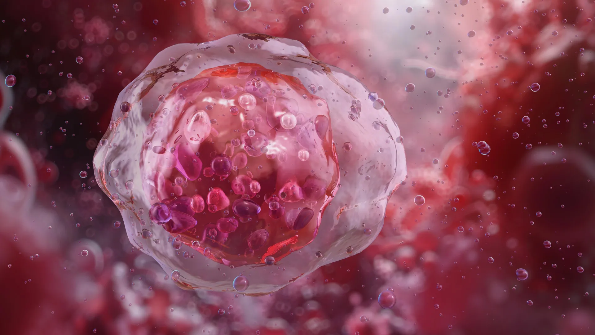
To comprehend the significance of the Weill Cornell Medicine discovery, it is essential to examine the fundamental biology of stem cells. Across virtually all mammalian tissues, stem cells act as the foundational reserve. In their natural, unperturbed environment, the vast majority of adult blood stem cells reside within the bone marrow in a state of deep quiescence, or dormancy. In this slow-dividing state, they conserve energy, protect their genomic integrity from mutative stressors, and maintain a steady, minimal output of blood cells sufficient for daily physiological maintenance.
However, when a major physiological stressor occurs—such as severe blood loss, systemic infection, tissue injury, or myeloablative conditioning prior to a transplant—these dormant cells must undergo a rapid transformation. They switch into an activated, highly proliferative state, multiplying rapidly and differentiating into mature, functional blood and immune cells.
Until now, the exact molecular triggers governing this crucial transition remained incompletely understood. While scientists knew that signals from the surrounding microenvironment—specifically the vascular niche composed of specialized endothelial cells lining blood vessels—played a role, the precise intracellular switch controlling stem cell readiness and adaptability had eluded researchers.
Uncovering the Role of FLI-1 Through Advanced Genomic Profiling
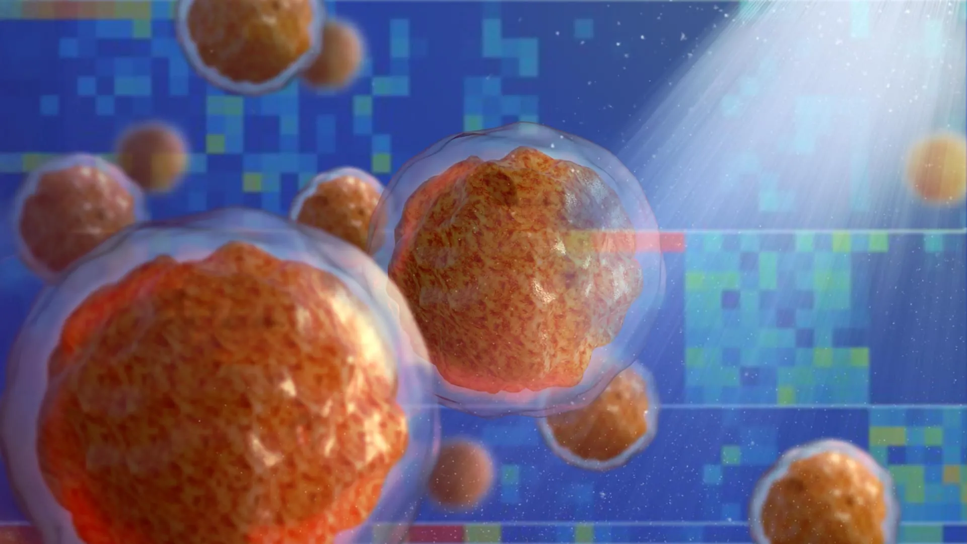
To decode the molecular mechanisms distinguishing quiescent blood stem cells from their active counterparts, the Weill Cornell research team deployed advanced single-cell profiling and high-throughput computational analyses. These techniques allowed the investigators to map differential gene activity at unprecedented resolution.
The analytical process ultimately zeroed in on FLI-1 (Friend leukemia integration 1 transcription factor), a master protein known to regulate the expression of thousands of individual genes. The investigators discovered that FLI-1 acts as the decisive molecular gatekeeper for stem cell activation. In the absence of adequate FLI-1 activity, blood stem cells remain locked in a quiescent state, largely severed from functional interactions with the surrounding bone marrow microenvironment, particularly the vascular niche.
Conversely, when FLI-1 activity is restored or stimulated, it bridges the communication gap between the stem cells and the endothelial vascular niche. This interaction re-establishes co-adaptability, prompting the stem cells to break their dormancy, expand exponentially in number, and dramatically increase their regenerative capacity.
Solving the Umbilical Cord Blood Puzzle
The discovery of FLI-1’s regulatory role also sheds light on a long-standing paradox in hematology. For decades, clinicians and researchers have observed that blood stem cells derived from human umbilical cords possess a markedly superior regenerative and engraftment potential compared to adult stem cells isolated from peripheral blood or bone marrow.
Through their comprehensive computational and biological analysis, the research team demonstrated that this enhanced potency is directly tied to naturally occurring differences in FLI-1 activity levels. Umbilical cord blood stem cells exhibit optimized baseline interactions with the regenerative vascular niche, a property mediated by this specific transcriptional pathway. By identifying the mechanics behind this natural advantage, the researchers have established a blueprint for artificially endowing adult stem cells with similar superior regenerative traits.
Overcoming Oncogenic Risks via Transient mRNA Delivery
A primary hurdle in targeting transcription factors like FLI-1 for therapeutic applications is their historical association with oncogenesis. Overactivity or genetic mutations resulting in the chronic overexpression of FLI-1 are known drivers in the pathogenesis of certain types of leukemia. Permanent genetic modifications or long-term stimulation of such a potent factor could theoretically trigger uncontrolled cellular proliferation and malignancy.
To circumvent this safety hazard, the Weill Cornell investigators engineered a novel, transient delivery method modeled after modified mRNA-based vaccine technologies. Rather than permanently altering the genome or sustaining high levels of the protein, the team utilized modified mRNA to transiently introduce FLI-1 into quiescent adult bone marrow stem cells for a window of only a few days.
This brief priming period is sufficient to coax the stem cells out of hibernation, drive their rapid expansion, and prepare them for successful transplantation, without triggering oncogenic pathways. In preclinical evaluations, stem cells primed using this temporary mRNA approach successfully and durably engrafted in recipient hosts, restoring complete blood cell production with no evidence of leukemia or abnormal tissue growth.
Clinical Implications for Transplants and Gene Therapies
The potential applications of this discovery span a wide spectrum of clinical hematology and oncology. Bone marrow transplants remain a cornerstone therapy for various forms of leukemia, lymphoma, and severe bone marrow failure syndromes. However, finding a matching donor can be exceptionally difficult, and harvested grafts often contain marginal quantities of viable blood stem cells, increasing the risk of graft failure.
Furthermore, autologous transplants—where patients receive their own previously harvested stem cells—frequently encounter hurdles when the harvested cells have been damaged or rendered stubborn by prior rounds of chemotherapy or radiation. The new mRNA priming technique could allow clinicians to rescue and robustly expand these compromised cells ex vivo prior to re-infusion.
The methodology is equally transformative for emerging gene therapies. Advanced treatments for monogenic blood disorders, such as beta-thalassemia and sickle cell disease, typically require extracting a patient’s hematopoietic stem cells, introducing a functional therapeutic gene vector into the cells within a laboratory setting, and subsequently expanding the population before returning them to the patient’s body. Because ex vivo manipulation often exhausts vulnerable stem cells, a safe, reliable molecular switch to enhance their regenerative capacity before re-infusion could significantly elevate the success rates and safety profiles of these complex genetic interventions.
Official Responses and Expert Insights
Reflecting on the broader impact of the findings, study senior author Dr. Shahin Rafii underscored the translational potential of the work. Dr. Rafii holds multiple prestigious leadership roles at Weill Cornell Medicine, serving as director of the Hartman Institute for Therapeutic Organ Regeneration and the Ansary Stem Cell Institute, chief of the division of regenerative medicine, and the Arthur B. Belfer Professor in Genetic Medicine, alongside appointments at the Englander Institute for Precision Medicine and the Sandra and Edward Meyer Cancer Center.
"The approach we outlined in this study could substantially improve the efficiency of marrow transplants and marrow-cell-targeted gene therapies, especially in cases where the donor has a very limited supply of viable blood stem cells," Dr. Rafii stated, emphasizing the urgency of translating these preclinical insights into human clinical trials.
The collaborative nature of the research was highlighted by co-first author Dr. Tomer Itkin, formerly an instructor of biology in medicine in the Rafii laboratory and currently director of Tel Aviv University’s Neufeld Cardiovascular Research Institute and assistant professor at the Sagol Center for Regenerative Medicine.
"The stem cells we prime with FLI-1 modified mRNA in this way wake up from hibernation, expand and functionally and durably engraft in the recipient host, without any evidence of cancer," Dr. Itkin noted, validating the safety profile achieved through transient administration.
Addressing the intricate signaling dynamics between stem cells and their physical environment, co-first author Sean Houghton, a senior bioinformatics analyst at the Englander Institute for Precision Medicine who served as a bioinformatics analyst in the Rafii lab during the study, emphasized the reciprocal nature of the regenerative niche.
"We showed that stem cell activity is not autonomous but also is not fully determined by endothelial cell vascular niche signals—it depends instead on signaling and adaptability between the two," Houghton explained. This nuanced understanding bridges gaps in tissue engineering by demonstrating that successful stem cell activation requires mutual biochemical dialogue rather than unilateral signaling.
Funding and Institutional Support
The rigorous multi-year investigation was made possible through substantial financial backing from several institutes within the National Institutes of Health (NIH), reflecting the high national priority placed on advancing stem cell technologies. Primary grant support was provided by the National Heart, Lung, and Blood Institute (NHLBI), the National Institute of Diabetes and Digestive and Kidney Diseases (NIDDK), and the National Institute of Allergy and Infectious Diseases (NIAID) under grant numbers R35HL150809, R01DK136327, and U01AI138329.
Additional philanthropic and institutional support was furnished by the Hartman Institute for Therapeutic Organ Regeneration, the Ansary Stem Cell Institute, and the Selma and Lawrence Ruben Daedalus Fund for Innovation at Weill Cornell Medicine. (Note: Dr. Shahin Rafii is listed as an unpaid co-founder of Angiocrine Bioscience, a biotechnology enterprise operating in a related field.)
Next Steps and Future Outlook
Building upon these successful preclinical milestones, the Weill Cornell research collective is actively engaged in scaling up and further refining their modified mRNA-based delivery protocols. The overarching objective of this ongoing development is to prepare the methodology for formal human clinical trials.
If validated in clinical settings, the ability to transiently manipulate the FLI-1 molecular switch could fundamentally alter the standard of care for patients requiring hematopoietic stem cell transplantation. By transforming scarce or exhausted stem cells into robust, highly regenerative populations capable of seamless vascular integration, medical science stands on the threshold of delivering safer, more efficient, and universally accessible treatments for an extensive array of life-threatening blood disorders and malignancies.