The Evolution of Neural Decoding
The quest to bridge the gap between biological neural signals and external reality has long been a "holy grail" of neuroscience. For decades, researchers have relied on functional Magnetic Resonance Imaging (fMRI) to map human brain activity. While effective at identifying which brain regions light up in response to specific stimuli, fMRI data is inherently limited by its reliance on blood-oxygen-level-dependent (BOLD) signals, which are relatively slow and provide low spatial resolution. These studies often required participants to view images for extended periods to gather enough data to reconstruct a single, blurry pixel-based image.
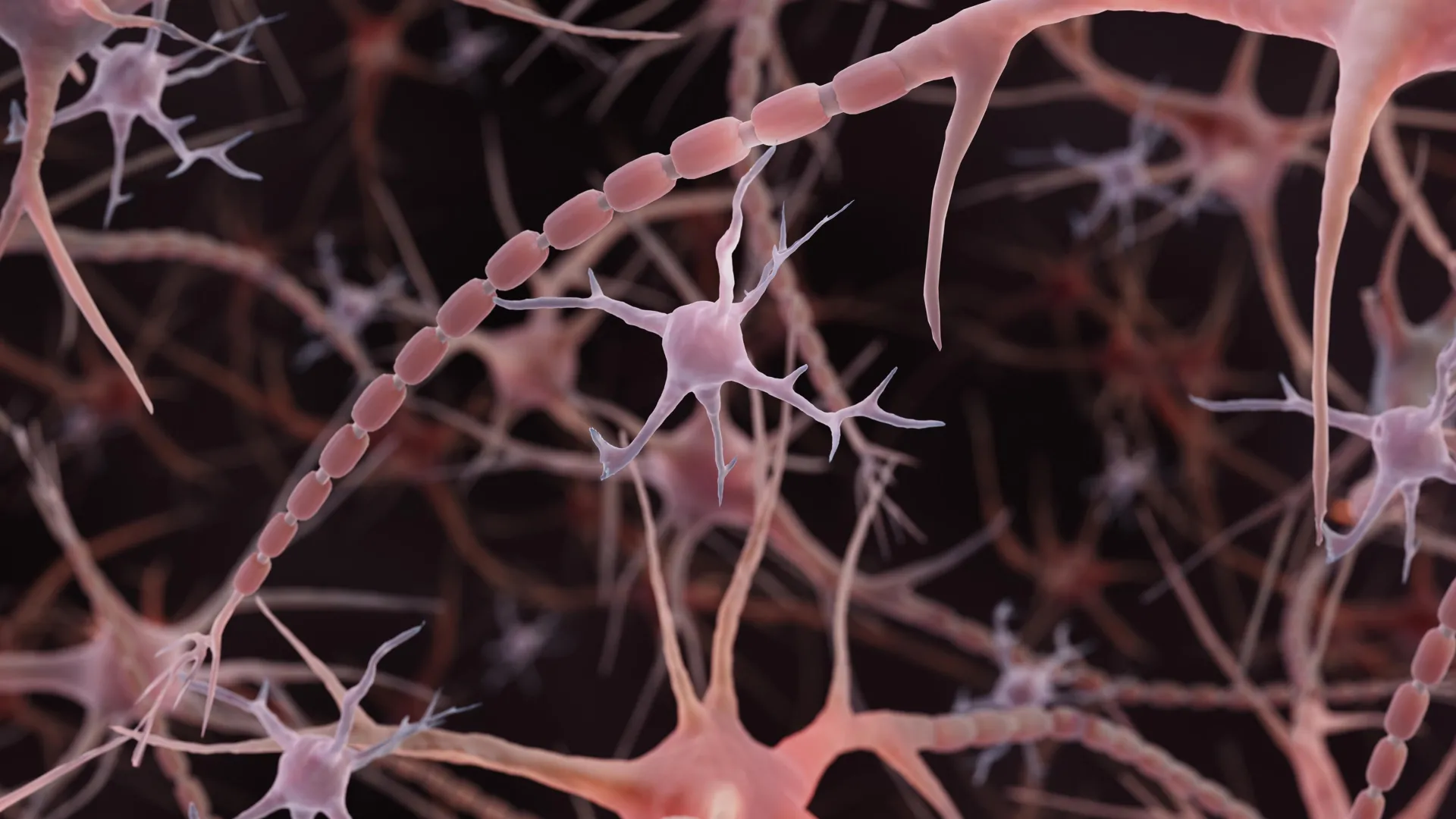
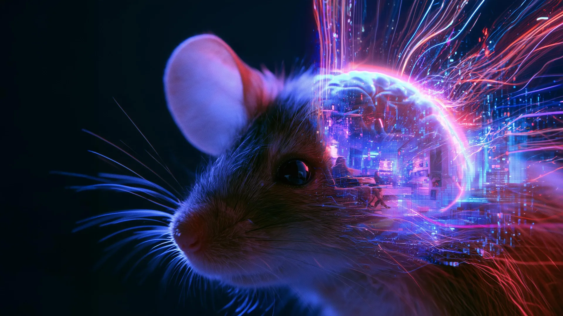
The UCL team, led by Dr. Joel Bauer at the Sainsbury Wellcome Centre, pivoted away from these broad, hemodynamic signals. Instead, they focused on single-cell recordings within the visual cortex of mice. By utilizing calcium imaging—a technique where microscopic changes in intracellular calcium levels act as a proxy for neural firing—the researchers gained access to the granular, millisecond-by-millisecond language of the brain. This methodology allows for an unprecedented level of precision, effectively "listening" to the individual neurons that translate photons into perception.
Chronology of the Breakthrough
The path to this discovery was paved by recent advancements in computational modeling and machine learning. The project’s timeline can be traced back to the 2023 Sensorium Competition, a global challenge aimed at predicting neural activity in the mouse visual cortex.
- Model Foundation (2023): Researchers utilized a dynamic neural encoding model originally designed to predict neuronal responses based on video input, animal movement, and pupil diameter.
- Refinement Phase (2023–2024): The UCL team adapted this model to function in reverse. Rather than predicting neuron activity from video, they developed an iterative algorithm that adjusted pixel values in a blank video until the predicted neural response matched the actual, recorded neural activity of the mouse.
- Validation Trial (2024): The model was tested on a "blind" dataset—a 10-second movie clip that the algorithm had never encountered during its training phase. The resulting reconstruction confirmed that the model was successfully inferring visual content rather than simply memorizing training data.
Technical Methodology: From Calcium Spikes to Cinema
The mechanism behind this reconstruction is a sophisticated iterative optimization process. The research team first established a baseline: how the mouse’s neurons behave in the absence of visual stimuli (a blank screen). By subtracting this baseline from the actual neural activity recorded while the mouse observed a movie, the researchers isolated the visual-specific signals.
The algorithm then systematically altered the pixel structure of a digital canvas. At each iteration, the model compared the predicted firing patterns of the neurons against the actual measured calcium activity. If the reconstruction was inaccurate, the algorithm adjusted the pixels to better align with the neuronal response. This loop continued until the synthetic video reflected the visual experience of the mouse with high fidelity. The inclusion of metadata, such as pupil diameter and body movement, was critical; by accounting for these variables, the researchers could filter out "noise" created by the mouse’s own physical behavior, ensuring the final video captured the external scene rather than the animal’s internal state.
Official Perspectives and Scientific Context
Dr. Joel Bauer, lead author of the study, emphasized that the goal was not merely to produce a video, but to understand the "translation" process inherent in biological vision. "The current methods of understanding what specific groups of neurons are representing are not very generalizable to situations which haven’t been specifically tested for," Bauer stated. "We wanted to develop a method that can capture what is being represented in the brain and compare that to reality."
Other neuroscientists in the field have noted that this work effectively challenges the "camera" theory of vision. If the brain were a simple recording device, the reconstructed video would be a perfect mirror of the original. However, the data reveals discrepancies, suggesting that the brain is actively curating sensory input. By highlighting specific features and filtering out others, the visual cortex creates a subjective internal representation of the world. This is not a failure of the biological system, but an adaptive feature that allows organisms to prioritize relevant environmental cues.
Quantitative Analysis and Implications
The efficacy of the reconstruction was measured using pixel correlation—a statistical metric that gauges how closely two images align on a pixel-by-pixel basis. While the current resolution shows room for improvement, the successful reconstruction of 10-second clips represents a major technical milestone. Data analysis indicated that the accuracy of the reconstructions scaled linearly with the number of neurons included in the dataset, reinforcing the theory that vision is a distributed process involving large, synchronized populations of brain cells.
The implications of this research are broad:
- Cross-Species Comparison: For the first time, researchers have a viable framework to compare how different species "see" the same environment. By running the same visual stimuli through different animal models, scientists can quantify how evolutionary history shapes sensory perception.
- Neuro-Prosthetics: While still in its infancy, the ability to decode neural visual signals could eventually inform the development of advanced neuro-prosthetics, potentially aiding in the creation of brain-computer interfaces for the visually impaired.
- Cognitive Mapping: By mapping the differences between the "input" (the movie) and the "representation" (the neural data), researchers can begin to isolate the exact areas of the brain responsible for high-level visual processing, such as motion tracking, object recognition, and depth perception.
Future Trajectories
The UCL team is already looking toward the next phase of research. The immediate objective is to increase the resolution of the reconstructed videos and expand the visual field covered by the model. Future iterations will likely incorporate more sophisticated neural network architectures capable of handling more complex, naturalistic stimuli.
Beyond technical optimization, the study invites a profound philosophical shift in how we interpret neuroscientific data. If the brain is indeed constantly "warping" reality to suit its own biological needs, then the reconstruction of neural activity is not just a technological challenge—it is a study of the architecture of consciousness. By decoding these signals, researchers are not just seeing what the mouse sees; they are beginning to uncover the hidden biases and shortcuts that define the biological experience of the world.
As this field matures, the distinction between "objective reality" and "internal representation" will likely become a primary focus of sensory neuroscience. With the ability to observe the brain in the act of interpretation, the scientific community is now better equipped than ever to ask how, and why, our minds construct the versions of reality we call our own.