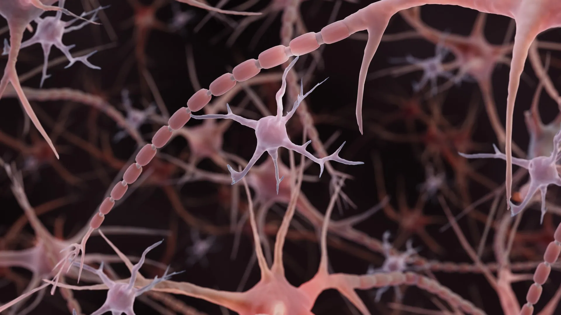
For over a century, the standard textbook illustration of a neuron has remained stubbornly consistent: a smooth, slender axon acting as a uniform conduit for electrical impulses, punctuated only by occasional, specialized synaptic boutons. This foundational image, taught to generations of medical students and neuroscientists, is now undergoing a radical revision. Recent findings from Johns Hopkins Medicine, published in the journal Nature Neuroscience in December 2024, suggest that the microscopic wiring of the brain is far more complex and dynamic than previously envisioned. Rather than static, featureless cables, many axons possess a "pearled" architecture—a repeating pattern of nanoscale bulges that are not signs of degradation, but rather integral components of normal, healthy neurological function.
A Paradigm Shift in Cellular Anatomy
The realization that axons are inherently beaded emerged from a methodological pivot in how researchers visualize brain tissue. Historically, electron microscopy required the chemical fixation and dehydration of tissue samples to withstand the vacuum of the microscope. This process, while standard, essentially turns a biological "grape" into a "raisin," potentially obscuring subtle morphological features.
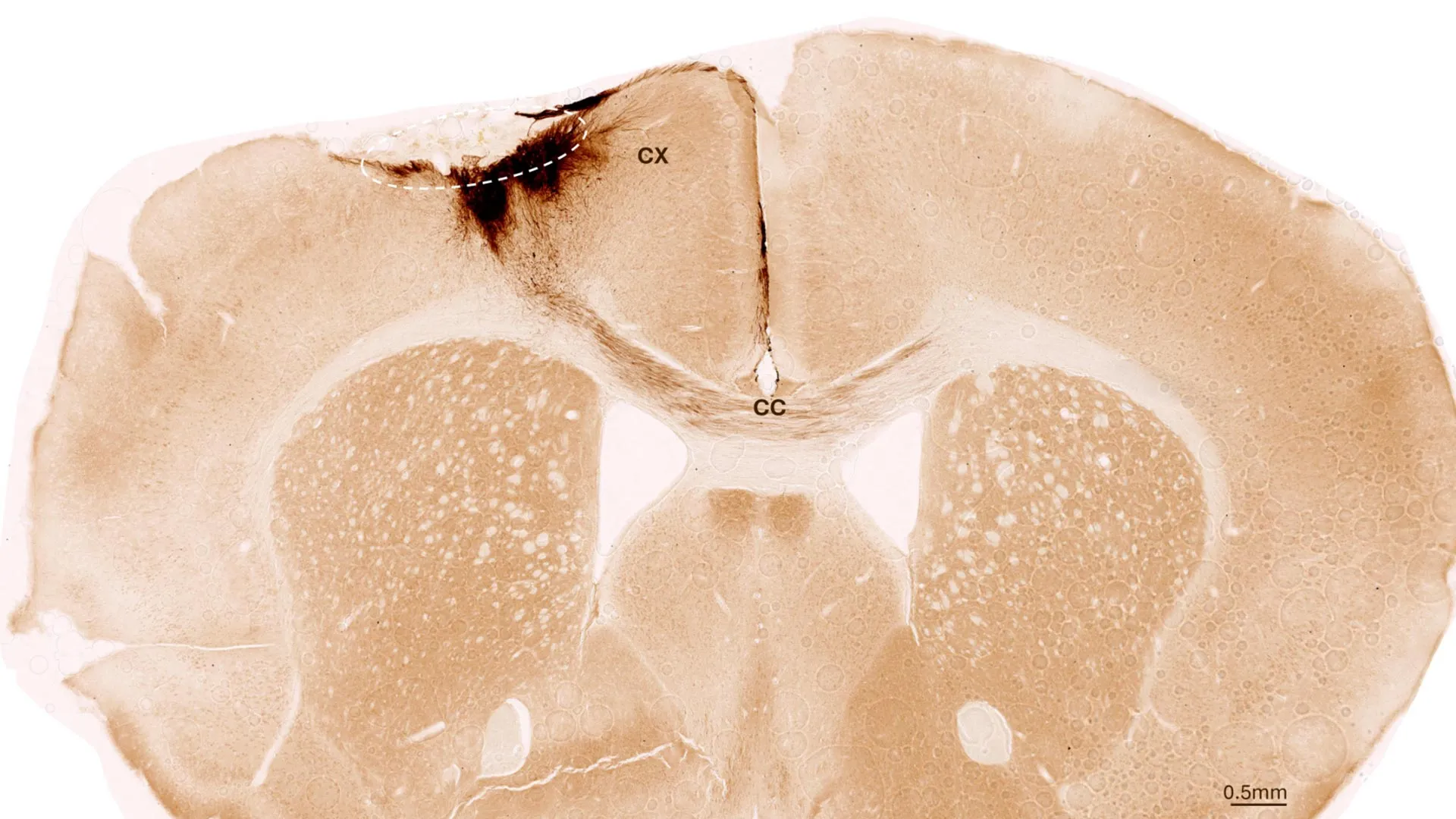
By employing high-pressure freezing—a technique that vitrifies tissue in milliseconds to preserve its near-native state—researchers at Johns Hopkins, led by associate professor of cell biology and neuroscience Shigeki Watanabe, Ph.D., observed a consistent, repeating pattern of small, non-synaptic varicosities. These structures, roughly 100 times smaller than the width of a human hair, were not the large, irregular swellings historically associated with neurodegenerative pathologies like Parkinson’s disease or axonal injury. Instead, they appeared as a uniform, structural property of the axons themselves.
This discovery challenges the traditional view of the axon as a passive wire. If these pearls are a default architectural feature, it implies that the physical geometry of the axon plays a more active, regulatory role in the conduction of electrical signals than current models account for.
The Chronology of a Discovery
The investigative path leading to this revelation was iterative, beginning with initial observations in model organisms. Watanabe’s curiosity was piqued by reports of repeating structures along the axons of worms. A collaborative dialogue with Swiss scientist Dr. Graham Knott spurred a more rigorous examination of what might underpin this morphology.
Initial hypotheses centered on the axon’s cytoskeleton, the internal protein scaffolding that provides structural integrity. However, experiments conducted by lead author and graduate student Jacqueline Griswold—which involved attempting to disrupt this internal framework—demonstrated that the pearling persisted even when the cytoskeleton was altered. This led the team to look toward the physics of the cell membrane itself. By partnering with theoretical biophysicist Dr. Padmini Rangamani of the University of California San Diego, the researchers began to treat the axon not as a rigid structure, but as a fluid-filled membrane system subject to the laws of surface tension and membrane mechanics.
The findings moved from the laboratory to the clinical setting over the following two years. In November 2025, a significant study led by Chelsy R. Eddings and colleagues—which included Watanabe—was published in the journal Neuron, confirming the presence of these pearled axons in human cortical tissue harvested during epilepsy surgeries. By 2026, the scientific community began to view this not as a curiosity limited to mice, but as a potential universal feature of neuronal signaling across species.
The Physics of Signaling: Membrane Tension and Traffic
The mathematical models developed by the research team suggest that the pearled shape is a product of membrane physics. The researchers discovered that the dimensions of these "pearls" are sensitive to the chemical environment of the neuron. When the concentration of sugar in the surrounding solution was increased, the bulges shrank. Similarly, increasing membrane tension resulted in a reduction of the pearl size.
Perhaps most critically, the team identified cholesterol as a key variable in this structural integrity. Cholesterol acts as a modulator of membrane stiffness; removing it made the membrane more fluid, which in turn altered the pearled structure and hindered the velocity of electrical signals. This phenomenon points to a "traffic" model of neural signaling. According to Watanabe, the "wide spaces" within the beaded axon may function as zones that allow ions to flow with less resistance, preventing the signal degradation that could occur in a narrower, more constricted tube.
This is not a static benefit, however. The researchers found that after high-frequency electrical stimulation, the pearls expanded in both length and width by roughly 8% and 17%, respectively. This change persisted for at least 30 minutes. Consequently, the speed of electrical transmission slowed following this period of intense activity. This suggests that the axon is an adaptive, "smart" cable that can physically reshape itself in response to the volume of traffic it carries, essentially acting as a biological rheostat for neural communication.
Implications for Neurodegenerative Research
The distinction between "normal" pearling and "pathological" beading is now a critical frontier in neuroscience. For decades, the presence of beads on an axon was treated as a diagnostic red flag for cell death, membrane rupture, or the breakdown of internal transport mechanisms. The Johns Hopkins findings necessitate a more nuanced diagnostic framework.
Distinguishing between healthy, functional varicosities and those that signal neurodegeneration is essential for understanding the progression of diseases like Alzheimer’s, Parkinson’s, and Amyotrophic Lateral Sclerosis (ALS). If clinicians cannot differentiate between the two, they may misinterpret structural adaptations as signs of irreparable damage. Conversely, understanding the mechanisms that maintain healthy pearling—such as the role of cholesterol and membrane tension—could open new therapeutic pathways for diseases where axonal transport is compromised.
Expanding the Scope: Multi-Institutional Efforts
The broader implications of these findings have attracted significant interest from the National Institute of Mental Health (NIMH), which has awarded a Multiple Principal Investigator grant to Watanabe and Rangamani. This funding is dedicated to developing sophisticated computational models that predict how a neuron’s physical properties dictate its signaling efficiency.
Parallel efforts outside of the Johns Hopkins group are further validating the importance of axon geometry. In July 2026, researchers at the University of Edinburgh utilized high-throughput automated imaging in zebrafish to screen nearly 900 chemical compounds for their ability to alter axon diameter. Their identification of 33 compounds that could effectively modify the width of these extensions provides a new toolkit for researchers. While these studies are distinct from the pearling research, they converge on a central theme: the diameter and physical shape of an axon are not fixed, but are instead variables that the brain uses to tune its own processing speed.
A New Chapter for Neuroscience Textbooks
The shift from viewing the axon as a passive, uniform conduit to a dynamic, pearled membrane structure represents a significant maturation of our understanding of brain physiology. It moves the conversation beyond mere synaptic activity—the communication between neurons—and toward the importance of the physical infrastructure that connects them.
As the scientific community continues to map these structures in human tissue, the focus will likely turn toward how this architecture varies across different brain regions and developmental stages. If the brain’s wiring is indeed a series of pearls that shift in response to activity, it suggests that "learning" or "memory" may have a structural component at the level of the axon that we are only beginning to quantify.
The work of Watanabe and his colleagues serves as a reminder that even in the most fundamental aspects of biology, long-held assumptions are subject to the rigors of modern, high-precision observation. The "smooth tube" model of the axon, while sufficient for the last century of neuroscience, is now being replaced by a more complex, rhythmic, and adaptive vision of the brain’s internal network. Future research will determine whether these nanoscale pearls are the key to understanding the speed, efficiency, and vulnerability of the human mind, but for now, they have firmly established that the brain’s wiring is as dynamic as the thoughts it carries.