Groundbreaking collaborative research from the University of Michigan and the University of California San Diego has uncovered a critical biological mechanism that explains how estrogen receptor-positive (ER-positive) breast cancer cells manage to survive targeted therapies for years or even decades in the human body. Published in the Journal of Clinical Investigation under the title "Breast cancers that disseminate to bone marrow acquire aggressive phenotypes through CX43-related tumor-stroma tunnels," the study details how dormant cancer cells exploit healthy bone marrow cells to evade destruction, adapt to drug treatments, and ultimately trigger devastating relapses.
The findings offer a profound shift in how oncologists understand cancer dormancy and metastasis. By mapping the microscopic channels that connect cancer cells to healthy tissue, the research team has not only identified a primary culprit behind late-stage breast cancer recurrence but also highlighted novel therapeutic targets that could one day prevent these deadly resurgences.
Understanding the Scope of ER-Positive Breast Cancer Relapse
Estrogen receptor-positive breast cancer represents the most prevalent subtype of the disease, accounting for approximately 70 to 80 percent of all breast cancer diagnoses. While standard treatments—including surgery, radiation, and hormone-blocking therapies such as Tamoxifen or aromatase inhibitors—achieve high initial success rates, the threat of long-term recurrence remains a persistent shadow for millions of survivors.
Clinical data indicates that approximately 40 percent of patients diagnosed with ER-positive breast cancer will experience a recurrence. What makes this statistic particularly alarming is the timeline involved. Unlike aggressive triple-negative breast cancers that typically recur within the first two to three years post-treatment, ER-positive cancers possess a unique capability to enter a state of prolonged dormancy. Disseminated tumor cells can lie undetected in the microenvironment of the bone marrow for five, ten, or even fifteen years after a patient has been declared in remission.
When these dormant "sleeper cells" finally reawaken, the resulting secondary disease is rarely benign. Recurrences often manifest as aggressive bone cancer, causing severe structural damage, debilitating bone fractures, and dangerous medical emergencies such as hypercalcemia, a condition marked by abnormally high calcium levels in the blood. Furthermore, these reactivated cells frequently spread beyond the skeletal system to vital organs including the liver, lungs, and brain. At this metastatic stage, current medical science can manage symptoms and extend life, but the disease is widely considered incurable.
The Biological Smuggling Ring: How Cancer Cells Exploit Bone Marrow
To address this enduring clinical challenge, the research teams led by Dr. Gary Luker at the University of Michigan and Dr. Pradipta Ghosh at the University of California San Diego set out to investigate the ecological interactions between disseminated breast cancer cells and the native cells of the bone marrow microenvironment.
For years, scientists recognized that the bone marrow acts as a sanctuary for tumor cells, shielding them from circulating chemotherapeutic drugs. However, the exact mechanics of how these cells sustained themselves over extended periods without a blood supply or active replication remained poorly understood.
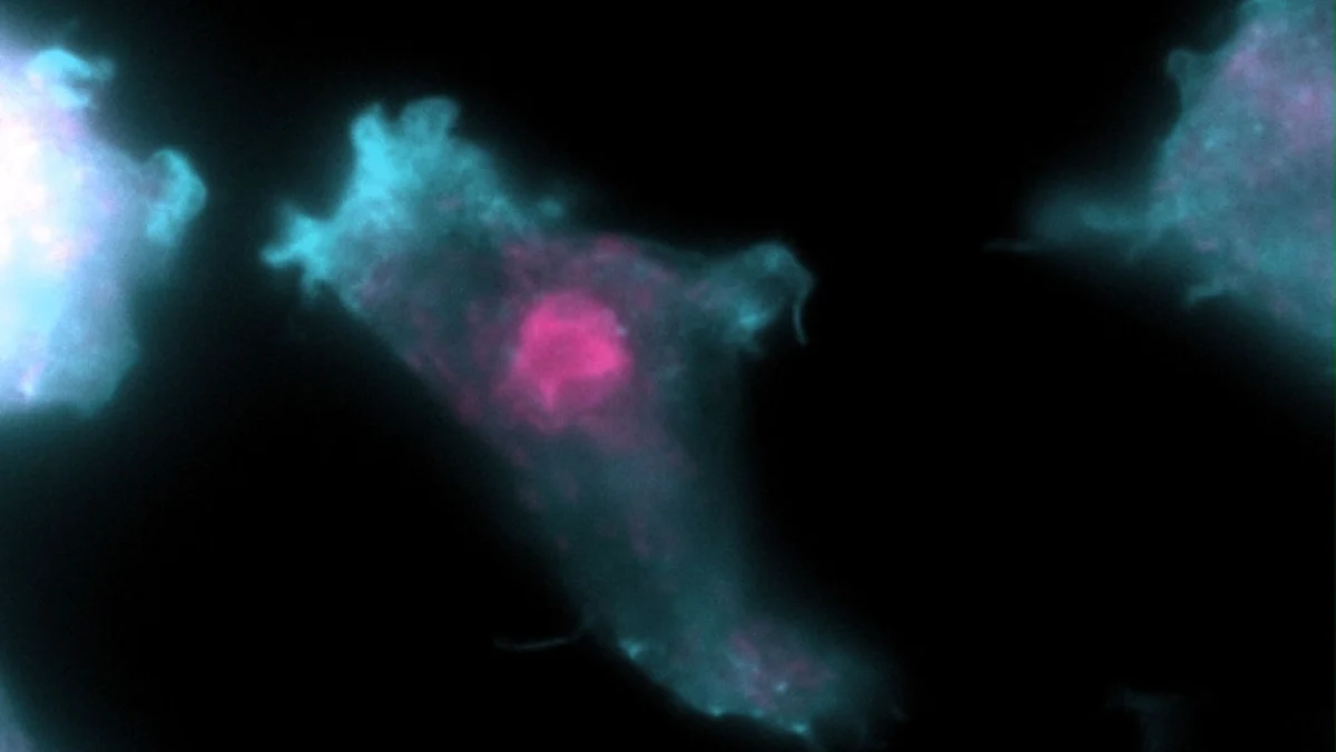
The breakthrough came when investigators discovered that surviving breast cancer cells establish physical, microscopic connections with mesenchymal stem cells—a type of adult stem cell found in bone marrow that normally plays a crucial role in repairing skeletal and connective tissues. Through specialized cellular tunnels facilitated by connexin-43 (CX43), a protein that forms gap junctions between cells, the cancer cells forge a direct pipeline to their healthy neighbors.
"We discovered that the breast cancer cells require direct contact with mesenchymal stem cells," explained Dr. Gary Luker, head of the Luker Lab within the Center for Molecular Imaging at the University of Michigan and senior author on the study. "The cancer cells physically borrow molecules—proteins, messenger RNA—directly from the mesenchymal stem cells. Essentially the mesenchymal stem cells act as very generous neighbors in donating things that make the cancer cells more aggressive and drug resistant."
This illicit transfer functions much like a biological smuggling ring. By siphoning essential structural and functional proteins from the stem cells, the dormant cancer cells bypass the metabolic energy and genetic mutations typically required to develop drug resistance. Instead of evolving resistance independently, the cancer cells simply acquire it from their microenvironment, altering the expression profiles of hundreds of proteins simultaneously.
The Central Role of GIV in Chemoresistance
Through comprehensive laboratory experiments and proteomic analyses, the research team sought to pinpoint which specific transferred or induced proteins were most critical for the survival and adaptation of the breast cancer cells. Their investigation zeroed in on GIV, also known as Girdin, a multifaceted protein heavily implicated in cancer progression.
Existing oncological literature establishes that GIV acts as a master regulator of cellular signaling pathways associated with tumor cell migration, invasion, and resistance to chemotherapy. When breast cancer cells engage in cross-talk with mesenchymal stem cells via CX43 tunnels, the expression and activity of GIV surge.
This upregulation of GIV specifically hardens the cancer cells against estrogen-targeted therapies. Drugs like Tamoxifen are designed to starve ER-positive cancer cells of the estrogen signals they need to proliferate. However, the acquisition of GIV pathways provides the cancer cells with alternative survival signals, allowing them to remain viable even in the presence of therapeutic agents. Consequently, when a patient ceases hormone therapy after the standard five-year regimen, these drug-tolerant sleeper cells remain fully equipped to wake up and drive a lethal relapse.
Implications for Future Cancer Therapeutics
The publication of this study marks a significant milestone in translational oncology, shifting the research focus from attacking the primary tumor to dismantling the supportive microenvironment that harbors metastatic cells.
Dr. Pradipta Ghosh, a professor in the Departments of Medicine and Cellular and Molecular Medicine at the UC San Diego School of Medicine and a co-author of the study, emphasized the clinical urgency of these findings. "Sleeper cells can be reawakened and cause estrogen receptor positive breast cancers to relapse years—in some cases as long as a decade—after patients were believed to be in remission," Dr. Ghosh noted.
Because the survival of these disseminated cells depends entirely on the physical bridges they build with mesenchymal stem cells, researchers now possess a highly specific therapeutic vulnerability to target. By developing pharmacological agents designed to block CX43-related tumor-stroma tunnels or inhibit the specific smuggled proteins like GIV, future therapies could effectively cut off the lifeline keeping dormant cancer cells alive.
Medical Oncologists and Industry Response
The broader oncology community has greeted the findings with cautious optimism, viewing the research as a crucial roadmap for preventing metastatic disease rather than merely treating it after the fact. While current clinical trials frequently test new drugs on advanced, symptomatic metastases, this study underscores the necessity of intercepting cancer cells during their dormant, pre-symptomatic phase in the bone marrow.
Pharmaceutical researchers are already evaluating the feasibility of repurposing existing gap-junction inhibitors or developing novel biologics that specifically disrupt cell-to-cell communication between tumors and stroma. However, experts caution that translating these laboratory discoveries into approved clinical treatments will require extensive pre-clinical safety testing and phased clinical trials. Because mesenchymal stem cells perform vital regenerative functions in healthy tissue, any targeted therapy must selectively disrupt the tunnels utilized by cancer cells without harming the normal physiological roles of bone marrow stem cells.
Broader Impact on Cancer Research and Patient Outcomes
The implications of the U-M and UC San Diego study extend far beyond breast cancer. Many epithelial cancers, including prostate, lung, and multiple myeloma, frequently metastasize to the bone marrow and exploit similar stromal niches to achieve dormancy and drug resistance. The identification of CX43-mediated tunneling as a universal mechanism of microenvironmental parasitism may provide a foundational framework for studying recurrence across a broad spectrum of oncological diseases.
For the hundreds of thousands of individuals living with a history of ER-positive breast cancer, the research offers a clearer understanding of a disease process that has historically felt unpredictable and terrifying. By shedding light on the hidden biology of bone marrow dormancy, science is steadily closing the escape routes that cancer cells have used for decades to evade detection.
As the research teams advance toward the next phases of drug discovery, the ultimate goal remains clear: to transform a late-stage, incurable relapse into a preventable medical event, ensuring that patients who achieve remission remain cancer-free for life.