For decades, the foundational principles of cellular biology have taught a consistent, unwavering lesson: glucose is the primary cellular battery. As the principal source of metabolic energy across nearly all living eukaryotic cells, this simple sugar was understood to exist for one fundamental purpose—to be catabolized, broken down through glycolysis and oxidative phosphorylation, and stripped of its chemical bonds to release adenosine triphosphate (ATP), the currency of cellular power. However, a landmark study conducted by researchers at Stanford Medicine has shattered this long-standing biochemical dogma. According to findings published in the journal Cell Stem Cell, glucose plays a profound, previously unrecognized dual role as a master regulator of tissue differentiation, governing the complex process by which uncommitted stem cells transform into the specialized cell types that constitute the tissues and organs of the human body.
This paradigm-shifting discovery reveals that glucose achieves this regulatory feat not by being burned for energy, but rather by interacting directly in its intact form with hundreds of intracellular proteins. By binding to these targets, glucose actively modulates their function and influences which genes are transcribed into proteins and when. The implications of this revelation stretch far beyond basic cell biology, offering fresh avenues for understanding metabolic disorders such as diabetes, where blood glucose levels are chronically elevated, and oncology, where malignancies are frequently characterized by a failure of cells to properly differentiate.
The Serendipitous Road to Discovery
The path to uncovering glucose’s undercover double life was as unexpected to the researchers as the results themselves. When senior author Paul Khavari, MD, PhD, chair of dermatology and the Carl J. Herzog Professor in Medicine, and lead author Vanessa Lopez-Pajares, PhD, a research scientist in dermatology, initiated their investigation, glucose was not even on their radar.
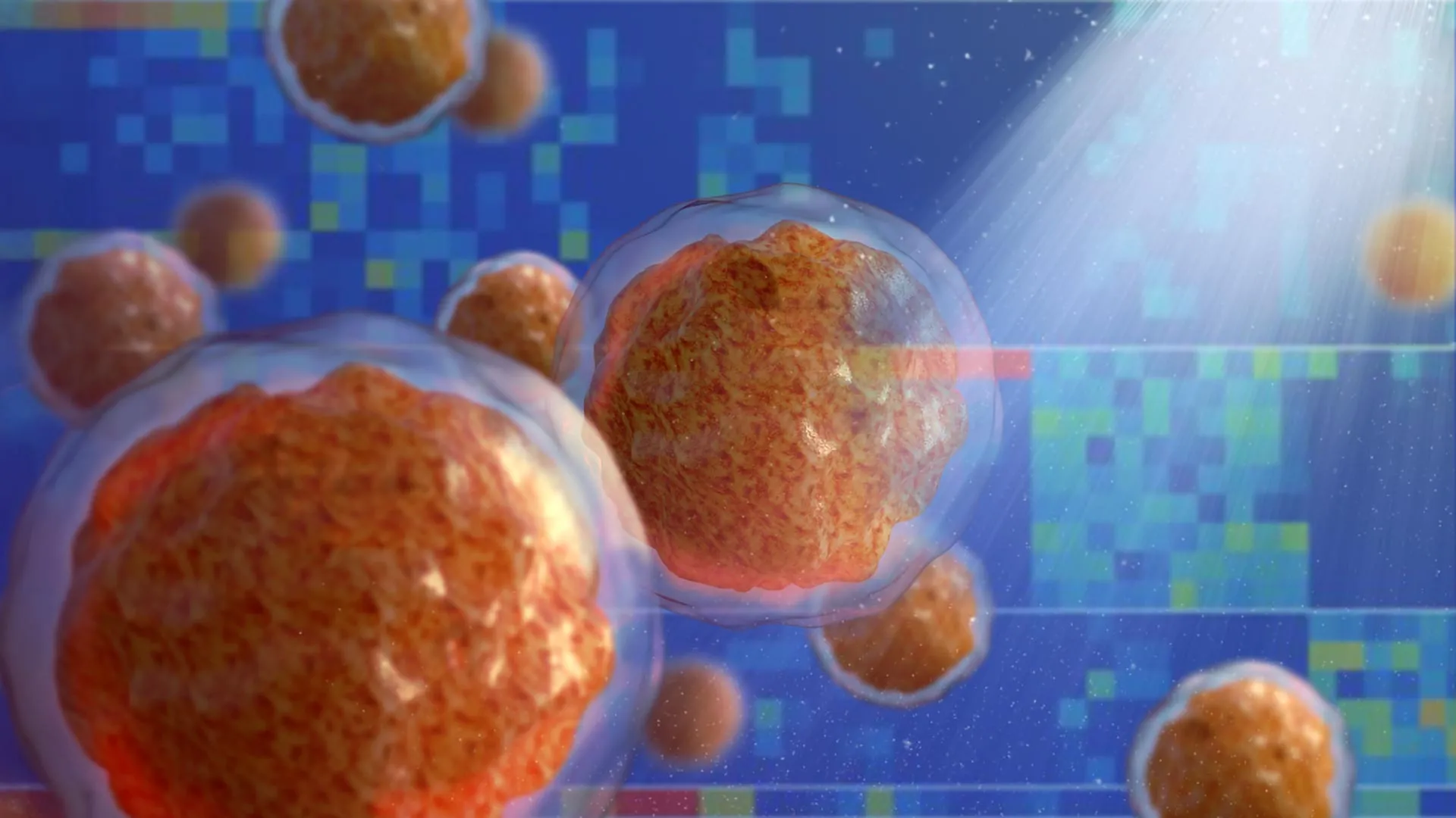
The research team set out to identify the molecular drivers of cellular differentiation using human skin stem cells. Their experimental design paired advanced mass spectrometry with high-throughput screening technologies to track the abundance of thousands of distinct biomolecules over time. Specifically, they monitored human epidermal stem cells as they naturally transitioned into mature keratinocytes—the primary cell type comprising the stratum corneum and outermost layers of human skin. Their working hypothesis was straightforward: molecules that increased significantly in abundance during this developmental window likely played a causal role in driving the cellular transition.
As the data compiled, the researchers identified 193 suspect molecules. Many of these were already known to science as regulators of cellular differentiation. However, it was the identity of the second-most elevated molecule that brought the laboratory work to a standstill.
"When we saw glucose at the top of that list, we were stunned," Khavari recalled. "We had expected glucose levels to decrease during differentiation because the cells begin to divide less rapidly, and their energy requirements are less. They are on the path to senescence and death. Yet glucose levels in the cells increase significantly as they move from epidermal stem cells to differentiated keratinocytes."
So counterintuitive was the finding that the research team spent several years validating and re-validating their data before they felt confident enough to publish. "At first we just didn’t believe it," Khavari admitted. "Pero the results of extensive follow-up experiments were clear: Glucose interacts with hundreds of proteins throughout the cell and modulates their function to promote differentiation."
Chronology of Validation and Cross-Tissue Confirmation
Following the initial mass spectrometry screen, the Stanford team embarked on a rigorous multi-year campaign to confirm the phenomenon across multiple experimental models and biological systems.
To ensure the rising glucose levels were not an artifact of their initial cell culture protocol, the researchers deployed fluorescent and radioactive glucose analogs. They also integrated biological sensors designed to emit green or red luminescence in the presence of physiologically relevant glucose concentrations. As cellular differentiation progressed in the human skin samples, these internal sensors glowed with increasing intensity, providing visual confirmation of the sugar’s accumulation.
Seeking to determine whether this was a localized quirk of human keratinocytes or a universal biological principle, the investigators expanded their scope. They examined developing fat cells (adipocytes), bone cells (osteocytes), and white blood cells (leukocytes). Furthermore, they utilized genetically engineered murine models expressing fluorescent glucose sensors in vivo. Across every single tissue examined, the results were consistent: intracellular glucose levels rose systematically as cells underwent differentiation.
Subsequent metabolic assays revealed that this intracellular accumulation was driven by a dual mechanism—an upregulation of glucose import proteins combined with a simultaneous downregulation of glucose export. Crucially, metabolic tracing confirmed that this massive influx of sugar was not accompanied by an increase in glycolysis or downstream metabolic catabolism. The glucose was accumulating not to be consumed as fuel, but to act locally.
To test the functional necessity of this sugar, the team utilized human skin organoids—sophisticated three-dimensional engineered skin tissues grown in liquid media that accurately mirror the cellular architecture and physiological organization of native human skin. When cultured in environments with abnormally low glucose levels, the organoids suffered a catastrophic failure in differentiation. Transcriptomic profiling revealed that the expression of more than 3,000 genes was altered under these low-glucose conditions, with many of the impacted genes directly responsible for skin maturation.
Remarkably, when these glucose-starved organoids were supplemented with non-metabolizable glucose analogs—sugars structurally identical to glucose that the cell’s metabolic machinery cannot break down—differentiation resumed normally. These analogs supported cellular development just as effectively as standard glucose.
"That was really the biggest shock," Khavari noted, "because we were stuck in the mindset that glucose is an energy source and nothing else. But these glucose analogs support differentiation just as well as regular glucose."
Mechanism of Action: The Cellular Broadcast Signal
With the physiological role established, the researchers dove deeper into the intracellular machinery to map out precisely how intact glucose exerts its regulatory influence.
Lopez-Pajares, Khavari, and their colleagues discovered that the localized surge in intracellular glucose is facilitated by an increased production of specific membrane transport proteins that ferry the sugar across the plasma membrane. Once inside the cytoplasm and nucleus, glucose does not drift aimlessly; it engages directly with hundreds of distinct proteins.
Among these interacting proteins is IRF6 (Interferon Regulatory Factor 6), a transcription factor known to play a critical role in tissue development and skin barrier formation. When intact glucose molecules bind to IRF6 and other target proteins, they induce a conformational change—a physical alteration in the protein’s three-dimensional shape. This structural shift modifies the protein’s functional capacity, specifically enhancing its ability to bind to DNA sequences and upregulate the expression of genes tied to differentiation.
Khavari likened this sweeping molecular interaction to an emergency warning system rather than a targeted digital message. "We’re seeing glucose acting like a broadcast signal in the cell, in contrast to the highly specific signaling cascades that drive many cellular functions," he explained. "When glucose levels rise in a cell, they rise everywhere, all at once. It’s like a fire alarm going off in a firehouse. Everyone in the firehouse activates in response."
Broader Implications for Diabetes and Oncology
The discovery of glucose’s non-metabolic signaling role provides a compelling mechanistic framework for several long-standing clinical observations that previously lacked clear molecular explanations.
For decades, clinicians have observed that embryonic stem cells—which possess the remarkable pluripotency to differentiate into any cell type in the human body—lose this developmental plasticity and maintain an undifferentiated, proliferative state when cultured in environments with elevated glucose concentrations. Conversely, patients suffering from diabetes mellitus, who experience chronic hyperglycemia, frequently exhibit severely impaired wound healing, delayed tissue regeneration, and compromised epithelial barrier repair. The new Stanford findings suggest that chronic elevations in circulating sugar may chronically saturate or disrupt the delicate signaling thresholds required for orderly cellular differentiation in healing tissues.
Furthermore, the research holds profound implications for the field of cancer therapeutics. Malignancies are fundamentally diseases characterized by a blockade in cellular differentiation, wherein immature, highly proliferative cells fail to mature and instead continue to divide uncontrollably. Interestingly, various non-metabolizable glucose analogs have previously shown therapeutic promise in preclinical and clinical trials as potential anticancer treatments. While these compounds were originally developed under the assumption that they would starve cancer cells by blocking energy metabolism, the Stanford study suggests an alternative or complementary mechanism: these analogs may act as differentiation agents, forcing malignant, undifferentiated cancer cells to mature, exit the cell cycle, and halt their rampant proliferation.
Future Horizons in Metabolic Research
As the scientific community digests these findings, the Stanford team is already looking toward the next phase of investigation. Researchers plan to explore how this newly identified signaling pathway operates across a wider array of diseased and healthy tissues, mapping out the precise protein interactome responsive to glucose-induced conformational changes.
"This finding is a springboard for research on dysregulation of glucose levels, which affects hundreds of millions of people," Khavari stated. "But it’s also likely to be important in cancer development because cancer is a disease of failed differentiation. This is an entirely new and growing field. People have thought that small biomolecules like glucose were quite passive in the cell. This is another piece of evidence to pay close attention to other roles these molecules might play."
The study was supported by grants from the National Institutes of Health (R01AR043799, AR045192, K01AR070895, and P30CA124435) alongside funding from the U.S. Department of Veterans Affairs Office of Research and Development. By demonstrating that one of biology’s most ubiquitous molecules possesses a hidden life as a master developmental architect, this research marks the opening chapter of a fundamentally new textbook in cellular and molecular biology.