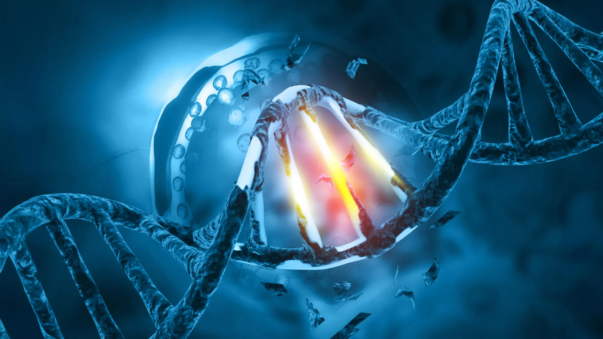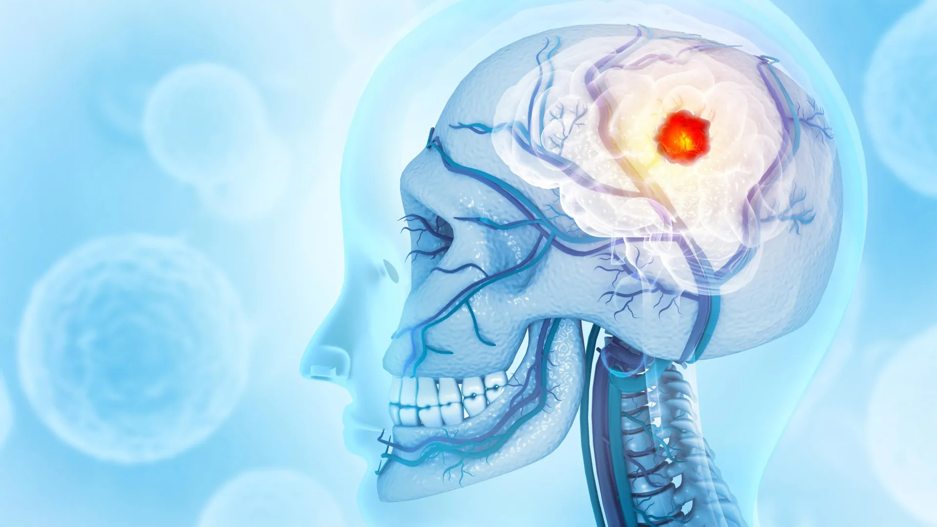
Researchers at the Montefiore Einstein Comprehensive Cancer Center (MECCC) and the Albert Einstein College of Medicine have fundamentally shifted the medical community’s understanding of glioblastoma—the most aggressive and lethal form of brain cancer—by proving that its destructive reach extends far beyond localized brain tissue. Published in the October 3 issue of the prestigious journal Nature Neuroscience, a comprehensive new study reveals that glioblastoma actively erodes the skull, alters the cellular composition of the bone marrow encased within it, and systematically disrupts the body’s broader immune defense mechanisms. Furthermore, the findings sound an alarm regarding standard osteoporosis medications, demonstrating that certain drugs prescribed to prevent bone loss can inadvertently accelerate tumor growth and neutralize the efficacy of modern immunotherapies.
This paradigm-shifting discovery introduces an entirely new lens through which oncologists must view neuro-oncology. Historically treated as an isolated, localized disease contained entirely within the cranium, glioblastoma is now revealed to be a systemic condition characterized by complex biochemical cross-talk between the brain tumor and the skeletal and immune systems. Experts believe this systemic interaction explains why conventional therapies—which have long focused exclusively on surgical resection, focal radiation, and localized chemotherapy—have historically yielded such dismal long-term survival statistics.
The Grim Statistics and Limitations of Current Care
Glioblastoma remains one of the most terrifying diagnoses in modern medicine. According to data compiled by the National Cancer Institute (NCI), approximately 15,000 individuals are diagnosed with glioblastoma in the United States each year. Despite aggressive intervention involving maximal safe surgical resection followed by concurrent temozolomide chemotherapy and localized radiation therapy—a regimen established nearly two decades ago—the prognosis remains starkly unfavorable. The median survival time for patients undergoing this current standard of care hovers at a mere 15 months, with five-year survival rates remaining in the single digits.
For decades, neuro-oncologists have grappled with the inevitability of recurrence. Even when surgeons successfully remove the primary tumor mass, microscopic cancer cells invariably infiltrate surrounding healthy brain tissue, rendering recurrence nearly universal. The revelation by the MECCC and Einstein research teams that the tumor actively remodels the cranial bone and co-opts the skull’s bone marrow provides a compelling biological explanation for this treatment resistance. By manipulating the body’s immune factories directly adjacent to the brain, glioblastoma creates a protected, self-sustaining microenvironment that thwarts standard therapeutic approaches.
Unraveling the Cranial Connection: A Matter of Marrow
The genesis of this breakthrough research lies in a fundamental anatomical reassessment of the skull. Traditionally viewed merely as a protective helmet for the brain, the skull—like the femur, pelvis, and sternum—contains active bone marrow responsible for generating vital immune and blood cells. Recent anatomical discoveries have demonstrated that the skull and the brain are physically interconnected via a network of extremely thin, microscopic vascular and lymphatic channels. These channels facilitate a continuous molecular and cellular exchange between the cranial bone marrow and the central nervous system.
Capitalizing on these insights, the research team—led by corresponding author Dr. Jinan Behnan, assistant professor in the Leo M. Davidoff Department of Neurological Surgery and the Department of Microbiology & Immunology at Einstein, and a member of MECCC—embarked on a rigorous investigation using advanced, high-resolution imaging technologies.
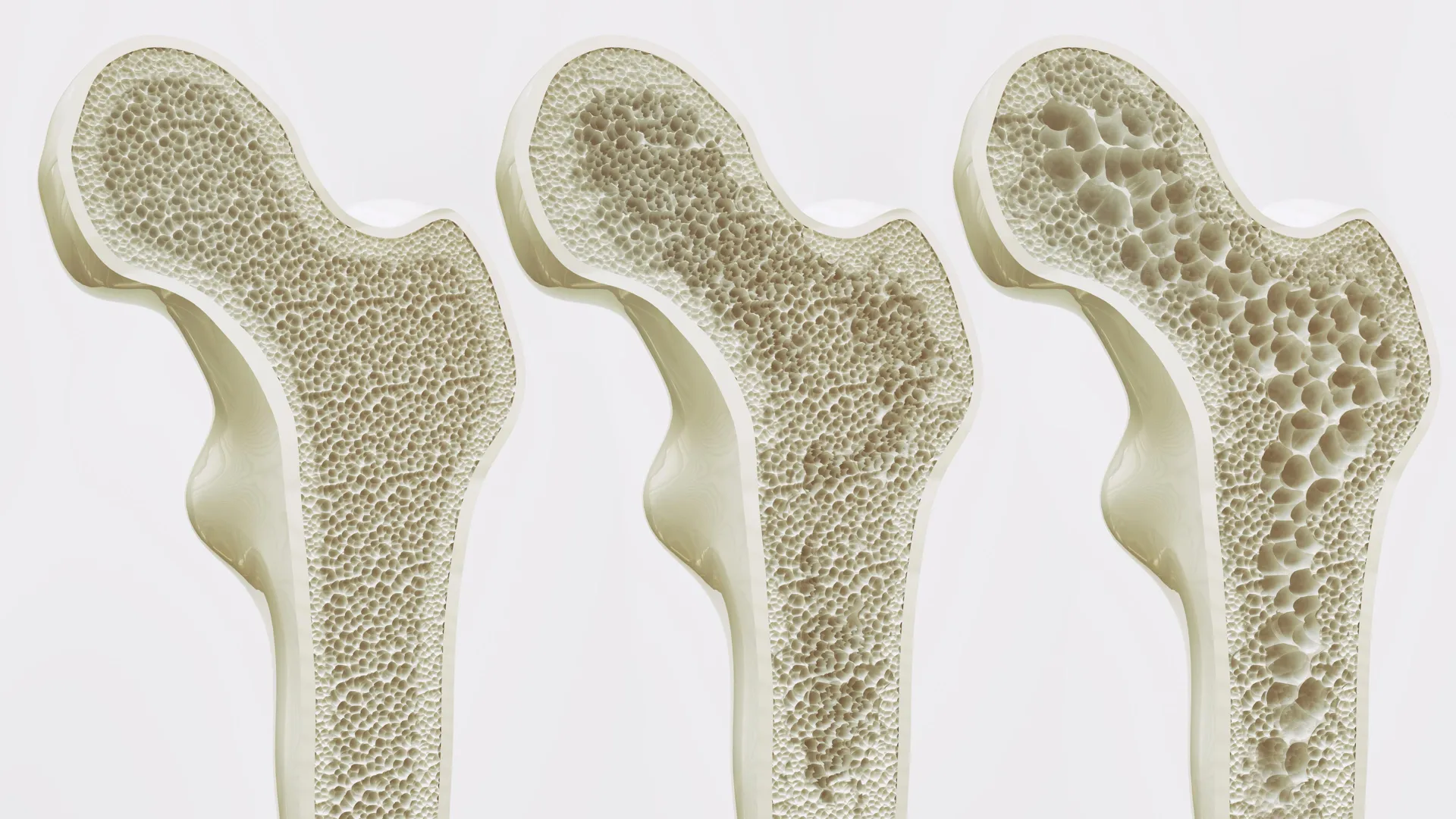
The researchers analyzed murine (mouse) models engineered to develop two distinct variants of glioblastoma. The results were striking: the presence of glioblastoma induced pronounced erosion of the skull bones, localized specifically along the cranial sutures where the individual plates of the skull fuse together. Crucially, this localized bone degradation appeared to be uniquely driven by aggressive primary brain tumors. Control mice subjected to ischemic strokes, traumatic brain injuries, or metastatic cancers originating outside the central nervous system exhibited no such cranial erosion. Subsequent computed tomography (CT) scans performed on human patients diagnosed with glioblastoma confirmed parallel patterns of localized skull thinning in identical anatomical regions, validating the translational relevance of the animal models.
As the skull bones eroded in the murine models, the physical channels linking the skull to the brain underwent significant structural alterations, increasing in both number and cross-sectional diameter. The researchers hypothesized that these widened conduits serve as biological superhighways, allowing tumor-derived molecular signals to migrate directly into the skull marrow and fundamentally reprogram its microenvironment.
Immune Polarization and the Microenvironmental Shift
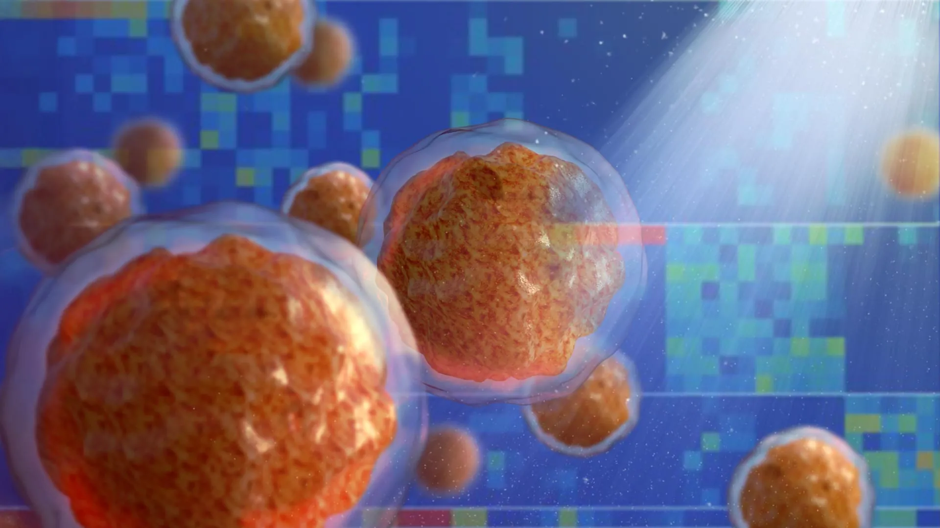
To decode the precise nature of this reprogramming, the research team employed single-cell RNA sequencing—a high-resolution molecular technique capable of analyzing gene expression profiles in individual cells. The genomic data revealed a dramatic, pathological skewing of the immune-cell repertoire within the skull marrow.
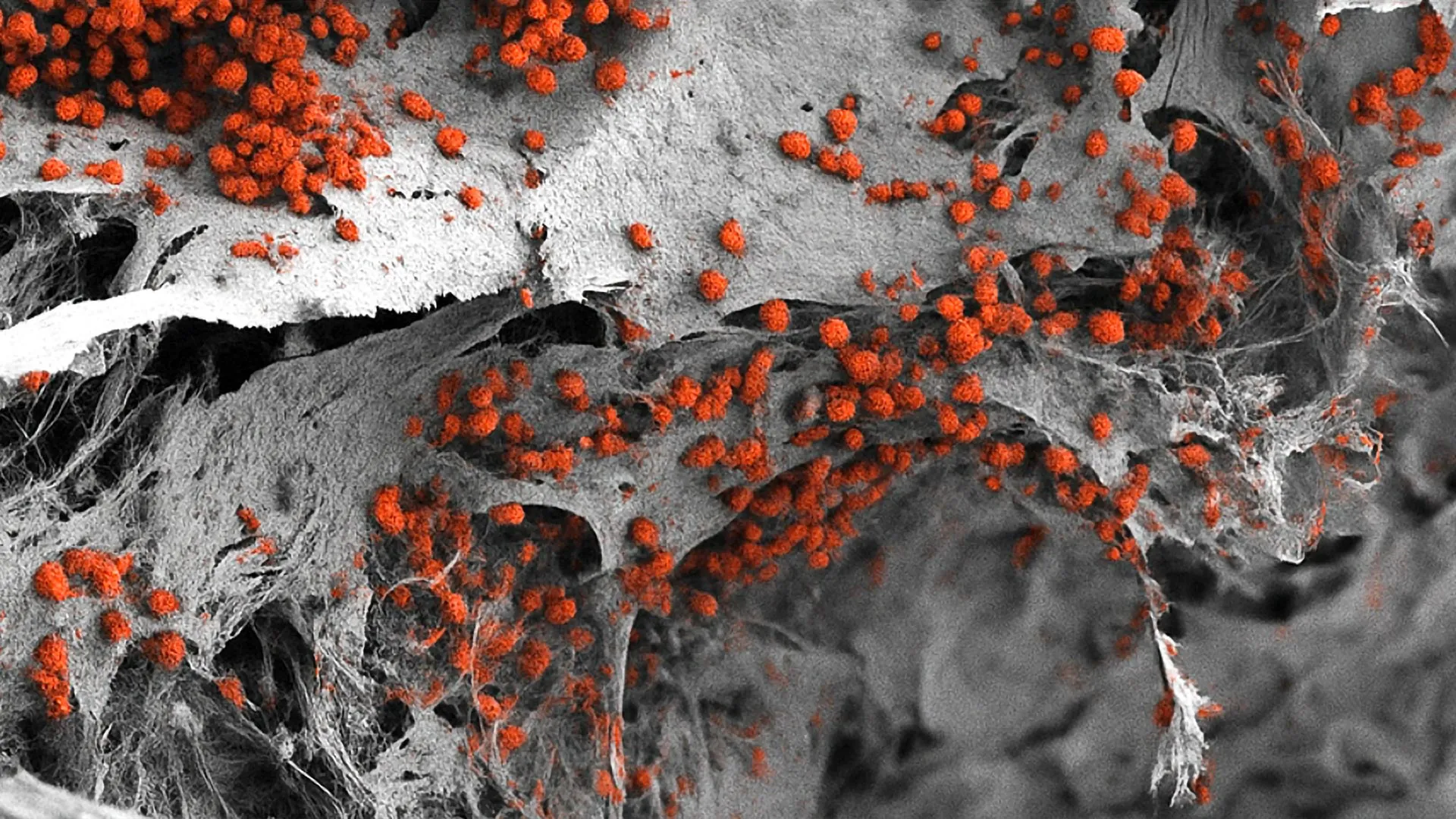
Specifically, glioblastoma drove an immense proliferation of pro-inflammatory myeloid cells. The concentration of inflammatory neutrophils within the skull marrow nearly doubled. Concurrently, the tumor actively depleted several vital populations of B cells, including antibody-producing subtypes essential for humoral immunity.
"The skull-to-brain channels allow an influx of these numerous pro-inflammatory cells from the skull marrow to the tumor, rendering the glioblastoma increasingly aggressive and, all too often, untreatable," noted study co-author Dr. E. Richard Stanley, professor of developmental and molecular biology at Einstein. "This indicates the need for treatments that restore the normal balance of immune cells in the skull marrow of people with glioblastoma. One strategy would be suppressing the production of pro-inflammatory neutrophils and monocytes while at the same time restoring the production of T and B cells."
The study also unveiled a fascinating dichotomy that further underscores glioblastoma’s systemic nature. While the skull marrow reacted to the tumor by upregulating genes associated with the hyper-production of inflammatory immune cells, the bone marrow of distant bones—such as the femur—exhibited the exact opposite response. In the femur marrow, glioblastoma suppressed the genetic pathways required to manufacture standard immune cell populations, illustrating that the tumor exerts a systemic, highly localized regulatory effect on the body’s hematopoietic organs.
The Danger of Osteoporosis Therapeutics
Perhaps the most clinically urgent finding of the study involves the unexpected interaction between glioblastoma and common bone-preserving medications. Recognizing that glioblastoma drives localized bone erosion in the skull, the researchers investigated whether administering anti-osteoporosis drugs could mitigate cranial damage and potentially alter the course of the disease.
The team administered two distinct Food and Drug Administration (FDA)-approved medications used to treat human osteoporosis—zoledronic acid and denosumab—to mice bearing glioblastoma tumors. While both pharmaceutical agents successfully halted the destructive erosion of the skull bone, their effects on the tumor itself diverged dangerously. Zoledronic acid was found to actively fuel tumor progression in a specific variant of glioblastoma, accelerating disease severity. Furthermore, both zoledronic acid and denosumab inadvertently abrogated the therapeutic benefits of anti-PD-L1 checkpoint immunotherapy, a class of cancer drug designed to invigorate tumor-fighting T cells.
This discovery carries immediate safety implications for clinical oncology. Many cancer patients, particularly older adults, suffer from concurrent osteoporosis or bone loss resulting from cancer treatments like glucocorticoids or androgen deprivation therapy. The finding that specific bisphosphonates can promote glioblastoma growth and counteract immunotherapy suggests that clinicians must exercise extreme caution when prescribing bone-density medications to patients harboring aggressive brain tumors.
Broader Implications and Future Treatment Paradigms
The publication of this study in Nature Neuroscience marks a watershed moment in neuro-oncology, establishing a clear imperative to redesign therapeutic strategies for glioblastoma. By proving that the disease operates as a multi-systemic pathology involving the cranium and the immune system, the research opens up entirely new avenues for drug development and clinical intervention.
Future treatment protocols may focus on dual-pronged approaches: simultaneously targeting the intracranial tumor mass while modulating the immune environment of the skull marrow. Therapies designed to curb the overproduction of pro-inflammatory neutrophils in the cranial bone marrow—while rescuing depleted T and B cell populations—could synergize with existing immunotherapies to overcome the profound treatment resistance that has defined glioblastoma for decades.
As clinical trials and follow-up investigations are planned, the findings offer renewed hope to researchers and patients alike. By looking beyond the confines of the brain tissue and understanding the intricate biological dialogue between cancer, bone, and immunity, medical science moves one step closer to dismantling one of oncology’s most formidable adversaries.