The landscape of regenerative medicine and hematology may be on the verge of a significant transformation following a groundbreaking preclinical study led by investigators at Weill Cornell Medicine. Published in the February 25 issue of Nature Immunology, the research identifies a single molecular switch that plays an essential role in compelling blood-forming stem cells to transition from a dormant state into an active, highly regenerative condition. This discovery centers on a DNA transcription-regulating protein known as FLI-1, which researchers found can be transiently stimulated to boost the expansion and transplantation success of blood stem cells. The findings offer a promising pathway toward overcoming persistent hurdles in bone marrow transplants, gene therapies, and the treatment of various refractory blood disorders.
Understanding the Mechanics of Stem Cell Dormancy and Activation
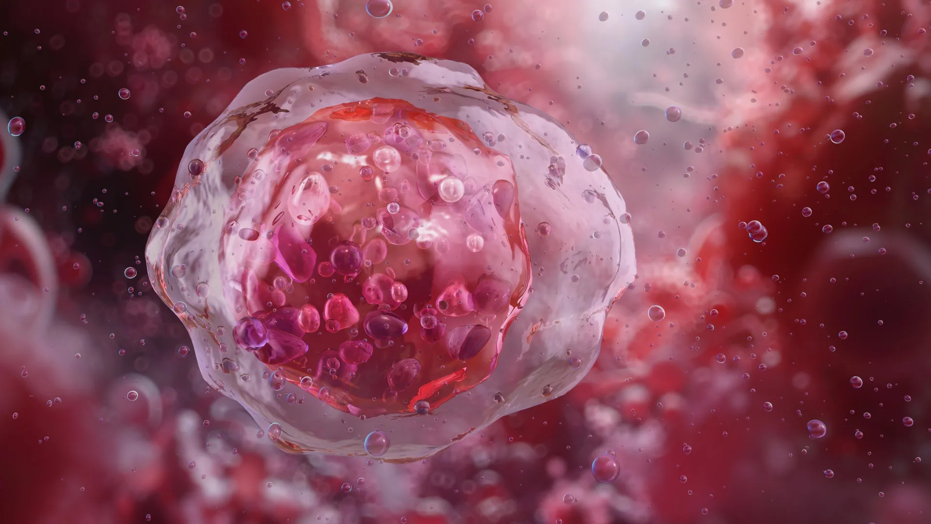
In human and mammalian physiology, stem cells serve as the fundamental building blocks of tissue maintenance and repair. Across virtually all organ systems, these immature cells reside in a primarily quiescent, or slowly dividing, state. This hibernation protects them from genomic damage and metabolic exhaustion over an organism’s lifespan. However, following physical trauma, systemic injury, or clinical mobilization, these cells must rapidly alter their behavior. They switch from dormancy to an activated state, proliferating exponentially and differentiating into mature, functional cell types required to restore damaged tissue.
For hematopoietic—or blood-producing—stem cells, this dynamic is largely centered within the bone marrow. These specialized cells generate every component of the circulatory and immune systems, including red blood cells, platelets, and white blood cells. Under normal conditions, they remain anchored inside specific microenvironments within the marrow. When stimulated, however, they mobilize into the bloodstream or expand locally to rebuild depleted cellular populations.
Pinpointing the Role of FLI-1 in Stem Cell Plasticity
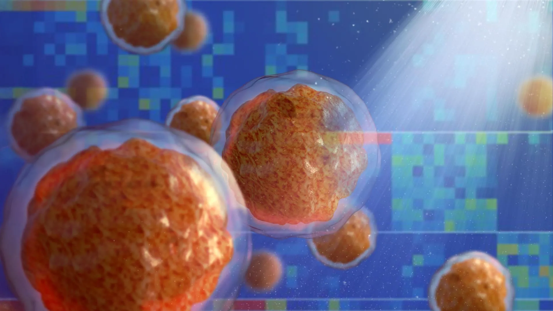
To uncover the precise mechanisms governing this cellular awakening, the Weill Cornell Medicine team utilized advanced single-cell profiling and high-resolution computational analyses. By comparing the gene activity profiles of quiescent blood stem cells with those of their actively proliferating counterparts, the researchers mapped out the regulatory networks driving the transition.
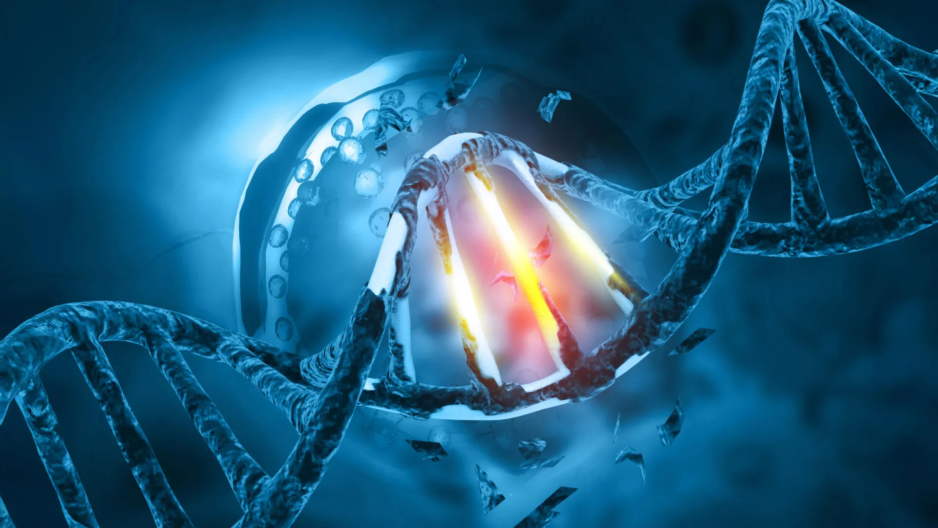
Their investigation zeroed in on FLI-1, a transcription factor protein capable of modulating the activity of thousands of distinct genes simultaneously. The study revealed that FLI-1 acts as a critical gatekeeper for regenerative capacity. When FLI-1 is absent or insufficiently expressed, blood stem cells remain locked in a deep, unresponsive state of quiescence, effectively severing their functional communication with the surrounding marrow infrastructure.
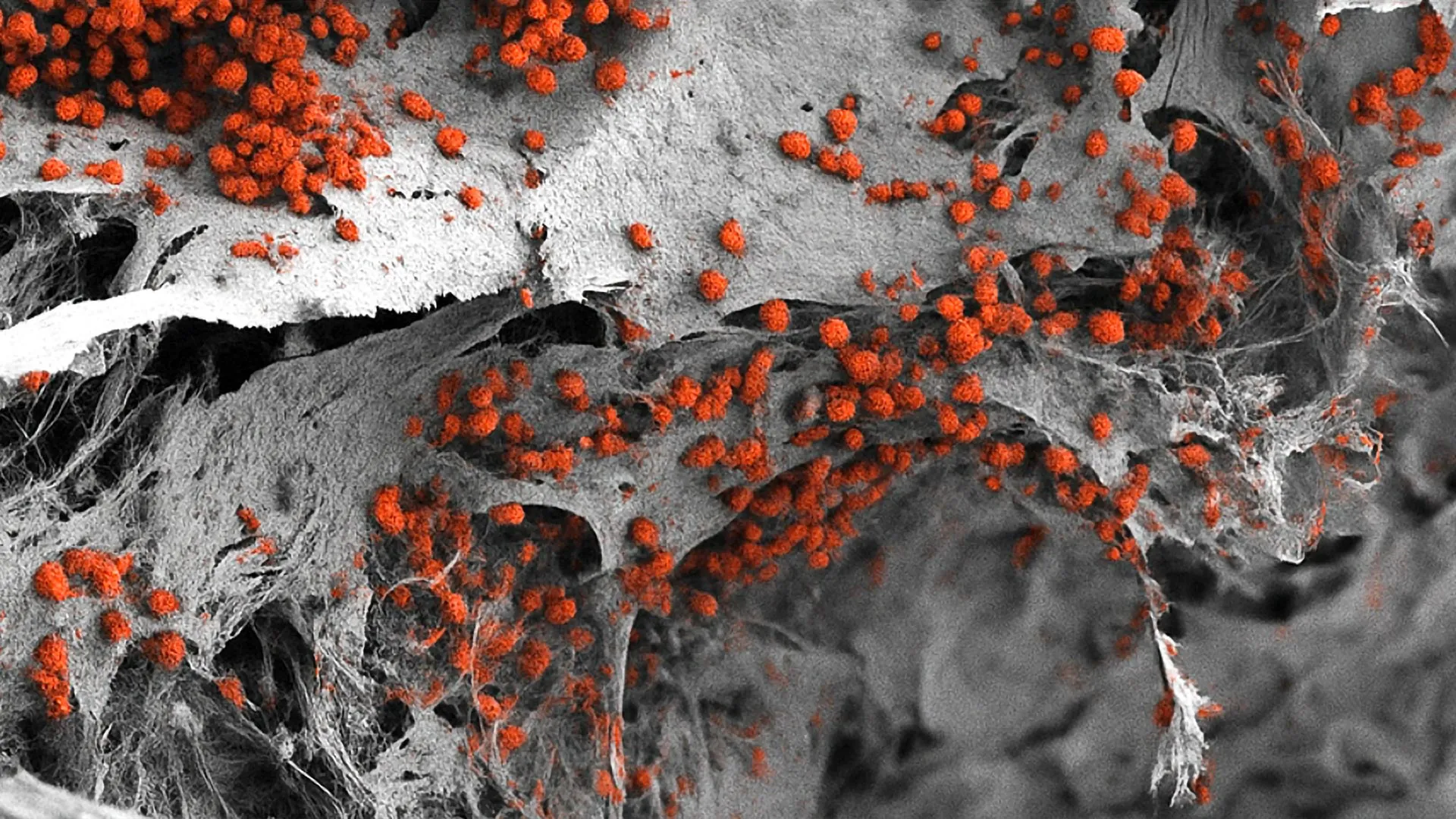
Conversely, the targeted introduction of FLI-1 revitalizes these cells, re-establishing their critical interactions with specialized endothelial cells that line the local blood vessels—a localized microenvironment known as the vascular niche. This molecular dialogue between the stem cell and its vascular niche pushes the hematopoietic cells into a robust, regenerative state. Consequently, the cells dramatically enhance their capacity to proliferate, expand in number, and successfully engraft into a new host environment.
A Novel, Vaccine-Inspired Delivery Method
A primary challenge in manipulating transcription factors like FLI-1 has historically been their association with oncogenesis. Overactivity or genetic mutations driving chronic expression of FLI-1 are well-documented contributors to the development of certain types of leukemia. To circumvent this risk, the research team developed a transient, highly controlled delivery mechanism.
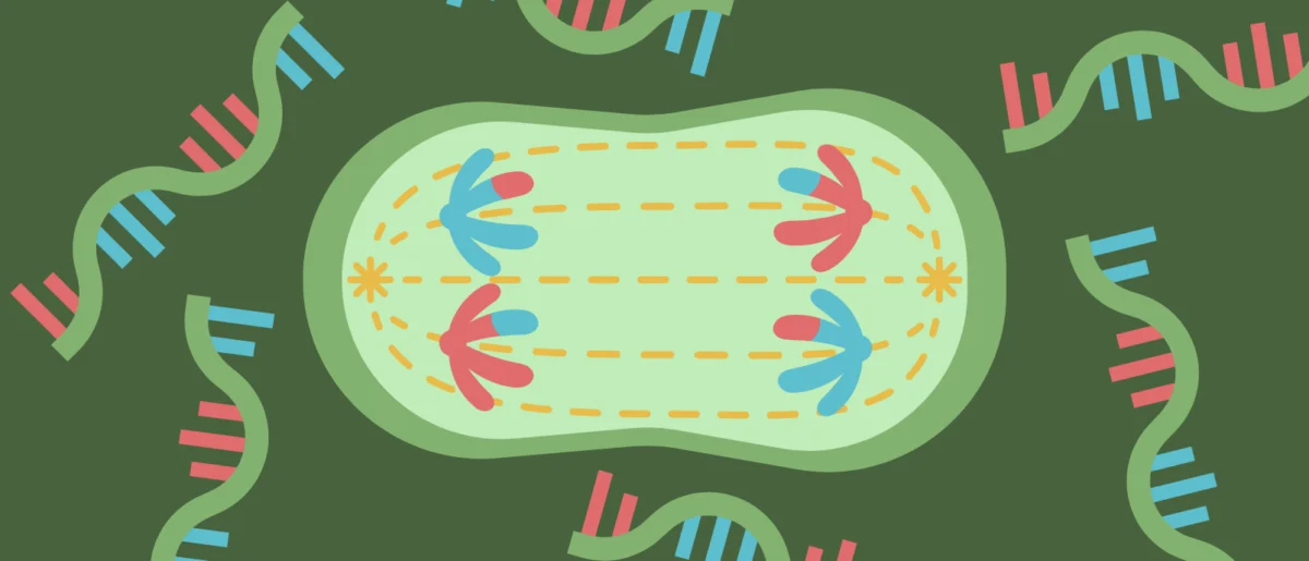
Drawing inspiration from the technological framework underpinning modified mRNA-based vaccines, the investigators engineered a method to introduce FLI-1 into adult mobilized bone marrow stem cells for only a brief, predetermined window of time—lasting merely a few days. This temporary pulse is sufficient to wake the stem cells from hibernation and prompt rapid expansion without triggering unchecked cellular division or malignant transformation.
Dr. Tomer Itkin, co-first author of the study and former instructor of biology in medicine in the Rafii laboratory—now serving as director of Tel Aviv University’s Neufeld Cardiovascular Research Institute and an assistant professor at the Sagol Center for Regenerative Medicine—highlighted the safety profile of the technique. The stem cells primed with FLI-1 modified mRNA wake up from hibernation, expand, and functionally and durably engraft in the recipient host, without any evidence of cancer, he noted.
Solving a Long-Standing Biological Puzzle
Beyond its immediate therapeutic applications, the study successfully resolved a long-standing mystery within the field of stem cell biology. For years, researchers have observed that human umbilical cord-derived blood stem cells possess a vastly superior regenerative potential compared to adult stem cells isolated from peripheral blood or bone marrow.
The Weill Cornell team demonstrated that this discrepancy is directly associated with baseline differences in FLI-1 activity. Specifically, umbilical cord stem cells maintain an optimal level of FLI-1 that dictates their heightened potency and adaptability when interacting with the vascular niche. By decoding this signaling axis, the researchers bridged a major knowledge gap regarding how systemic cues and niche-specific environments coordinate to regulate stem cell self-renewal.
Bioinformatics analyst and co-first author Sean Houghton, currently a senior bioinformatics analyst for the Englander Institute for Precision Medicine at Weill Cornell Medicine, emphasized the collaborative nature of cellular regulation revealed by the computational models. The team showed that stem cell activity is not autonomous, but also is not fully determined by endothelial cell vascular niche signals, Houghton explained. Instead, it depends fundamentally on bidirectional signaling and adaptability between the two cellular populations.
Implications for Bone Marrow Transplants and Gene Therapies
The clinical implications of this discovery are far-reaching, particularly for patients suffering from hematological malignancies, severe anemias, and inherited genetic disorders. Bone marrow transplants are a cornerstone of modern oncology, utilized to replenish compromised immune and blood cell populations following intensive chemotherapy or radiation regimens.
However, standard transplantation protocols frequently encounter significant bottlenecks. Donors may have an extremely limited supply of viable blood stem cells, or patients requiring autologous transplants—where their own healthy stem cells are harvested prior to cancer treatment—may yield cells that are sluggish and resistant to activation due to prior exposure to cytotoxic therapies. Furthermore, emerging gene therapies targeting severe blood disorders such as beta-thalassemia rely on harvesting a patient’s stem cells, inserting therapeutic genes into the genetic code, and expanding these fragile cells in a laboratory setting before re-infusing them into the body.
Dr. Shahin Rafii, senior author of the study, director of the Hartman Institute for Therapeutic Organ Regeneration and the Ansary Stem Cell Institute, chief of the division of regenerative medicine, and the Arthur B. Belfer Professor in Genetic Medicine at Weill Cornell Medicine, emphasized the transformative potential of the research. The approach outlined in this study could substantially improve the efficiency of marrow transplants and marrow-cell-targeted gene therapies, especially in cases where the donor has a very limited supply of viable blood stem cells, Dr. Rafii stated. By providing a safe, reliable method to switch quiescent blood stem cells into an activated regenerative state, clinicians could drastically reduce engraftment failures and expand the eligibility pool for complex cellular therapies.
Future Directions and Clinical Translation
Following the successful preclinical validation of the transient FLI-1 stimulation method, the research team is moving forward with plans to scale up the technology. Future phases of development will focus on refining the modified mRNA delivery platforms to meet rigorous clinical manufacturing standards, with the ultimate objective of advancing the approach into human clinical trials.
If proven safe and effective in clinical settings, this strategy could establish a new standard of care across a wide spectrum of hematological disorders. By enabling efficient, long-term, and stable blood cell production from limited cellular sources, the discovery represents a vital step forward in translating fundamental molecular biology into life-saving medical interventions.
The research was supported by grants from the National Institutes of Health, including the National Heart, Lung, and Blood Institute, the National Institute of Diabetes and Digestive and Kidney Diseases, and the National Institute of Allergy and Infectious Diseases (under award numbers R35HL150809, R01DK136327, and U01AI138329). Additional financial backing was provided by the Hartman Institute for Therapeutic Organ Regeneration, the Ansary Stem Cell Institute, and the Selma and Lawrence Ruben Daedalus Fund for Innovation at Weill Cornell Medicine. (Note: Dr. Shahin Rafii is an unpaid co-founder of Angiocrine Bioscience.)