
The landscape of cellular biology has long been defined by a fundamental trade-off: the desire to see the internal "machinery" of life in high definition versus the requirement to keep that life undisturbed. For decades, fluorescence microscopy has served as the gold standard, allowing researchers to tag specific proteins or organelles with glowing markers to track their movements. However, this reliance on exogenous labels has inherent limitations—namely phototoxicity, the potential for labels to interfere with biological function, and the rapid degradation of signal known as photobleaching. Now, a team at Stanford University has introduced a paradigm-shifting solution. By developing Interferometric Image Scanning Microscopy (iISM), researchers have successfully bypassed the need for fluorescent tags while maintaining the high-resolution imaging required to observe the minute, complex interactions that drive cellular life.
The breakthrough, published in the Nature portfolio journal Light: Science and Applications, represents a synthesis of classical optical physics and modern computational power. By achieving a spatial resolution of approximately 120 nanometers, the Stanford system allows scientists to peer into the "cellular box" without the intrusive preparation methods that have characterized biological imaging since the late 20th century.
The Evolution of Imaging: From Fluorescence to Scattering
To understand the significance of this development, one must look at the historical trajectory of microscopy. Fluorescence microscopy, which earned W.E. Moerner, the Harry S. Mosher Professor of Chemistry at Stanford, a share of the 2014 Nobel Prize, revolutionized the field by enabling researchers to see beneath the diffraction limit of light. By using molecules that emit light when excited by lasers, scientists could pinpoint the location of specific proteins.
Yet, this method is fundamentally subtractive in its nature. It requires the cell to be engineered or stained, a process that can alter the very behavior being studied. Furthermore, the high-intensity light required to excite these fluorophores can lead to photodamage, essentially stressing or killing the specimen under observation.
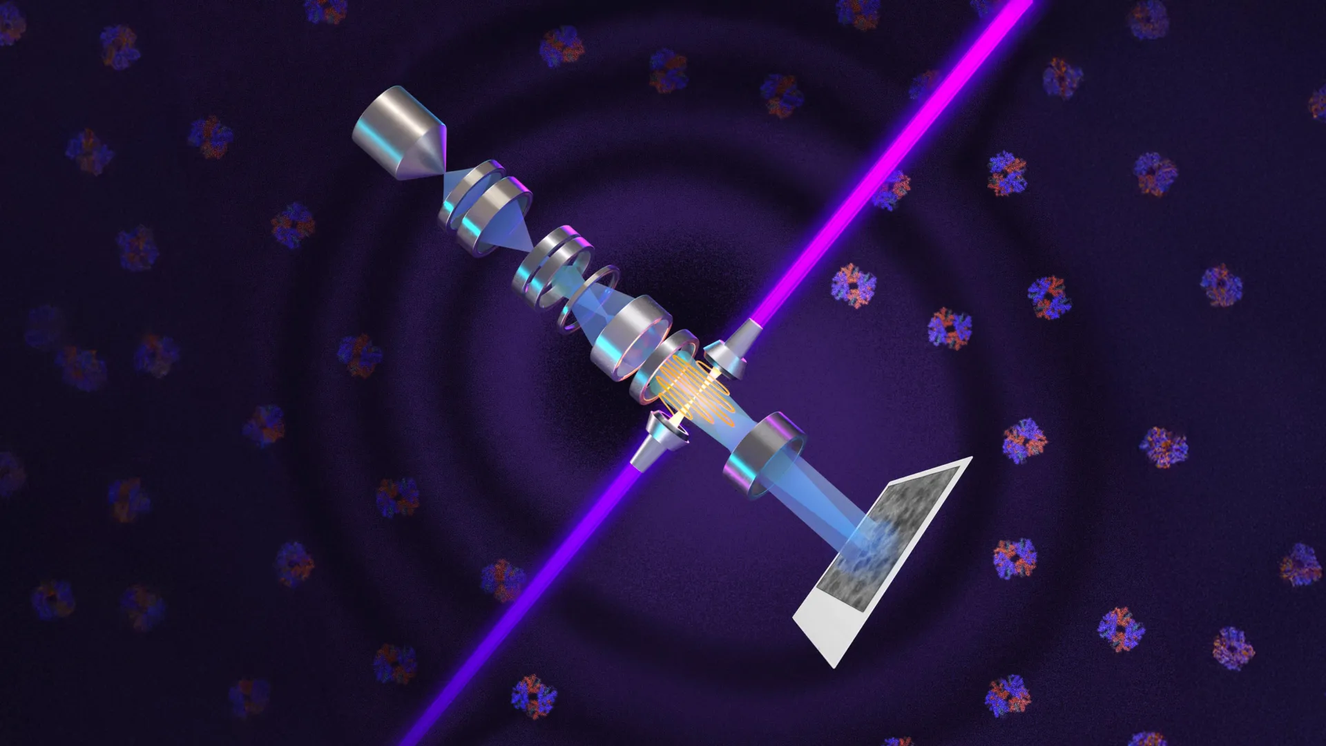
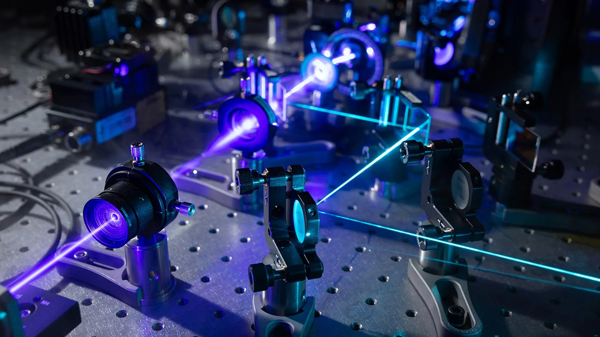
The Stanford team’s new approach relies on a different physical principle: interferometric scattering. This method leverages the natural interaction between light and the internal structures of a cell. As light passes through a specimen, it is scattered by cellular organelles and structures. While this scattered light is typically too faint to be useful on its own, the researchers developed an interferometric system that uses a reference laser beam to amplify these weak signals. By combining this with an advanced detector array—a technology borrowed from high-end confocal microscopy—they have created a tool that captures the "natural" image of the cell with high contrast and without the need for external labels.
Chronology of the Innovation
The development of iISM was not an overnight success but the result of a deliberate, multi-year collaboration between senior laboratory leadership and specialized postdoctoral expertise.
- 2014–2018: Following his Nobel win, Professor W.E. Moerner focused on addressing the persistent bottlenecks in super-resolution imaging. He identified a need for label-free methods that could match the resolution of fluorescence microscopy.
- 2019–2021: Michelle Kueppers, whose doctoral work focused on interferometric scattering, joined the Moerner laboratory. Her integration into the team provided the necessary technical bridge to combine scattering-based imaging with advanced signal processing.
- 2022: The team successfully integrated an array detector into the interferometric scattering setup, replacing the traditional pinhole-and-single-detector architecture that had limited previous label-free efforts.
- 2023: The prototype underwent rigorous validation, testing its ability to resolve cellular structures down to the 120-nanometer threshold.
- 2024: The results were published, marking the transition from an experimental concept to a viable tool for broad biological research.
Technical Mechanics: How iISM Bridges the Gap
The ingenuity of the Stanford system lies in its computational approach to signal collection. Traditional confocal microscopes rely on a pinhole to reject out-of-focus light, a process that inherently sacrifices much of the available signal. The Stanford team replaced this with an array detector, which collects hundreds of views of the same sample simultaneously.
This array-based collection functions analogously to the human visual system, which uses two offset eyes to build a depth-aware, high-contrast representation of the world. By aggregating hundreds of these perspectives, the researchers’ computational algorithms generate a sharper, more precise image than any single detector could produce. Because the microscope requires significantly lower illumination power than comparable high-contrast systems, the risk of phototoxicity is drastically reduced. This "gentle" imaging modality allows for the observation of cells over longer periods, enabling the capture of biological processes—such as organelle transport or structural remodeling—that are often missed during the short windows allowed by traditional fluorescent imaging.
Implications for Modern Biomedical Research
The potential applications for iISM are vast, extending into several critical domains of human health and environmental science. Current collaborations at Stanford illustrate the breadth of the technology’s utility.
In the realm of infectious disease, researchers are investigating the physical mechanics of malaria. By watching how red blood cells deform and change in real time when exposed to the malaria parasite, scientists hope to identify the specific mechanical failure points that occur during the infection cycle. This could lead to new diagnostic markers or therapeutic interventions that target the physical structural integrity of the cell.
In oncology, the microscope is being deployed to track the entry of therapeutic agents into cancer cells. Because iISM does not require the drug to be tagged with a label—which might change its chemical properties or its ability to penetrate a cell—researchers can observe the "native" interaction between the drug and the cellular membrane or internal targets. This offers a more accurate representation of how a medication actually performs in a physiological environment.
Finally, in agricultural and environmental biology, the technology is being used to observe the symbioses between plant cells and microorganisms. Understanding how these organisms communicate and trade nutrients across their boundaries is essential for developing more resilient crop varieties, and the ability to watch these exchanges in an unperturbed state provides a level of clarity that has previously remained out of reach.
A Complementary Future
Despite the excitement surrounding iISM, both Moerner and Kueppers are quick to emphasize that this technology is not intended to replace fluorescence microscopy. Instead, they envision a future of "multimodal imaging," where the two techniques work in tandem.
"Every method has its advantages and disadvantages, and we believe in a complementary implementation in the future," Kueppers noted. The current strategy for the laboratory is to combine the molecular specificity of fluorescence—which is excellent for identifying specific proteins—with the label-free, high-speed, and low-damage context of iISM. By overlaying these datasets, researchers can gain a comprehensive "map" of the cell that includes both specific chemical identities and the broader, dynamic structural context.
Future Outlook and Scientific Community Impact
As the team continues to refine the iISM instrument, the goal is to standardize the technology for wider adoption. The complexity of current super-resolution microscopes often limits them to specialized physics or optics laboratories. By simplifying the computational requirements and streamlining the optical architecture, the Stanford researchers hope to make this tool a staple of biology departments worldwide.
The broader implications of this development touch upon the very definition of biological inquiry. By removing the "labels" that have acted as a filter between scientists and their subjects, iISM provides a clearer, more honest view of the cell. As the scientific community begins to adopt these label-free methods, it is likely that we will uncover new classes of biological activity that were previously obscured or misrepresented by the invasive nature of fluorescent markers.
The work of Moerner and Kueppers serves as a testament to the power of interdisciplinary research. By taking a concept rooted in the optics of the sky—the scattering of light—and applying it to the smallest units of life, they have opened a new window into the complex, rhythmic, and highly organized world inside the cell. As this technology matures, it will undoubtedly contribute to a more nuanced understanding of the mechanics of disease, the nature of cellular growth, and the delicate balance of life at the nanoscale.