
In a landmark achievement for developmental biology, researchers have successfully reconstructed a comprehensive family tree of a developing mouse embryo, capturing the life history of cells from the moment of the first post-fertilization division. This study, published October 8, 2026, in the journal Science, introduces a sophisticated "DNA Typewriter" technology that functions as a biological ledger, recording the division and diversification of cells directly into their own genetic material. The research provides an unprecedented view of the first 13.5 days of mammalian gestation, offering a roadmap of how a single fertilized egg transforms into a complex organism with distinct tissues and organs.
The research was spearheaded by Dr. Jay Shendure, a professor of genome sciences at the University of Washington School of Medicine and a Howard Hughes Medical Institute Investigator, in collaboration with Dr. Chengxiang Qiu, a molecular and systems biologist at Dartmouth College. By bridging the gap between computational biology and genomic engineering, the team has addressed one of the most persistent hurdles in the life sciences: observing the continuous, real-time lineage decisions of mammalian cells without disrupting their natural developmental trajectory.
The Mechanics of the DNA Typewriter
The core innovation behind this study is the DNA Typewriter, a synthetic biological system designed by Dr. Shendure and Junhong Choi, currently of the Sloan Kettering Cancer Center. Unlike traditional lineage tracing methods, which often rely on retrospective analysis or destructive labeling techniques, the DNA Typewriter acts as a persistent, sequential recorder.
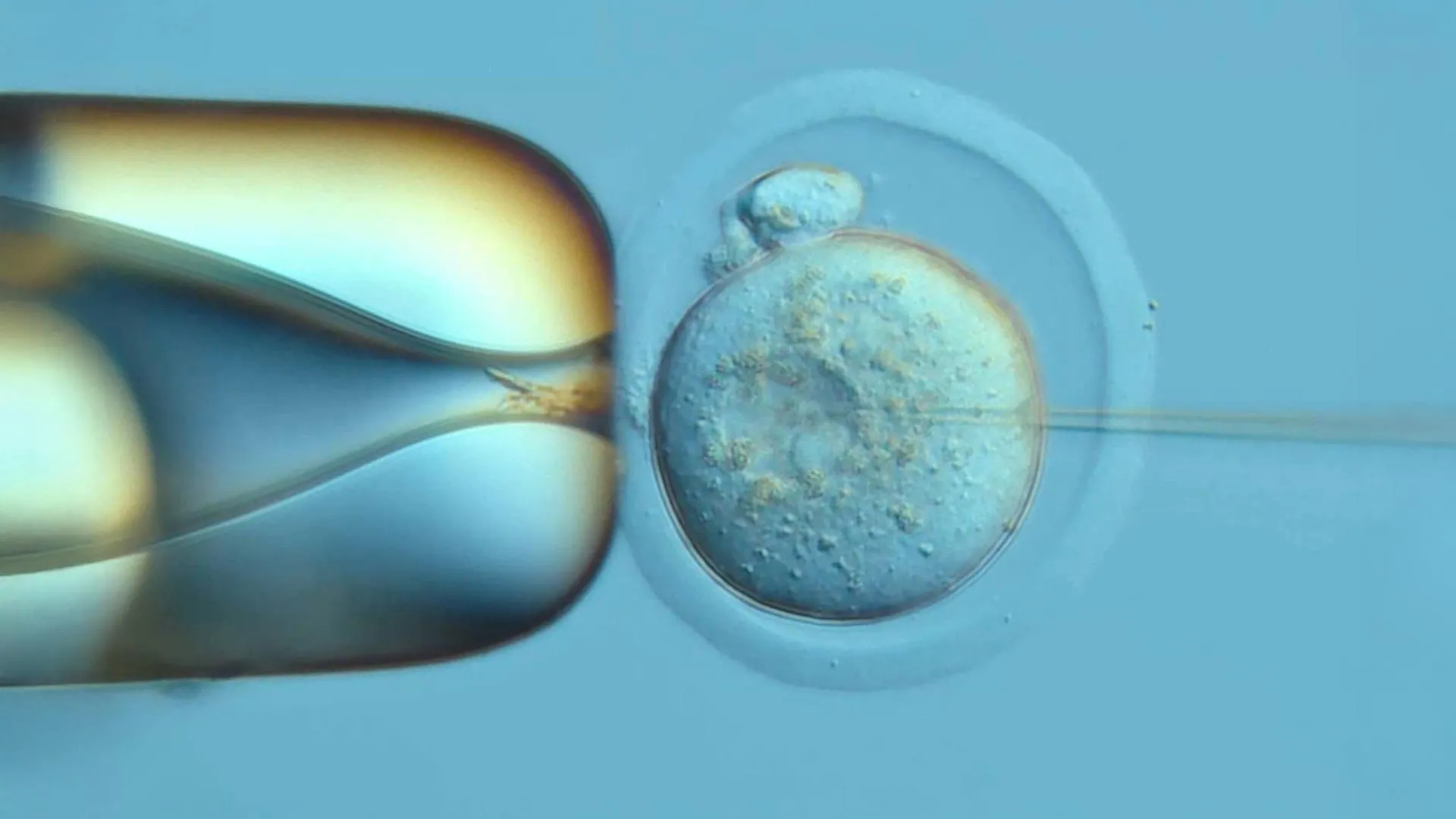
The researchers integrated a specialized recording sequence—metaphorically described as a "tape"—into the genome of a fertilized mouse egg. As the cell undergoes mitosis, the system records each division event by inscribing a unique genetic "character" onto this tape. Because the recording process is strictly sequential and does not overwrite existing data, the resulting DNA sequence provides a permanent, chronological logbook of the cell’s ancestral history.
Haedong Kim, a postdoctoral scientist at UW Medicine and a co-first author of the study, noted that the primary challenge was not just the writing, but the extraction of the data. "Writing the record is only half the problem," Kim stated. "You also have to read it back out of each cell, so we redesigned the tape to make that easier." This refinement allowed the team to recover high-resolution data from individual cells, enabling the reconstruction of branches in the cellular family tree that were previously obscured by the sheer complexity of mammalian growth.
Chronology of the Experiment
The study involved an initial cohort of 100 fertilized mouse eggs, with 10 embryos successfully reaching the stage required for high-depth genomic analysis. The researchers identified "Embryo No. 3" as the primary subject for the study, as its recording apparatus remained stable and highly legible throughout the observation period.
The analysis focused on the critical 13.5-day window, a significant portion of the total 19-to-21-day gestation period for mice. By examining the DNA signatures left by the very first cleavage of the fertilized egg, researchers were able to trace almost every cell in the embryo back to one of the two founding cells. This fundamental observation revealed a fascinating asymmetry: while one founding cell produced a larger number of descendants than the other, both cells contributed to the embryo’s eventual tissue types in remarkably similar proportions. This finding suggests that the initial division of an embryo involves a robust regulatory mechanism that ensures balanced development, even when cellular proliferation rates differ.
Advancing Beyond Traditional Lineage Tracing
Developmental biology has long relied on observational techniques that are either limited by the organism’s complexity or by the invasive nature of the markers used. For decades, the nematode Caenorhabditis elegans served as the gold standard for lineage mapping due to its transparency and limited cell count—totaling only 959 cells in its adult form. Mammals, however, present a significantly greater challenge, characterized by millions of cells, high levels of plasticity, and rapid, multi-layered differentiation.
Traditional methods, such as CRISPR-based "scarring" of DNA, have historically struggled with accuracy and durability. These methods often involve severing DNA strands, which can inadvertently damage the cell or delete information about previous generations. Furthermore, because these older systems do not necessarily record events in a fixed, linear order, scientists have had to rely on complex statistical modeling to infer the sequence of events. The DNA Typewriter bypasses these limitations by maintaining a steady, non-destructive recording pace, allowing for a resolution that spans the entire developmental timeline.
Mapping the Decision Points of Cell Fate
A major component of the study was determining exactly when cells commit to specific roles. During embryonic development, cells transition from "pluripotent" states—where they have the potential to become any cell type—to "committed" states, where their future is restricted to specific tissues like bone, muscle, or neurons.
The DNA Typewriter data confirmed that this commitment is not a synchronized event across the entire embryo. Instead, the team observed a staggered timeline of differentiation. For instance, the lineage pathways leading to blood cells and retinal cells were established relatively early in the development of the embryo. In contrast, the cells destined for the outer layers of the skin maintained their flexibility for a longer duration before committing to their final function.
This hierarchical and asynchronous decision-making process highlights the sophisticated nature of developmental biology. It suggests that different tissues follow distinct regulatory clocks, a finding that could fundamentally shift our understanding of how organs maintain their specific identities during the turbulent period of organogenesis.
Medical and Therapeutic Implications
The potential applications of this technology extend far beyond the laboratory mouse. By establishing a clear map of how a single cell gives rise to a functioning body, scientists are now better equipped to study the "errors" in this process. Congenital malformations, for instance, are often the result of minor deviations during these early, high-stakes cellular decisions. Similarly, the study of cancer—often described as a breakdown in the regulatory mechanisms that govern cell growth and specialization—stands to benefit from this deeper understanding of lineage.
Dr. Shendure emphasized the broader implications of the work: "This kind of large-scale developmental recording gives scientists a map of how one cell becomes a whole body—which helps explain how organs form correctly, how that process can go wrong in birth defects, and how abnormal cell growth drives cancer."
Furthermore, as the field of regenerative medicine matures, the ability to track lineage becomes critical for stem cell engineering. If scientists can understand the precise sequence of events required for a stem cell to become a healthy, functioning specialized cell, they can better guide these cells for therapeutic applications, such as repairing damaged cardiac tissue or replacing insulin-producing cells in patients with diabetes.
Collaborative Support and Future Outlook
The scale and complexity of the research required an extensive interdisciplinary effort. In addition to the University of Washington and Dartmouth College, the study was supported by a consortium of major scientific institutions, including the Allen Institute, the Howard Hughes Medical Institute, and the Brotman Baty Institute for Precision Medicine. Funding was provided by a diverse group of organizations, including the National Institutes of Health, the Damon Runyon Cancer Research Foundation, and the Swiss National Science Foundation.
The research team, which included co-first authors Qi Yu and Sophie Seidel, represents a growing movement within synthetic biology to treat the genome as an information-storage system. As the technology matures, the ability to "write" into the DNA of living systems promises to provide a deeper understanding of the biological history of organisms, potentially ushering in a new era of precision medicine. While the current study remains a fundamental proof-of-concept, it provides a blueprint for future explorations into the mechanisms of life itself, offering a detailed, chronological record of the events that turn a single cell into a complex, sentient being.
This research marks a significant shift from observing the results of development to witnessing the process of development in real-time, setting the stage for future studies that could eventually incorporate more complex organisms and even human cell models in controlled environments. As the scientific community continues to refine the DNA Typewriter, the once-opaque "black box" of embryonic development is slowly being opened, revealing the hidden logic of life.