
For decades, the field of structural biology has been haunted by a fundamental question: how do viruses, which lack the central nervous system or complex machinery to direct their own growth, consistently construct intricate, perfectly symmetrical protein shells? This mystery has finally begun to unravel. Researchers at the University of Oxford have successfully observed, for the first time, virus-like particles assembling themselves one molecule at a time. The findings, published in the journal Nature, represent a watershed moment in nanotechnology and virology, revealing that the complex architecture of a virus is governed by surprisingly elegant, self-correcting physical rules.
The study, led by a team from the Department of Chemistry and the Kavli Institute for Nanoscience Discovery, provides the first direct, real-time observation of this biological process. By leveraging a high-precision imaging technique known as mass photometry, the researchers have effectively "filmed" the construction of a virus, offering a level of granular detail that was previously thought to be impossible to capture.
The Complexity of Viral Architecture
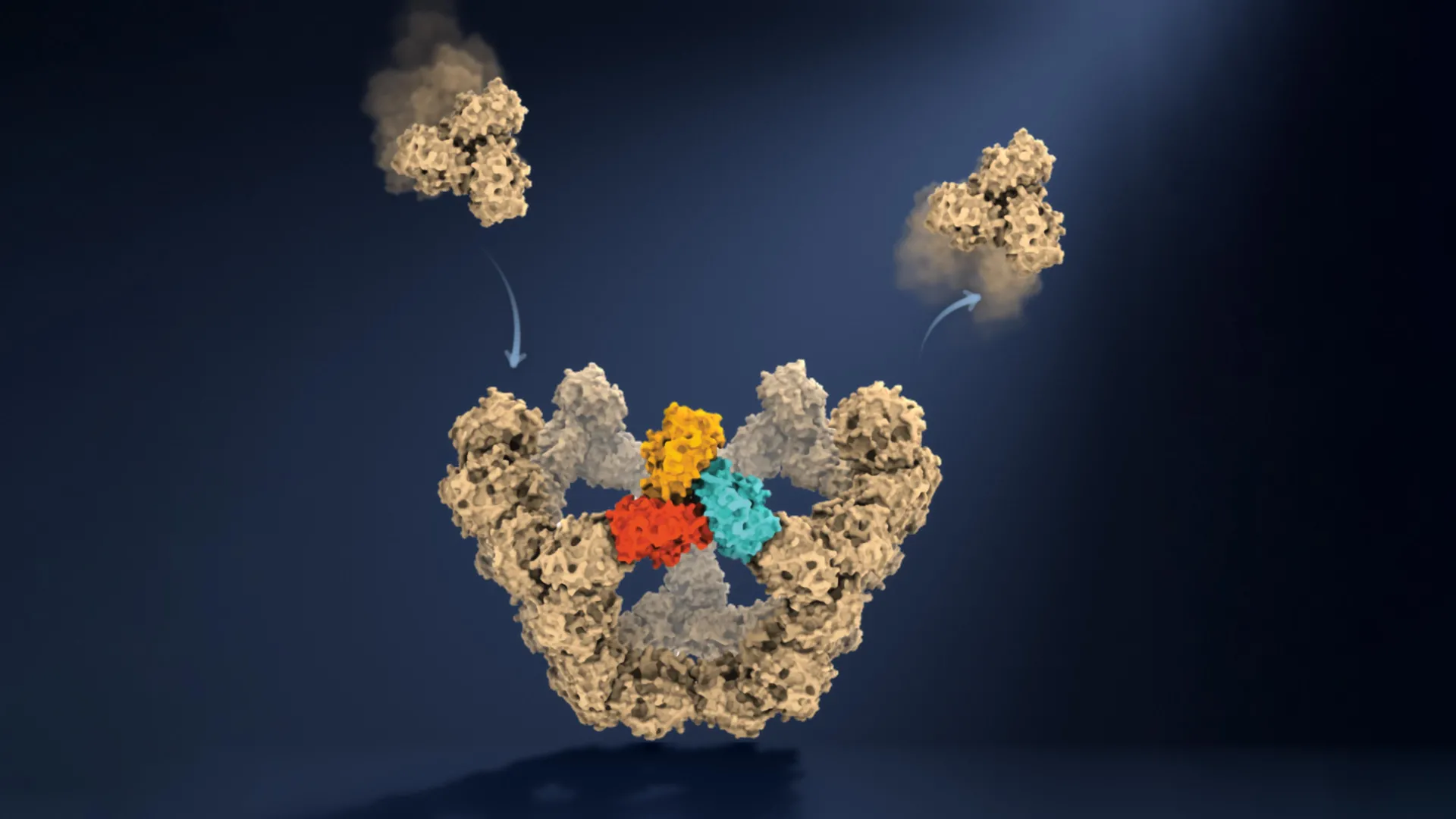
Viruses store and safeguard their genetic material—whether DNA or RNA—within a protective protein container known as a capsid. These shells can range from relatively simple structures consisting of 60 protein subunits to gargantuan, highly complex arrangements involving thousands of individual components. Despite this variation, the outcome is remarkably consistent: the proteins almost always snap together into the exact same, functional, closed-loop geometry.
Historically, the difficulty in observing this process stemmed from the ephemeral nature of the assembly intermediates. These intermediate structures exist only for fractions of a second and often in quantities too low to be detected by traditional imaging methods like cryo-electron microscopy, which typically provides static "snapshots" of the starting and ending points. The "middle" of the assembly process—the critical stage where the architecture is determined—remained a theoretical black box.
A Technological Breakthrough: Mass Photometry
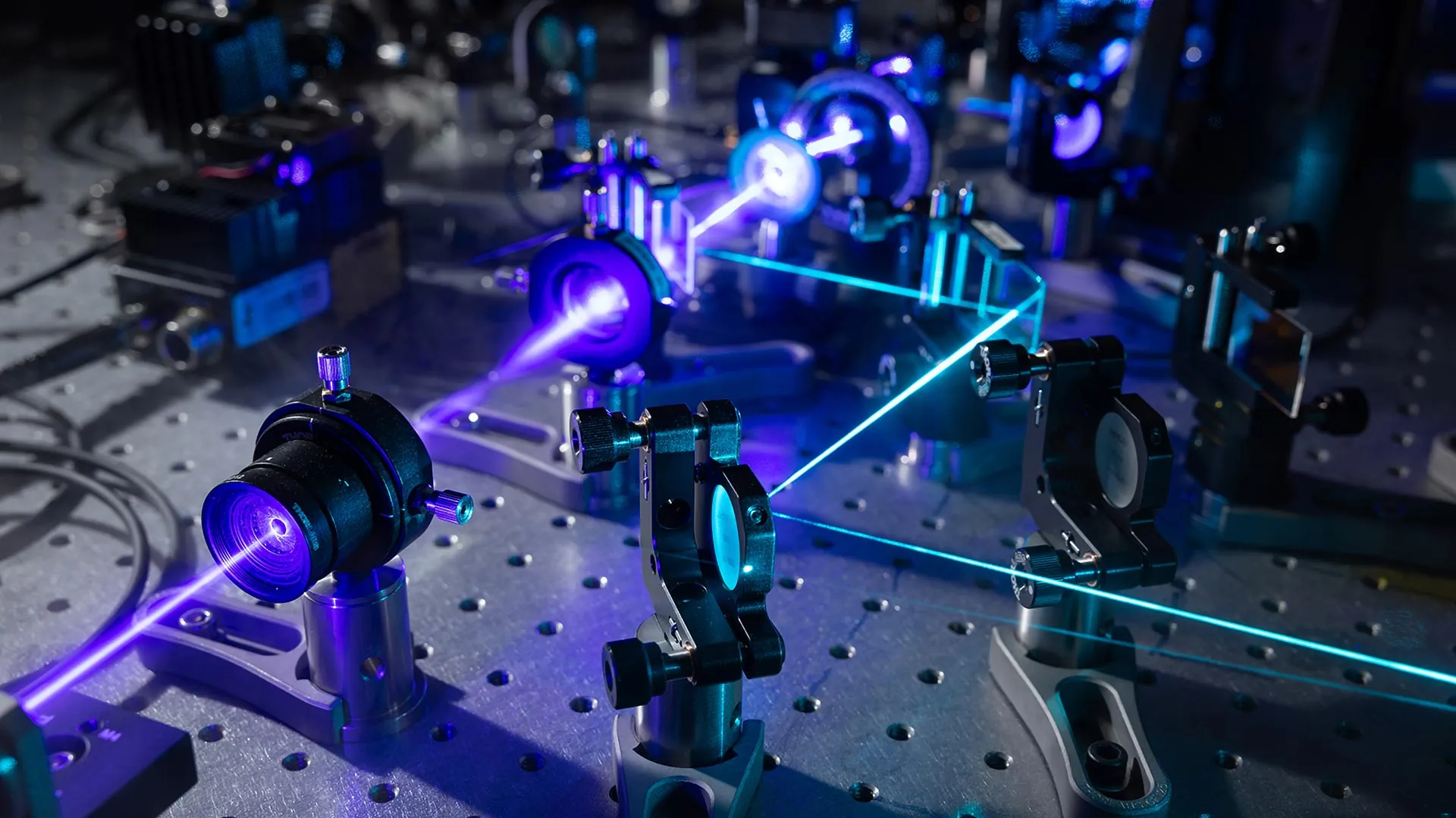
To bridge this observational gap, the Oxford team utilized mass photometry, a proprietary technology developed at the University of Oxford. This method measures the mass of individual molecules by quantifying the light they scatter as they move through a solution. When coupled with a novel micro-confinement strategy that held individual particles in a localized space, the researchers were able to track the precise addition of building blocks to a growing capsid over time.
"Until now, much of our understanding of how these structures assemble has had to be reconstructed from snapshots or theoretical models," said Professor Philipp Kukura, co-lead author of the study. "Being able to both quantify the underlying interactions and follow one particle as it grows changes that. We can see the important intermediate structures appear and deduce a detailed model of the construction process."
Chronology of Discovery: Navigating the Molecular Maze
The research team’s analysis revealed that viral assembly is less like a predetermined construction project and more like a stochastic, high-stakes game of trial and error. The team identified three distinct phases in the assembly of the 60-protein particle:
- The Nucleation Phase: This is the most precarious stage. Initially, protein components collide and form weak, reversible connections. Because these bonds are unstable, they frequently break apart. This "failure" is, paradoxically, the key to the system’s efficiency. It allows the molecules to discard incorrect or suboptimal configurations, preventing the formation of "dead-end" structures that would otherwise waste resources.
- The Checkpoint Milestone: The researchers discovered that assembly accelerates significantly once a "closed pentagonal ring"—consisting of five protein blocks—is formed. This pentagonal structure acts as the first highly stable intermediate, or "molecular checkpoint." Once this threshold is crossed, the geometric constraints of the structure favor the rapid addition of subsequent units.
- The Growth Phase: Once the stable checkpoint is established, the assembly process becomes significantly more robust. Each subsequent protein addition is guided by the established geometry, reducing the number of possible pathways and funneling the particle toward its final, fully enclosed, and highly stable state.
PhD student and co-first author Dan Loewenthal summarized the simplicity of the observation: "This method lets us study the assembly process directly; we just take a video!"
Implications for Antiviral Therapeutics
The discovery that viral assembly relies on a limited number of stable intermediate stages provides a new target for pharmaceutical intervention. If scientists can identify the exact "checkpoints" that viruses must pass to achieve stability, they may be able to develop small-molecule drugs designed to jam these checkpoints or disrupt the intermediate structures.
"The ability to understand these processes at the molecular level could have important applications in vaccine development and antivirals that disrupt viral assembly," noted co-author Dr. Jack Tan of the MRC Weatherall Institute of Molecular Medicine.
Current antiviral treatments often focus on inhibiting enzymes or proteins after the virus has already entered a cell and begun replicating. A drug that targets the assembly process itself—effectively forcing the virus to build a "broken" shell that cannot protect its genetic material—could offer a new class of broad-spectrum antivirals. Furthermore, these insights are invaluable for synthetic biology; researchers working on bio-engineered vaccines or targeted drug-delivery systems (which often use virus-like particles as "containers") can now design their components with a deeper understanding of how to encourage or discourage specific assembly pathways.
Broader Scientific Context
The significance of this work extends beyond virology. Self-assembly is a fundamental mechanism of life, responsible for everything from the organization of the cytoskeleton within human cells to the formation of complex protein complexes involved in metabolic regulation.
By demonstrating that complex biological structures can be decoded as a series of simple physical rules and molecular checkpoints, the Oxford team has provided a framework that could be applied to a variety of other biological systems. The ability to observe these processes at the single-molecule level effectively shifts the field from descriptive biology to predictive engineering.
"A virus has to solve an extraordinary construction problem," explained Dr. Roi Asor, co-lead author. "Its components somehow have to find the right arrangement among a huge number of possibilities, without a blueprint or machinery directing the process. We can now watch what happens molecule by molecule and see the physical rules that make it possible."
Future Outlook
As the scientific community digests these findings, the focus will likely shift toward applying this methodology to more complex, multi-component viruses and assessing how environmental factors—such as pH levels, temperature, or the presence of specific ions—influence the assembly "maze."
The Oxford study serves as a potent reminder that biological complexity often hides a foundation of simple, elegant physical laws. By capturing the moment of "locking in" a structure, the team has not only solved a long-standing mystery but has also provided a new toolkit for the next generation of precision medicine. As research continues, the ability to manipulate these molecular checkpoints may well become a cornerstone of future pandemic preparedness and therapeutic innovation.