
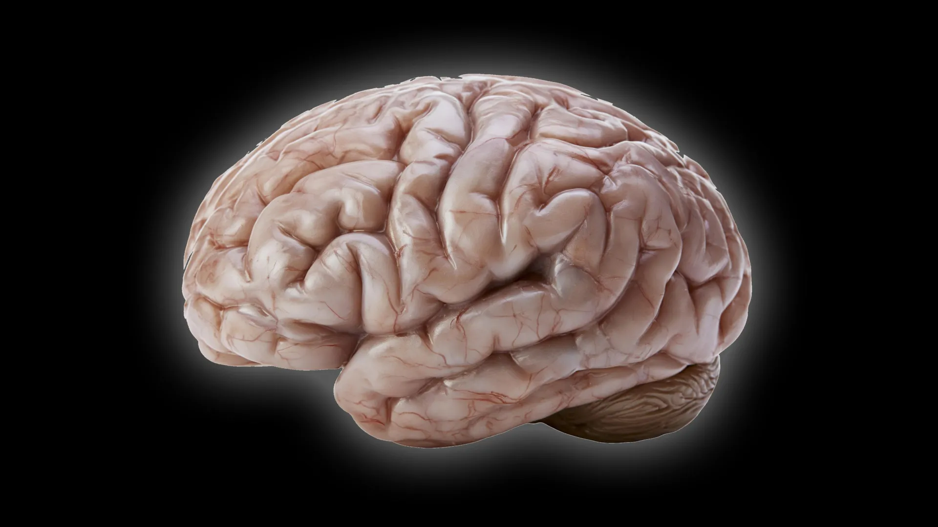
The architecture of the primate brain is one of biology’s most profound puzzles, characterized by a striking disparity between the highly convoluted, walnut-like cerebral cortex of humans and the remarkably smooth surface found in smaller species like the common marmoset (Callithrix jacchus). While evolutionary biology has long theorized that brain folding, or gyrencephaly, serves as an essential mechanism to increase surface area for cognitive processing, the precise cellular choreography that dictates these structural differences has remained elusive. A landmark study published in the journal Science Advances by researchers at the German Primate Center (DPZ) in Göttingen has now illuminated the cellular mechanisms that govern this divergence, providing a blueprint for how primate brain morphology is sculpted during early development.
The Mechanics of Cortical Folding
The human cerebral cortex is a masterpiece of biological engineering, packed with billions of neurons that facilitate complex executive function, memory, and language. This vast neural real estate is made possible by the process of gyrification, which creates the characteristic folds and grooves—gyri and sulci—that allow a large surface area to fit within the confines of the cranium. Conversely, the common marmoset, a small South American monkey increasingly vital to biomedical research, possesses a lissencephalic, or smooth, brain.
For decades, the prevailing scientific consensus suggested that the common ancestor of modern primates likely possessed a moderately sized, partially folded brain. The transition from that ancestral state to the diversity observed today—ranging from the highly folded brains of humans and great apes to the smooth brains of marmosets—suggests that evolutionary pressures favored different developmental strategies. The DPZ research team, led by Michael Heide, head of the Junior Research Group for Brain Development and Evolution, sought to move beyond descriptive anatomy to identify the specific cellular behaviors that throttle brain growth in marmosets.
A Chronology of Discovery: From Organoids to Fetal Tissue
The research effort spanned several years, utilizing advanced biotechnological models to observe the nascent stages of brain formation. The methodology relied heavily on the development of brain organoids—three-dimensional, self-organizing clusters of cells derived from stem cells that mimic the early stages of brain development in a petri dish.
By 2021, the team had successfully established a protocol to grow organoids from both human and common marmoset cells, creating a comparative model that allowed for real-time observation of neurogenesis. A critical milestone in the study occurred when the researchers successfully mapped the developmental timeline of these organoids against actual biological tissue. They determined that a 50-day-old marmoset brain organoid aligns with the developmental stage of a natural marmoset brain at 90 days. This calibration was essential, as it provided the temporal accuracy required to conclude that the differences in brain structure were not merely incidental, but the result of programmed variations in cell behavior.
The study identified that the divergence in brain architecture is not governed by a single mutation but by a cumulative "slow-down" effect. According to Lidiia Tynianskaia, one of the study’s first authors, the early stages of marmoset brain development actually resemble the onset of human brain growth. However, as the developmental process unfolds, the marmoset system engages specific biological brakes that limit the production of neurons, thereby preventing the expansion and subsequent folding of the cortical plate.
Cellular Factors Limiting Neural Expansion
The research identified three primary cellular deviations that restrict the marmoset’s neural expansion:
- Reduced Proliferation Rates: The progenitor cells responsible for generating neurons in marmosets exhibit significantly slower division cycles compared to their human counterparts. In the human brain, these cells undergo rapid, high-frequency division, creating a massive pool of neurons. In the marmoset, the extended cycle duration limits the total number of cells available to populate the cerebral cortex.
- Structural Simplification: The progenitor cells in marmosets display a less complex morphological structure. They possess fewer processes—the cellular extensions that allow for communication and structural support—which renders them less efficient at proliferating. This structural simplicity correlates directly with a diminished capacity for the massive cellular expansion required for folding.
- Truncated Growth Windows: The chronological window during which these progenitor cells remain active and capable of rapid division is significantly shorter in marmosets than in humans. By prematurely exiting the cycle of rapid proliferation, the marmoset brain effectively "shuts down" production before it can reach the volume threshold necessary to trigger the physical folding of the cortex.
César Mateo Bastidas Betancourt, co-first author of the study, emphasized that these findings demonstrate how incremental changes in timing and cellular structure can lead to monumental evolutionary outcomes. "By analyzing these cells in isolation, we were able to pinpoint that it is a combination of slower division and shorter growth windows that essentially ‘cap’ the marmoset brain’s potential size," Betancourt noted.
Validating the Miniature Model
To ensure the findings were not artifacts of laboratory culture, the DPZ team performed a rigorous validation process. They replicated key experiments using fetal brain tissue from marmosets, confirming that the organoids accurately reflected the cellular events occurring in vivo. This "dual-method" approach—combining the statistical power of organoids with the biological confirmation of fetal tissue—represents a new standard in developmental neurobiology.
The use of organoids also addressed a significant ethical and logistical hurdle in primate research. Performing longitudinal studies on primate fetal development is highly restricted and difficult due to the limited number of samples available. By successfully using organoids as a reliable proxy, the research team has opened a pathway for future studies to investigate brain development across other species without the need for extensive reliance on live animal models.
Implications for Human Health and Evolutionary Science
The impact of this study extends well beyond the taxonomy of monkeys. By mapping the regulatory mechanisms of neuron production, scientists are gaining a clearer picture of the processes that can go awry in human neurodevelopmental disorders. Conditions such as microcephaly (an abnormally small brain) and lissencephaly (a condition characterized by a smooth brain surface in humans) are fundamentally linked to the same pathways the DPZ researchers are investigating.
"Understanding the ‘off-switches’ of brain growth in primates provides us with a roadmap for identifying when those switches might be triggered prematurely in humans," the research team suggested in their summary. If a disorder causes progenitor cells to exit their division phase too early, the result is a brain with fewer neurons and impaired cognitive capacity. By identifying the specific proteins and signaling pathways that control these progenitor cells, the researchers hope to facilitate future therapies that could potentially mitigate the effects of such developmental disruptions.
Broader Context in Evolutionary Biology
From an evolutionary perspective, the marmoset’s smooth brain is not necessarily a sign of a "lesser" intelligence, but rather an example of a highly specialized evolutionary strategy. The energetic cost of maintaining a large, folded brain is immense. In the harsh, high-competition environments of the South American rainforest, the marmoset’s evolutionary trajectory may have favored a brain that is metabolically efficient, allowing for rapid reproduction and agile behavior over the high-calorie demands of a massive, folded cortex.
This research underscores the fluidity of brain evolution. It suggests that the transition from smooth to folded brains is a process that has been "tuned" over millions of years by slight adjustments in cell cycle timing. As the scientific community continues to analyze these findings, it is clear that the humble marmoset, once viewed simply as a biological curiosity, has become a key to deciphering the evolutionary trajectory of the human mind. The work conducted at the German Primate Center provides a firm, empirical foundation for future studies, ensuring that the study of brain development remains at the forefront of modern neuroscience.