
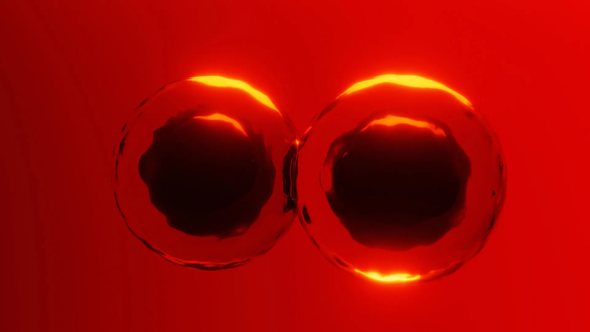
Cell division, the fundamental process by which life perpetuates, has long been understood through the lens of a contractile ring of actin filaments that constricts a cell into two. However, this widely accepted model has encountered significant challenges when explaining the earliest stages of embryonic development, particularly in oviparous (egg-laying) animals. For decades, scientists have grappled with how particularly large embryonic cells, such as those found in sharks, platypus, birds, and reptiles, manage this critical division. Now, groundbreaking research from the Brugués group at the Cluster of Excellence Physics of Life (PoL) at TUD Dresden University of Technology has unveiled a previously unrecognized mechanism that allows these colossal cells to divide without the necessity of a fully formed and continuously functional contractile ring. Published in the prestigious journal Nature, these findings not only challenge traditional biological dogma but also illuminate a sophisticated interplay between the cytoskeleton and the physical properties of the cell’s interior, operating through what the researchers term a ‘mechanical ratchet’ system.
The Enigma of Large Embryonic Cell Division
The conventional model of cell division, known as cytokinesis, relies on the formation of a contractile ring composed primarily of the protein actin. This ring assembles at the cell’s midpoint and tightens, much like a drawstring on a bag, to physically pinch the cell into two genetically identical daughter cells. This mechanism is highly effective in most somatic cells. However, the developmental context of early embryos, especially those of oviparous species, presents a formidable challenge to this paradigm.
In these animals, embryonic cells are often exceptionally large and are packed with a substantial amount of yolk. This yolk sac serves as a vital nutrient reserve for the developing embryo. Its sheer volume creates a significant geometric constraint, making it exceedingly difficult for a single, continuous actin ring to form and close effectively. The question that has perplexed researchers for years is how these oversized cells, burdened by their nutrient-rich yolk, manage to achieve the essential separation into distinct cellular units.
"With such a large yolk in the embryonic cell, there is a geometric constraint," explained Alison Kickuth, a recently graduated PhD student from the Brugués group and lead author of the study. "How does a contractile band, with loose ends, remain stable and generate enough force to divide these huge cells?" This fundamental question, at the heart of developmental biology, has now been addressed by the team’s meticulous experimental work.
Zebrafish as a Model for Unraveling Cellular Mechanics
To tackle this complex biological puzzle, the researchers turned their attention to the zebrafish embryo. Zebrafish are a favored model organism in developmental biology due to their rapid external development, optical transparency, and the presence of large, yolk-rich cells during their early embryonic stages, mirroring the conditions found in many other oviparous species. This made the zebrafish an ideal system for observing and manipulating the intricate processes of cell division.
The team employed advanced microscopy techniques and laser-based microsurgery to investigate the behavior of the actin ring. A crucial experiment involved precisely severing the actin band in developing zebrafish cells. The observation that followed was revelatory: even after being cut, the severed actin band continued to move inward. This indicated that the band was not solely anchored at its ends, as the traditional model might suggest, but was rather supported along its entire length.
Further investigation revealed the involvement of another critical component of the cytoskeleton: microtubules. These are filamentous protein structures that play diverse roles in cell shape, intracellular transport, and cell division. When the actin band was severed, the researchers observed that microtubules nearby bent and spread out. This suggested a stabilizing role for microtubules, acting as a kind of scaffolding that supported the actin ring as it attempted to constrict.
To confirm the significance of microtubules, the researchers experimentally disrupted them. They utilized two distinct methods: chemical depolymerization, which prevented the formation of new microtubules, and physical interference, where a tiny oil droplet was inserted as an obstacle to hinder microtubule assembly. In both scenarios, the actin band not only failed to constrict effectively but collapsed entirely. This provided compelling evidence that microtubules are not mere passive bystanders but actively contribute crucial mechanical support and signaling throughout the process of actin band formation and contraction.
The Dynamic Nature of Cytoplasmic Stiffness
The cytoskeleton undergoes continuous reorganization throughout the cell cycle, a highly regulated sequence of events that culminates in cell division. The cell cycle comprises distinct phases, including mitosis (M-phase), when the cell’s chromosomes are segregated, and interphase, a period of growth and DNA replication. During interphase, large microtubule structures known as asters expand from the poles of the separating chromosomes, radiating outwards into the cytoplasm. These asters are known to play a role in determining the site where the actin ring will eventually form, effectively marking the future division furrow.
The Brugués group hypothesized that these asters might influence cytoplasmic stiffness, thereby aiding in the stabilization of the actin band. To test this, they devised a method to measure the mechanical properties of the cytoplasm. This involved introducing microscopic magnetic beads into the zebrafish cells and applying a controlled magnetic force. By tracking the movement of these beads under the influence of the magnetic field, the researchers could quantitatively assess how the stiffness of the cytoplasm changed at different stages of the cell cycle.
Their findings revealed a remarkable temporal dynamic in cytoplasmic stiffness. They discovered that the cytoplasm becomes significantly stiffer during interphase. This increased stiffness is attributed to the extensive network of microtubules, including the expanding asters, which collectively form a more rigid internal scaffold. This stiffened cytoplasm then provides essential support to the actin band, preventing premature collapse. Conversely, during M-phase, when the cell is actively dividing, the cytoplasm becomes more fluid. This increased fluidity allows the actin band to move inward more readily, facilitating the physical constriction of the cell. These precisely timed shifts between stiffness and fluidity are fundamental to enabling efficient cell division, particularly in large cells.
A Mechanical Ratchet: Gradual Division Over Multiple Cycles
While the role of microtubules and dynamic cytoplasmic stiffness explained much of the process, a lingering question remained: how does the actin band avoid complete collapse during M-phase, when the cytoplasm is most fluid and the band is inherently unstable?
By meticulously tracking the ends of the actin band over extended periods, the researchers observed that while the band does become unstable and shows signs of partial retraction during M-phase contraction, it does not disintegrate. Instead, this transient instability is "rescued" by the rapid pace of the early embryonic cell cycles.
The key insight is that division is not necessarily a single, rapid event in these large cells. Instead, it is a stepwise process. As the cell progresses through M-phase, the actin band ingresses, or moves inward, to a certain extent. Then, as the cell re-enters interphase, the asters reform, the cytoplasm stiffens once again, and the actin band is re-stabilized. This cycle of partial ingression, followed by stabilization in the next interphase, repeats across several cell cycles. Each cycle contributes incrementally to the overall division process.
This gradual, cyclical advancement of the division furrow functions much like a mechanical ratchet. It allows the cell to make progress towards division without requiring a perfectly formed and continuously functional contractile ring at any single moment. Rather than achieving complete separation in one go, the cell achieves it through a series of small, controlled steps, leveraging the alternating physical states of its interior.
"The temporal ratchet mechanism fundamentally alters our view of how cytokinesis works," stated Jan Brugués, the corresponding author of the study. The researchers propose that this elegant mechanism provides a highly effective solution for very large embryonic cells that divide rapidly and cannot rely on the conventional, single-cycle contractile ring model.
Alison Kickuth elaborated on the significance of this finding, stating, "Zebrafish are a fascinating case, as cytoplasmic division in their embryonic cells is inherently unstable. To overcome this instability, their cells divide rapidly, allowing ingression of the band over several cell cycles by alternating between stability and fluidization until division is complete."
Broader Implications and Future Directions
This pioneering work introduces a new conceptual framework for understanding cell division in large, yolk-rich embryonic cells. The implications extend far beyond the zebrafish, suggesting that this ‘mechanical ratchet’ mechanism may be conserved across a wide range of egg-laying species, including many that are of significant ecological or evolutionary interest.
The study also powerfully underscores the critical importance of precisely timed changes in the material properties of the cytoplasm. These dynamic shifts in stiffness and fluidity are not merely passive consequences of the cell cycle but are active regulators of fundamental cellular processes. Understanding these mechanical cues opens new avenues for research into cell morphogenesis, tissue development, and even the cellular basis of disease.
The findings could reshape how scientists approach the study of early development across diverse organisms. By moving beyond the singular focus on the actin ring, researchers can now explore the intricate interplay of cytoskeletal elements and intracellular mechanics in a broader context. This could lead to a more nuanced and comprehensive understanding of developmental trajectories, evolutionary adaptations in cell division, and the potential for therapeutic interventions targeting cellular mechanics.
Funding and Acknowledgements
This research was generously supported by the Deutsche Forschungsgemeinschaft (DFG, German Research Foundation) through Germany’s Excellence Strategy, specifically under grant number EXC-2068-390729961, as part of the Cluster of Excellence Physics of Life at TU Dresden. Additional support was provided by the Volkswagen Foundation through its ‘Life’ grant program, under number 96827. The collaborative efforts of numerous researchers, technicians, and support staff were instrumental in achieving these significant scientific breakthroughs. The study’s publication in Nature signifies the high impact and scientific rigor of this multidisciplinary investigation.


Leave a Reply