

For decades, the clinical hallmark of Alzheimer’s disease has been the progressive erosion of memory and cognitive function. However, emerging evidence suggests that the pathological foundations of the disease are laid years, or even decades, before a patient struggles to recall a name or find their way home. One of the most persistent yet poorly understood early symptoms is the decline in the sense of smell, a condition known as hyposmia. New research published in the journal Nature Communications has identified a specific immunological mechanism that explains why this sensory deficit occurs. Scientists from the German Center for Neurodegenerative Diseases (DZNE) and Ludwig-Maximilians-Universität München (LMU) have discovered that the brain’s own immune system may mistakenly destroy the nerve fibers responsible for processing odors during the earliest stages of the disease.
The study, led by Dr. Lars Paeger and Prof. Dr. Jochen Herms, indicates that microglia—the primary immune cells of the central nervous system—play a central role in this sensory decline. Rather than protecting the brain, these cells appear to receive "eat-me" signals from hyperactive neurons, leading them to prune away essential connections between the olfactory bulb and the locus coeruleus. This discovery provides a biological roadmap for one of Alzheimer’s earliest warning signs and offers a potential window for medical intervention long before irreversible cognitive damage occurs.
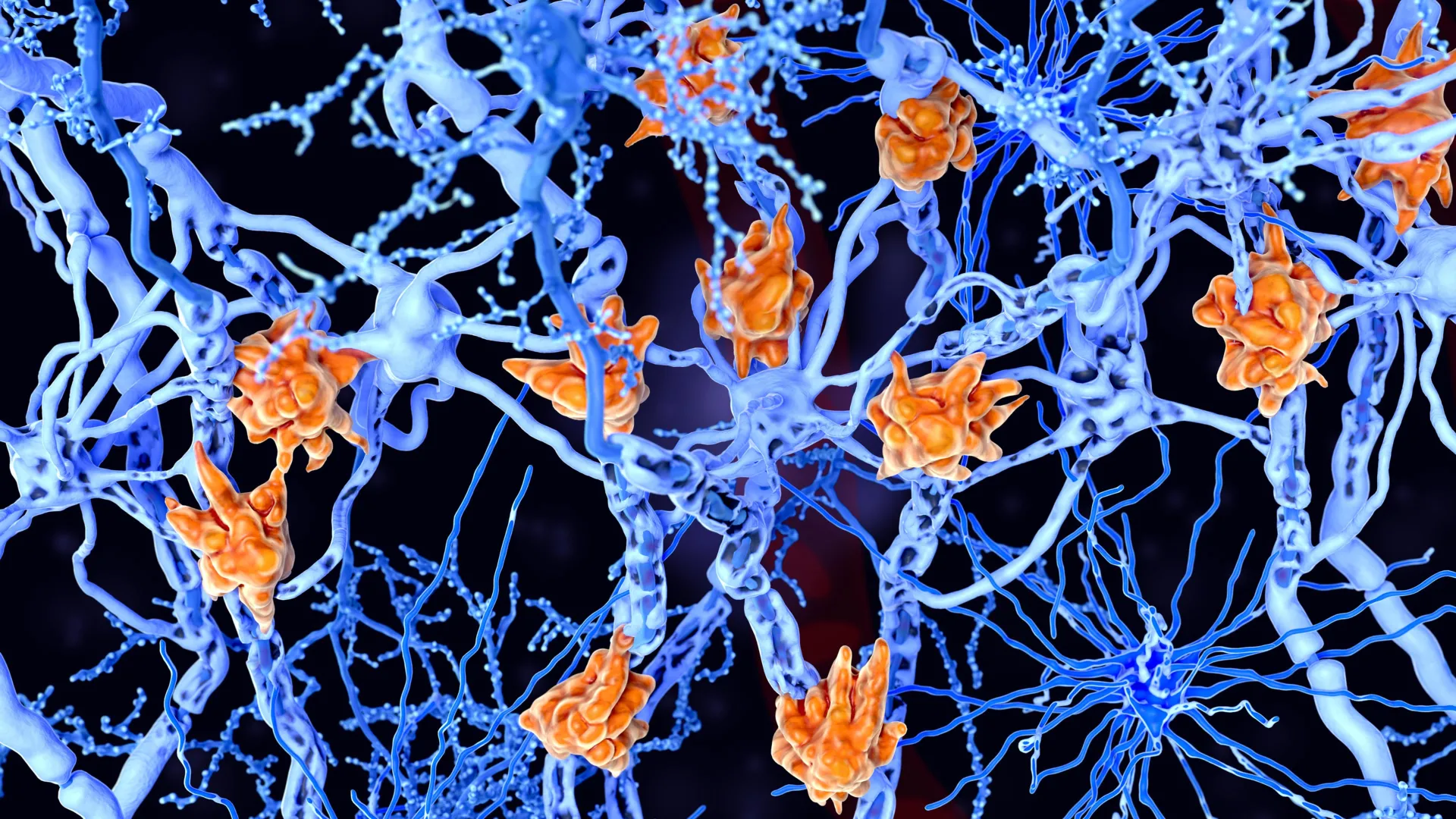
The Biological Mechanism: Microglia and the "Eat-Me" Signal
To understand why the sense of smell fades, the research team focused on the communication pathways between two distinct brain regions: the olfactory bulb and the locus coeruleus. The olfactory bulb, located in the forebrain, acts as the primary processing center for sensory data received from the nose. The locus coeruleus, situated in the brainstem, is a small but vital nucleus responsible for physiological responses to stress and panic, as well as the regulation of attention, sleep-wake cycles, and sensory processing.
Under healthy conditions, the locus coeruleus sends long nerve fibers to the olfactory bulb to modulate how we perceive scents. However, in the early stages of Alzheimer’s, these fibers undergo a localized distress response. The researchers found that the membranes of these nerve fibers experience a biochemical shift. Specifically, a fatty molecule called phosphatidylserine, which is normally tucked away on the inner side of the cell membrane, migrates to the outer surface.
In the language of the brain’s immune system, the presence of phosphatidylserine on the exterior of a cell acts as a potent "eat-me" signal. Microglia, which function as the brain’s specialized waste-removal and defense crew, detect this signal. While this process—known as synaptic pruning—is essential during brain development to remove weak or unnecessary connections, its activation in the adult Alzheimer’s brain is catastrophic. The microglia begin to break down the nerve fibers linking the locus coeruleus to the olfactory bulb, effectively "disconnecting" the brain’s ability to properly regulate and process smell.
Evidence from Multi-Modal Research
The strength of the study lies in its comprehensive methodology, which combined three distinct lines of evidence to validate the findings. The research team utilized animal models, post-mortem human brain tissue analysis, and advanced neuroimaging in living patients.
In the first phase, the scientists studied transgenic mice engineered to exhibit features of Alzheimer’s disease. These animal models allowed the researchers to observe the real-time interactions between microglia and nerve fibers. They confirmed that the "eat-me" signal appeared on the fibers before significant amyloid-beta plaques—the traditional markers of Alzheimer’s—had fully accumulated in those regions.
The second phase involved the examination of brain tissue from deceased patients who had been diagnosed with Alzheimer’s. This analysis confirmed that the structural degradation observed in mice was also present in human pathology. The researchers identified a significant reduction in the density of nerve fibers connecting the brainstem to the olfactory centers, accompanied by high concentrations of activated microglia.
Finally, the team turned to Positron Emission Tomography (PET) scanning to observe these processes in living humans. By analyzing PET scans from individuals with mild cognitive impairment (MCI) and confirmed Alzheimer’s, the researchers were able to correlate the loss of olfactory function with increased microglial activity and the degradation of the locus coeruleus pathways. This "triangulation" of data provides a high level of confidence that the mechanism identified in the lab is actively occurring in human patients.
The Role of Neuronal Hyperactivity
A critical question addressed by the study was why the "eat-me" signal appears in the first place. Dr. Lars Paeger and his colleagues found that the shift of phosphatidylserine to the outer membrane is likely triggered by neuronal hyperactivity.
In the early stages of Alzheimer’s, neurons in certain circuits often become overactive, firing more frequently than normal. This "abnormal firing" creates metabolic stress on the nerve fibers. "We assume that the shift in membrane composition is triggered by hyperactivity of the affected neurons due to Alzheimer’s disease," Paeger explained. This suggests that the immune system’s attack is not a random malfunction but a misguided response to the physiological stress of the neurons themselves. The microglia perceive the hyperactive fibers as defective or superfluous and move in to "clean up" the site, inadvertently worsening the patient’s sensory symptoms.
Historical Context and the Evolution of Alzheimer’s Research
The link between smell and neurodegeneration is not entirely new, but the "how" and "why" have remained elusive for decades. As early as the 1980s, clinicians noted that patients with Parkinson’s and Alzheimer’s often lost their sense of smell years before the onset of motor or cognitive symptoms. Historically, research focused heavily on the "Amyloid Hypothesis," which posited that the accumulation of amyloid-beta plaques was the primary driver of the disease.
However, recent failures of many plaque-clearing drugs have led the scientific community to broaden its focus. There is now a growing emphasis on neuroinflammation and the role of the brain’s innate immune system. This LMU and DZNE study fits into a new era of research that views Alzheimer’s not just as a protein-folding disorder, but as a complex immunological event. By identifying the locus coeruleus as a primary site of early degradation, the study aligns with other recent findings suggesting that this small region of the brainstem is one of the very first to show signs of tau protein accumulation, another hallmark of the disease.
Chronology of Disease Progression
The findings allow for a more detailed timeline of how Alzheimer’s manifests in the brain:
- The Pre-Clinical Phase: Neuronal hyperactivity begins in the locus coeruleus and associated pathways. Amyloid-beta begins to aggregate at microscopic levels.
- The Immunological Trigger: Hyperactivity causes the "eat-me" signal (phosphatidylserine) to flip to the outside of the nerve fiber membranes.
- Sensory Decline (Hyposmia): Microglia begin pruning the fibers connecting to the olfactory bulb. The patient may notice a decreased ability to identify common scents like coffee, roses, or citrus.
- The Prodromal Phase: As the locus coeruleus is further compromised, sleep-wake cycles and mood regulation may be affected. Mild cognitive impairment (MCI) begins to manifest.
- Clinical Alzheimer’s: Widespread plaque and tangle formation lead to significant cell death in the hippocampus and cortex, resulting in the well-known symptoms of memory loss and disorientation.
Implications for Early Diagnosis and New Therapies
The most significant implication of this research is its potential to improve early diagnosis. Currently, Alzheimer’s is often diagnosed only after significant cognitive decline has occurred, at which point much of the brain’s hardware has already been lost.
"Our findings could pave the way for the early identification of patients at risk of developing Alzheimer’s," says Prof. Dr. Joachim Herms. If a standardized, highly sensitive smell test could be coupled with PET scans or biomarkers that detect microglial activity, doctors could identify at-risk individuals in the "pre-clinical" stage.
This early window is vital for the effectiveness of new treatments. Recently, the medical community has seen the emergence of amyloid-beta antibodies, such as lecanemab (Leqembi) and donanemab. These drugs are designed to clear plaques from the brain, but clinical trials have shown they are most effective when administered at the very beginning of the disease. By the time a patient has severe memory loss, the removal of plaques may do little to restore function. Identifying patients during the "smell-loss" phase would allow these therapies to be deployed when they have the highest chance of slowing the disease’s progression.
Expert Reactions and Future Directions
The scientific community has reacted with cautious optimism to the DZNE and LMU findings. Independent researchers note that while the study identifies a clear mechanism in mice and correlated evidence in humans, the challenge remains to develop a non-invasive way to measure this specific microglial "pruning" in a standard clinical setting.
Future research is expected to investigate whether drugs that inhibit the "eat-me" signal or modulate microglial activity could prevent the destruction of these nerve fibers. If the immune system’s attack can be halted, it might not only preserve the sense of smell but also protect the locus coeruleus’s other vital functions, potentially delaying the onset of more severe cognitive symptoms.
Furthermore, this study highlights the importance of the locus coeruleus as a "hub" for neurodegeneration. Because this region influences everything from blood flow to the sleep-wake cycle, protecting its integrity could have cascading benefits for overall brain health in aging populations.
Conclusion
The study published in Nature Communications marks a pivotal shift in our understanding of the sensory symptoms of Alzheimer’s disease. By shifting the focus from the "trash" (plaques) to the "janitors" (microglia), Dr. Paeger, Prof. Herms, and their team have uncovered a dynamic process of immune-mediated destruction that precedes memory loss.
As the global population ages, the prevalence of Alzheimer’s is expected to rise significantly, placing an immense burden on healthcare systems. Insights into the immunological roots of early symptoms like smell loss offer a glimmer of hope. They suggest that the disease is not a sudden collapse, but a gradual process that provides us with early warnings—if only we know how to listen to, or in this case, smell them. The transition from reactive treatment to proactive intervention depends on such breakthroughs, moving medicine closer to a future where Alzheimer’s can be managed or even halted before it takes its toll on the human mind.


Leave a Reply